Triceps Surae: Difference between revisions
Shejza Mino (talk | contribs) No edit summary |
Shejza Mino (talk | contribs) No edit summary |
||
| Line 11: | Line 11: | ||
The anatomical details of the individual muscles are listed below but in general, the main function of the triceps surae is to perform plantar flexion of the foot at the ankle joint, allowing the heel to elevate against gravity. This results in generation of the propulsion force required for actions such as walking, jumping or leaping. Furthermore, the gastrocnemius plays a subtle role in producing leg flexion at the knee joint. | The anatomical details of the individual muscles are listed below but in general, the main function of the triceps surae is to perform plantar flexion of the foot at the ankle joint, allowing the heel to elevate against gravity. This results in generation of the propulsion force required for actions such as walking, jumping or leaping. Furthermore, the gastrocnemius plays a subtle role in producing leg flexion at the knee joint. | ||
Additionally, there is a vertical center of gravity line anterior to the ankle that results in a "natural tendency" for the human body to lean forward. To counteract this natural pull due to the effect of gravity, the soleus muscle plays a significant role in generating a force posterior to the ankle joint in order for the human body to maintain stability. This is attributed to the "fact" that the soleus muscle is constantly contracting during periods of walking and standing. | |||
== Anatomy == | === Anatomy === | ||
The muscles that comprise the triceps surae (gastrocnemius, soleus and plantaris) are part of the posterosuperficial compartment of the calf <ref name=":0" />. | The muscles that comprise the triceps surae (gastrocnemius, soleus and plantaris) are part of the posterosuperficial compartment of the calf <ref name=":0" />. | ||
| Line 44: | Line 44: | ||
== Clinical Significance == | == Clinical Significance == | ||
The triceps surae muscle is connected to the S1 nerve root. Compression of this nerve root may occur with a disc herniation or vertebral fracture. Signs & symptoms that are typical include gluteal and posterior leg pain as well as a diminished or absent achilles tendon reflex (S1 reflex) <ref name=":4">Sendic G. Triceps surae muscle [Internet]. Kenhub. 2020 [cited 8 January 2021]. Available from: https://www.kenhub.com/en/library/anatomy/triceps-surae-muscle</ref>. | |||
= | Functional limitation of the triceps surae muscle unit is common due to being outweighed by the muscles of the anterior leg. What is know as "talipes calcaneus' is present in these patients (''talipes calcaneus'' is defined as "a deformity due to weakness or absence of the calf muscles in which the axis of the calcaneus becomes vertically oriented")<ref name=":4" /><ref>www.dictionary.com. 2021. Definition of talipes calcaneus | Dictionary.com. [online] Available at: <https://[https://www.dictionary.com/browse/talipes-calcaneus www.dictionary.com/browse/talipes-calcaneus]> [Accessed 14 January 2021].</ref>. | ||
The Achilles tendon is the most powerful tendon in the human body, with a load bearing capacity up to one tonne. Ruptures of the tendon are typically associated with prior damage. Specifically, microtrauma to the tendon disrupts it's vascular supply, decreasing the overall strength of the tendon. The Achilles tendon is supplied relatively poorly ~3-5 cm proximal to it's insertion onto the calcaneus, making this region more vulnerable <ref name=":4" />. | |||
The region about 3 to 5 centimeters proximal to the tendon insertion is particularly vulnerable as it is relatively poorly supplied already. In adolescents, rupture of the Achilles tendon is often accompanied with a fracture of the calcaneus bone <ref name=":4" />. | |||
== Resources == | == Resources == | ||
Revision as of 04:01, 1 February 2021
Original Editor - Shejza Mino
Top Contributors - Shejza Mino, Kim Jackson and Lucinda hampton
Introduction[edit | edit source]
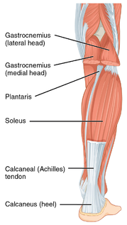
The triceps surae, also known as the muscle of the calf, is constructed by the soleus, the two-headed (medial & lateral) gastrocnemius and the plantaris muscles [1]. Research suggests that contracture of the triceps surae is correlated with various conditions that affect the forefoot and midfoot, therefore consideration of these muscles is valuable when evaluating and managing such conditions [2].
Function[edit | edit source]
The anatomical details of the individual muscles are listed below but in general, the main function of the triceps surae is to perform plantar flexion of the foot at the ankle joint, allowing the heel to elevate against gravity. This results in generation of the propulsion force required for actions such as walking, jumping or leaping. Furthermore, the gastrocnemius plays a subtle role in producing leg flexion at the knee joint.
Additionally, there is a vertical center of gravity line anterior to the ankle that results in a "natural tendency" for the human body to lean forward. To counteract this natural pull due to the effect of gravity, the soleus muscle plays a significant role in generating a force posterior to the ankle joint in order for the human body to maintain stability. This is attributed to the "fact" that the soleus muscle is constantly contracting during periods of walking and standing.
Anatomy[edit | edit source]
The muscles that comprise the triceps surae (gastrocnemius, soleus and plantaris) are part of the posterosuperficial compartment of the calf [1].
The soleus muscle and both heads of the gastrocnemius muscle, fuse to insert onto the calcaneus (heel bone) through the achilles tendon (also known as the calcaneal tendon). This structure is considered to be the strongest tendon in the human body [2].
It has been proposed that the plantaris muscle can either attach onto the calcaneus independently from the achilles tendon or may form a portion of the achilles tendon, accompany the tendon to insert onto the calcaneus. Interestingly, the plantaris tendon is commonly intact with rupture of the achilles tendon [3]. This plantaris muscle is proposed to be absent in 7-20% of the population [3].
- Innervation - tibial nerve, nerve roots S1, S2 [1].
- Blood Supply - posterior tibial artery
Gastrocnemius[edit | edit source]
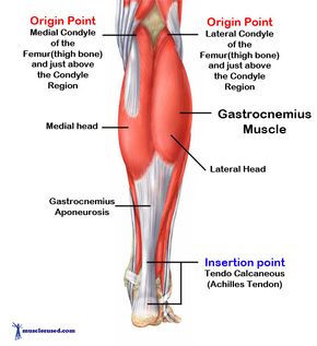
The gastrocnemius muscle consists of a lateral and medial head at it's origin, and make up the superficial portion of the triceps surae. Both the medial and lateral head insert into the calcaneus bone.
The gastrocnemius traverses three joints including the knee, ankle and subtalar joints [4].
- Origin - posterosuperior region of the corresponding femoral condyle, specifically:
- Medial head: arises from the posterior aspect of the femur, posterior to the medial supracondylar ridge and adductor tubercle. The medial head is thicker and wider than the lateral head [5].
- Lateral head: arises from the lateral aspect of the lateral femoral condyle. Proximal, we well as posterior, to the lateral epicondyle [5].
Additionally, the lateral and medial heads of the gastrocnemius muscle further attachments from the oblique popliteal ligament as well as from the posterior capsule of the knee joint.
- Function - the gastrocnemius muscle produces flexion of the leg at the knee joint and plantarflexor of the foot at the talocrural joint (ankle mortise). Further, the gastrocnemius is most effective when the knee is in an extended position and the ankle plantarflexed [5].
Soleus[edit | edit source]
The soleus muscle is situated deep to the gastrocnemius and crosses two joints including the ankle and subtalar joints [5].
- Origin - posterior aspect of the fibular head, medial border of the tibia (soleal line) and the interosseous membrane [5].
- Function - soleus muscle is shown to be most effective with the ankle in plantarflexion (similar to the gastrocnemius muscle) but with the knee in flexion (opposite of the gastrocnemius) [5].
Plantaris[edit | edit source]
The plantaris muscle is made up of a small muscle that has a slim and long tendon, ranging from 7 to 13 cm long [3].
- Origin - superior & medial to the lateral head of the gastrocnemius on the lateral supracondylar femoral line. It also has an origin in the posterior aspect of the knee from the oblique popliteal ligament [3].
- Function - similar action as the gastrocnemius muscle however the plantaris muscle is considered an insignficant knee flexor and ankle plantarflexor [3]. Because the plantaris muscle consists of a "high density of muscle spindles", it is considered to be an "organ of proprioceptive function" for the more powerful plantarflexor muscles [3].
Clinical Significance[edit | edit source]
The triceps surae muscle is connected to the S1 nerve root. Compression of this nerve root may occur with a disc herniation or vertebral fracture. Signs & symptoms that are typical include gluteal and posterior leg pain as well as a diminished or absent achilles tendon reflex (S1 reflex) [6].
Functional limitation of the triceps surae muscle unit is common due to being outweighed by the muscles of the anterior leg. What is know as "talipes calcaneus' is present in these patients (talipes calcaneus is defined as "a deformity due to weakness or absence of the calf muscles in which the axis of the calcaneus becomes vertically oriented")[6][7].
The Achilles tendon is the most powerful tendon in the human body, with a load bearing capacity up to one tonne. Ruptures of the tendon are typically associated with prior damage. Specifically, microtrauma to the tendon disrupts it's vascular supply, decreasing the overall strength of the tendon. The Achilles tendon is supplied relatively poorly ~3-5 cm proximal to it's insertion onto the calcaneus, making this region more vulnerable [6].
The region about 3 to 5 centimeters proximal to the tendon insertion is particularly vulnerable as it is relatively poorly supplied already. In adolescents, rupture of the Achilles tendon is often accompanied with a fracture of the calcaneus bone [6].
Resources[edit | edit source]
- bulleted list
- x
or
- numbered list
- x
References[edit | edit source]
- ↑ 1.0 1.1 1.2 Keith LM, Arthur FD, Anne MR. Clinically oriented anatomy. Clinically oriented anatomy. 2006.
- ↑ 2.0 2.1 Dalmau-Pastor M, Fargues-Polo B, Casanova-Martínez D, Vega J, Golanó P. Anatomy of the triceps surae: a pictorial essay. Foot and ankle clinics. 2014 Dec 1;19(4):603-35.
- ↑ 3.0 3.1 3.2 3.3 3.4 3.5 Spina AA. The plantaris muscle: anatomy, injury, imaging, and treatment. The Journal of the Canadian Chiropractic Association. 2007 Jul;51(3):158.
- ↑ Cohen JC. Anatomy and biomechanical aspects of the gastrocsoleus complex. Foot and ankle clinics. 2009 Dec 1;14(4):617-26.
- ↑ 5.0 5.1 5.2 5.3 5.4 5.5 Cohen JC. Anatomy and biomechanical aspects of the gastrocsoleus complex. Foot and ankle clinics. 2009 Dec 1;14(4):617-26.
- ↑ 6.0 6.1 6.2 6.3 Sendic G. Triceps surae muscle [Internet]. Kenhub. 2020 [cited 8 January 2021]. Available from: https://www.kenhub.com/en/library/anatomy/triceps-surae-muscle
- ↑ www.dictionary.com. 2021. Definition of talipes calcaneus | Dictionary.com. [online] Available at: <https://www.dictionary.com/browse/talipes-calcaneus> [Accessed 14 January 2021].


