Syndesmotic Ankle Sprains: Difference between revisions
No edit summary |
No edit summary |
||
| Line 1: | Line 1: | ||
<div class="noeditbox">Welcome to [[Texas State University Evidence-based Practice Project|Texas State University's Evidence-based Practice project space]]. This is a wiki created by and for the students in the Doctor of Physical Therapy program at Texas State University - San Marcos. Please do not edit unless you are involved in this project, but please come back in the near future to check out new information!!</div> <div class="editorbox"> | <div class="noeditbox">Welcome to [[Texas State University Evidence-based Practice Project|Texas State University's Evidence-based Practice project space]]. This is a wiki created by and for the students in the Doctor of Physical Therapy program at Texas State University - San Marcos. Please do not edit unless you are involved in this project, but please come back in the near future to check out new information!!</div><div class="editorbox"> | ||
'''Original Editors''' | '''Original Editors''' | ||
'''Lead Editors''' - Your name will be added here if you are a lead editor on this page. [[Physiopedia:Editors|Read more.]] | '''Lead Editors''' - Your name will be added here if you are a lead editor on this page. [[Physiopedia:Editors|Read more.]] | ||
</div> | </div> | ||
== Search Strategy == | == Search Strategy == | ||
[http://www.ncbi.nlm.nih.gov/pubmed? | Search Strategies: [http://www.ncbi.nlm.nih.gov/pubmed PubMed],[http://www.cochrane.org/ Cochrane],[http://www.ebscohost.com/cinahl/ CINAHL],[http://www.nlm.nih.gov/medlineplus/ MedlinePlus], [http://scholar.google.com/schhp?ie=UTF-8&hl=en&tab=ws Google Scholar]<br> | ||
Key Terms: syndesmotic ankle sprain, high ankle sprain, atheletes, physical therapy | |||
Time frame: June 12, 2011- July 20, 2011<br><br> | |||
== Definition/Description == | == Definition/Description == | ||
A syndesmotic, or ‘high’ ankle sprain is one that involves the ligaments binding the distal tibia and fibula. Injuries can occur with any ankle motion, but the most common motions are extreme external rotation or dorsiflexion of the Talus. The dome of the Talus is wider in the anterior than in the posterior, and these movements force apart the medial and lateral aspects of the mortise, respectively the tibial and fibular malleoli. Sufficient distraction of the distal fibula from the tibia can cause strain or rupture of one or more of the following ligaments: the anterior tibiofibular ligament, superficial posterior inferior tibiofibular ligament, transverse tibiofibular ligament.<ref name="Norkus">Norkus S, Floyd RT, The Anatomy and Mechanisms of Synedesmotic Ankle Sprains. J Athletic Training. 2001;36:68-73</ref> Rupture injuries also commonly present with concomitant fractures of the either malleolus (lateral being more common) or proximal fibular spiral fracture known as a Maissonneuve fracture. <ref name="Clanton">5. Clanton T. Syndesmotic ankle sprains in athletes. International SportMed Journal. 2003;4 (4):1-10.
6</ref><br> | A syndesmotic, or ‘high’ ankle sprain is one that involves the ligaments binding the distal tibia and fibula. Injuries can occur with any ankle motion, but the most common motions are extreme external rotation or dorsiflexion of the Talus. The dome of the Talus is wider in the anterior than in the posterior, and these movements force apart the medial and lateral aspects of the mortise, respectively the tibial and fibular malleoli. Sufficient distraction of the distal fibula from the tibia can cause strain or rupture of one or more of the following ligaments: the anterior tibiofibular ligament, superficial posterior inferior tibiofibular ligament, transverse tibiofibular ligament.<ref name="Norkus">Norkus S, Floyd RT, The Anatomy and Mechanisms of Synedesmotic Ankle Sprains. J Athletic Training. 2001;36:68-73</ref> Rupture injuries also commonly present with concomitant fractures of the either malleolus (lateral being more common) or proximal fibular spiral fracture known as a Maissonneuve fracture. <ref name="Clanton">5. Clanton T. Syndesmotic ankle sprains in athletes. International SportMed Journal. 2003;4 (4):1-10.
6</ref><br> | ||
== Epidemiology/Etiology == | == Epidemiology/Etiology == | ||
<br>Syndesmotic ankle sprains commonly occur to athletes participating in American football and downhill skiing. Football injuries are usually a result of forced external rotation of the foot while the athlete is prone, as in at the bottom of the pile. The injuries can also result from a blow to the lateral knee while the foot is planted and dorsiflexed, resulting in an eversion or external rotation moment at the talocrural joint. <br>In downhill ski racing, the boot does not allow dorsi- or plantar-flexion movement, which can result in excessive allowance of talocrural external rotation and injury to the anterior or posterior tibiofibular ligament, as well as the interosseous membrane.<br>Research has documented that syndesmosis injuries account for 1-11% of all injuries <ref name="Norkus" />. Incidence among professional American football players is reported to be much high, up to 29% as documented by Boytim et al (<ref name="91">Boytim MJ, Fischer DA, Neumann L. Syndesmotic ankle sprains. Am J Sports Med. 1991;19:294–298</ref>)<br><br> | <br>Syndesmotic ankle sprains commonly occur to athletes participating in American football and downhill skiing. Football injuries are usually a result of forced external rotation of the foot while the athlete is prone, as in at the bottom of the pile. The injuries can also result from a blow to the lateral knee while the foot is planted and dorsiflexed, resulting in an eversion or external rotation moment at the talocrural joint. <br>In downhill ski racing, the boot does not allow dorsi- or plantar-flexion movement, which can result in excessive allowance of talocrural external rotation and injury to the anterior or posterior tibiofibular ligament, as well as the interosseous membrane.<br>Research has documented that syndesmosis injuries account for 1-11% of all injuries <ref name="Norkus" />. Incidence among professional American football players is reported to be much high, up to 29% as documented by Boytim et al (<ref name="91">Boytim MJ, Fischer DA, Neumann L. Syndesmotic ankle sprains. Am J Sports Med. 1991;19:294–298</ref>)<br><br> | ||
== Characteristics/Clinical Presentation == | == Characteristics/Clinical Presentation == | ||
Observationally the Syndesmotic will show significantly less swelling than a lateral ankle sprain, as well as demonstrate a loss of full plantar flexion and an inability to bear weight<sup>2</sup>. Ecchymosis may appear several days post-injury due to the injury of the intereosseuos membrane. A difficulty or inability to toe walk are often noted. History includes chronic pain, prolonged recovery, recurrent sprains, and the formation of heterotopic ossification within the interosseous membrane. <ref name="Nussbaum et al.">Nussbaum E, Hosea T, Sieler S, Incremona B, Kessler D. Prospective Evaulation of Syndesmotic Ankle Sprains Without Diastasis. The American Journal of Sports Medicine. 2001;29 (1): 31-35.</ref>The most common MOI is when the foot is in external rotation with excessive dorsiflexion<ref name="Lin">Lin CF, Gross ML, Weinhold P. Ankle syndesmosis injuries: anatomy, biomechanics, mechanism of injury, and clinical guidelines for diagnosis and intervention. JOSPT. 2006; 36(6):372-384. 3. Fincher L. Early Recognition of Syndesmotic Ankle Sprain. Athletic Therapy Today. 1999; 42-42.</ref>. <br> | Observationally the Syndesmotic will show significantly less swelling than a lateral ankle sprain, as well as demonstrate a loss of full plantar flexion and an inability to bear weight<sup>2</sup>. Ecchymosis may appear several days post-injury due to the injury of the intereosseuos membrane. A difficulty or inability to toe walk are often noted. History includes chronic pain, prolonged recovery, recurrent sprains, and the formation of heterotopic ossification within the interosseous membrane. <ref name="Nussbaum et al.">Nussbaum E, Hosea T, Sieler S, Incremona B, Kessler D. Prospective Evaulation of Syndesmotic Ankle Sprains Without Diastasis. The American Journal of Sports Medicine. 2001;29 (1): 31-35.</ref>The most common MOI is when the foot is in external rotation with excessive dorsiflexion<ref name="Lin">Lin CF, Gross ML, Weinhold P. Ankle syndesmosis injuries: anatomy, biomechanics, mechanism of injury, and clinical guidelines for diagnosis and intervention. JOSPT. 2006; 36(6):372-384. 3. Fincher L. Early Recognition of Syndesmotic Ankle Sprain. Athletic Therapy Today. 1999; 42-42.</ref>. <br> | ||
== Differential Diagnosis == | == Differential Diagnosis == | ||
Because of the occult nature of the high ankle sprain during clinical evaluation it is important to rule out pathologies with a similar MOI. First and foremost an x-ray should be taken to rule out fx of the tibia, fibula and/or the talus <ref name="Norwig">Norwig JA. Injuring Management Update: Recognizing and Rehabilitating the High Ankle Sprain. Professional Jrnl of Athletic Trng &amp;amp;amp;amp;amp;amp; Ther. 1998, July; 12-13.</ref>. Secondly, the clinician should address concerns of a lateral ankle sprain as the mechanism of injury are between the two injuries are very similar. Norwig writes “Syndesmotic ankle sprains can usually be distinguished from inversion ankle sprains by a history of an external rotation component.” Other possible pathologies are medial ankle sprain, compartment syndrome, severe joint laxity<ref name="Clanton" />, severe contusion, dystrophic calcification, infection or tumor. These pathologies should be preferentially ruled out before tx of a syndesmotic ankle sprain begins. <br> | Because of the occult nature of the high ankle sprain during clinical evaluation it is important to rule out pathologies with a similar MOI. First and foremost an x-ray should be taken to rule out fx of the tibia, fibula and/or the talus <ref name="Norwig">Norwig JA. Injuring Management Update: Recognizing and Rehabilitating the High Ankle Sprain. Professional Jrnl of Athletic Trng &amp;amp;amp;amp;amp;amp;amp; Ther. 1998, July; 12-13.</ref>. Secondly, the clinician should address concerns of a lateral ankle sprain as the mechanism of injury are between the two injuries are very similar. Norwig writes “Syndesmotic ankle sprains can usually be distinguished from inversion ankle sprains by a history of an external rotation component.” Other possible pathologies are medial ankle sprain, compartment syndrome, severe joint laxity<ref name="Clanton" />, severe contusion, dystrophic calcification, infection or tumor. These pathologies should be preferentially ruled out before tx of a syndesmotic ankle sprain begins. <br> | ||
== Outcome Measures == | == Outcome Measures == | ||
| Line 28: | Line 32: | ||
*[[Foot and Ankle Disability Index|Foot and Ankle Disability Index]] (FADI) | *[[Foot and Ankle Disability Index|Foot and Ankle Disability Index]] (FADI) | ||
<br> | <br> | ||
== Examination == | == Examination == | ||
| Line 43: | Line 47: | ||
*'''<u>Special Test</u>:''' | *'''<u>Special Test</u>:''' | ||
<br>'''[[Image:ER of the foot.JPG|thumb|left | <br>'''[[Image:ER of the foot.JPG|thumb|left]]1. External Rotation''' '''Test''' (Kleiger’s Test) <ref name="Starkley">Starkley C, Ryan J. Orthopedic &amp;amp;amp;amp;amp;amp;amp;amp; Athletic Injury Evaluation Handbook. Phyladelphia, PA: Davis Company; 2003</ref><br> | ||
- Determines rotator damage to the deltoid ligament or the distal tibiofibular syndesmosis. | - Determines rotator damage to the deltoid ligament or the distal tibiofibular syndesmosis. | ||
- Performed by having the knee flexed by 90 degrees with the ankle in neutral position and appyling an external rotational force to the affected foot and ankle. <ref name="Norwig" /> | - Performed by having the knee flexed by 90 degrees with the ankle in neutral position and appyling an external rotational force to the affected foot and ankle. <ref name="Norwig" /> | ||
-(+) test: Pain in the anterolateral ankle. An indicator of deltoid ligament damage would be if there is a displacement of the talus away from the medial malleolus. | -(+) test: Pain in the anterolateral ankle. An indicator of deltoid ligament damage would be if there is a displacement of the talus away from the medial malleolus. | ||
- Interrater kappa= 0.75 (best)- <ref name="Alonso">Alonso A, Khoury L, Adams R. Clinical tests for Ankle Syndesmosis Injury: reliability and prediction of return to function. JOSPT. 1998; 27(4):276-84</ref>; <ref name="Clanton" /><br> | - Interrater kappa= 0.75 (best)- <ref name="Alonso">Alonso A, Khoury L, Adams R. Clinical tests for Ankle Syndesmosis Injury: reliability and prediction of return to function. JOSPT. 1998; 27(4):276-84</ref>; <ref name="Clanton" /><br> | ||
<br> | <br> | ||
[[Image:Squeeze Test.JPG|thumb|right | [[Image:Squeeze Test.JPG|thumb|right]]<br> | ||
'''2. [http://www.physio-pedia.com/index.php5?title=Squeeze_Test Squeeze Test]'''- separation of the tibia and fibula <ref name="Starkley" /><br>- Identifies a fibular fracture or syndesmosis sprain. | '''2. [http://www.physio-pedia.com/index.php5?title=Squeeze_Test Squeeze Test]'''- separation of the tibia and fibula <ref name="Starkley" /><br>- Identifies a fibular fracture or syndesmosis sprain. | ||
| Line 63: | Line 67: | ||
-(+) test: Pain will be reproduced along the fibular shaft if it’s a fibular fracture and the distal tibiofibular jt for syndesmosis sprain.<br>- interrater= 0.5 (moderate). <ref name="Alonso" /> | -(+) test: Pain will be reproduced along the fibular shaft if it’s a fibular fracture and the distal tibiofibular jt for syndesmosis sprain.<br>- interrater= 0.5 (moderate). <ref name="Alonso" /> | ||
<br>'''3. Cotton Test''' (Magee)<br>- Assess for syndesmosis instability with diastasis.<br>- Performed: steadying the distal leg with one hand while grasping the plantar heel with the opposite hand and moving the heel directly from side to side <ref name="Clanton" /><br>- (+) test: Any lateral translation would indicate syndesmotic instability <ref name="Magee">Magee D. Orthopedic Physical Assessment. 4th Edition. St. Louis, Missourie: Saunders Elsevier; 2006</ref><br> | <br>'''3. Cotton Test''' (Magee)<br>- Assess for syndesmosis instability with diastasis.<br>- Performed: steadying the distal leg with one hand while grasping the plantar heel with the opposite hand and moving the heel directly from side to side <ref name="Clanton" /><br>- (+) test: Any lateral translation would indicate syndesmotic instability <ref name="Magee">Magee D. Orthopedic Physical Assessment. 4th Edition. St. Louis, Missourie: Saunders Elsevier; 2006</ref><br> | ||
== Medical Management <br> | == Medical Management <br> == | ||
Imaging is still considered the diagnostic standard and should be sought as quickly as possible to rule out any expected fractures and to aid in restoring normal anatomy. A one millimeter lateral displacement of the fibula results in a reduction in the available of area of tibiotalar contact in weight bearing by 42%! One can easily see how such a “minor” yet misdiagnosed injury can lead to a lifetime of chronic sprains. Plain films are the bare minimum suggested, but due to the complexity of the structures and tissues, a CT scan is recommended for bony detail, MRIs give an accurate picture of the ligamentous injury, and they are the imaging gold standard for this injury. They are surpassed in accuracy only by arthroscopy 2. Images should be done in a bilateral fashion to better determine an injury from a natural joint gap or overlap. | Imaging is still considered the diagnostic standard and should be sought as quickly as possible to rule out any expected fractures and to aid in restoring normal anatomy. A one millimeter lateral displacement of the fibula results in a reduction in the available of area of tibiotalar contact in weight bearing by 42%! One can easily see how such a “minor” yet misdiagnosed injury can lead to a lifetime of chronic sprains. Plain films are the bare minimum suggested, but due to the complexity of the structures and tissues, a CT scan is recommended for bony detail, MRIs give an accurate picture of the ligamentous injury, and they are the imaging gold standard for this injury. They are surpassed in accuracy only by arthroscopy 2. Images should be done in a bilateral fashion to better determine an injury from a natural joint gap or overlap. | ||
<br> | <br> | ||
== Physical Therapy Management <br> == | == Physical Therapy Management <br> == | ||
[[Image:Calf Stretch Composite.jpg|thumb|center|569x256px]] 1.) Calf Stretch with Step 2.) Calf Strengthening Exercise 3.) Lunging Calf Stretch<br> | [[Image:Calf Stretch Composite.jpg|thumb|center|569x256px|Calf Stretch Composite.jpg]] 1.) Calf Stretch with Step 2.) Calf Strengthening Exercise 3.) Lunging Calf Stretch<br> | ||
<u>'''Goals:<br>'''</u> | <u>'''Goals:<br>'''</u> | ||
| Line 86: | Line 90: | ||
*'''<u></u>'''Surgeon/PT’s recommended weightbearing protocol | *'''<u></u>'''Surgeon/PT’s recommended weightbearing protocol | ||
*Caution against vigorous physical activity until full weight bearing and dynamic balance has normalized. <ref name="Dressendorfer" /> | *Caution against vigorous physical activity until full weight bearing and dynamic balance has normalized. <ref name="Dressendorfer" /> | ||
*Gait training with crutches or boot/brace <ref name="Clanton" /> | *Gait training with crutches or boot/brace <ref name="Clanton" /> | ||
*Fall risk <ref name="Dressendorfer" /> | *Fall risk <ref name="Dressendorfer" /> | ||
| Line 94: | Line 98: | ||
<u>'''Assistive Devices:<br>'''</u> | <u>'''Assistive Devices:<br>'''</u> | ||
*Crutches- must be maintained until normal, pain free gait is obtained <ref name="Fincher" /> | *Crutches- must be maintained until normal, pain free gait is obtained <ref name="Fincher" /> | ||
*Walking boot or stirrup brace for unstable injuries <ref name="Clanton" /> | *Walking boot or stirrup brace for unstable injuries <ref name="Clanton" /> | ||
| Line 101: | Line 105: | ||
<u>'''Modalities:<br>'''</u> | <u>'''Modalities:<br>'''</u> | ||
*RICE (rest, ice, compression, elevation) initially for 15 min 3x a day.<ref name="Harvard" />. However, Bleakley et al suggested that there is little evidence to support the use of RICE, although it is a widely accepted treatment | *RICE (rest, ice, compression, elevation) initially for 15 min 3x a day.<ref name="Harvard" />. However, Bleakley et al suggested that there is little evidence to support the use of RICE, although it is a widely accepted treatment | ||
*Non-steroidal anti-inflammatory drugs and comfrey ointment have been shown to improve short-term recovery following acute ankle sprain. <ref name="Dolan" /> | *Non-steroidal anti-inflammatory drugs and comfrey ointment have been shown to improve short-term recovery following acute ankle sprain. <ref name="Dolan" /> | ||
| Line 115: | Line 119: | ||
Ex: Single-leg stance, disk or balance pad training, aquatic therapy | Ex: Single-leg stance, disk or balance pad training, aquatic therapy | ||
*Progress to jogging, cycling, agility, jumping, and sport-specific drills. <ref name="Dressendorfer" /> | *Progress to jogging, cycling, agility, jumping, and sport-specific drills. <ref name="Dressendorfer" /> | ||
*Modify exercises to avoid hyperdorsiflexion (stresses mortise joint), subtalar eversion, and loaded external rotation. <ref name="Fincher" /> | *Modify exercises to avoid hyperdorsiflexion (stresses mortise joint), subtalar eversion, and loaded external rotation. <ref name="Fincher" /> | ||
| Line 122: | Line 126: | ||
<u>'''Manual Therapy:<br>'''</u> | <u>'''Manual Therapy:<br>'''</u> | ||
*Passive accessory movement of the talocrural and subtalar joints and passive stretching may help stiffness. <ref name="Dressendorfer" /> | *Passive accessory movement of the talocrural and subtalar joints and passive stretching may help stiffness. <ref name="Dressendorfer" /> | ||
*Green et al: those subjects who used RICE with manual therapy were more likely to reach this normal ROM within the first 2 weeks of the ankle sprain than those who received RICE alone. <ref name="Green">Green T, Refshauge K, Crosbie J, Adams A (2001) A randomized controlled trial of a passive accessory joint mobilization on acute ankle inversion sprains. Physical Therapy 81: 984-994</ref> | *Green et al: those subjects who used RICE with manual therapy were more likely to reach this normal ROM within the first 2 weeks of the ankle sprain than those who received RICE alone. <ref name="Green">Green T, Refshauge K, Crosbie J, Adams A (2001) A randomized controlled trial of a passive accessory joint mobilization on acute ankle inversion sprains. Physical Therapy 81: 984-994</ref> | ||
*Collins et al: Subjects showed immediate ROM gains when Mulligan’s movement with mobilization was applied in the sub-acute sprains after ankle sprains and in patients with recurrent sprains. <ref name="Collins">Collins N, Teys P, Vicenzino B (2004) The initial effects of a Mulligan’s movilisation with movement technique on dorsiflexion and pain in subacute ankle sprains. Manual Therapy 9: 77-82.</ref> | *Collins et al: Subjects showed immediate ROM gains when Mulligan’s movement with mobilization was applied in the sub-acute sprains after ankle sprains and in patients with recurrent sprains. <ref name="Collins">Collins N, Teys P, Vicenzino B (2004) The initial effects of a Mulligan’s movilisation with movement technique on dorsiflexion and pain in subacute ankle sprains. Manual Therapy 9: 77-82.</ref> | ||
| Line 134: | Line 138: | ||
|} | |} | ||
<br><br>''' **The recovery for Syndesmotic Ankle Sprain is often twice that of a typical ankle sprain!**<br>'''[[Image:Theraband Ankle Composite.jpg|thumb|center|320x240px | <br><br>''' **The recovery for Syndesmotic Ankle Sprain is often twice that of a typical ankle sprain!**<br>'''[[Image:Theraband Ankle Composite.jpg|thumb|center|320x240px]] 1.) Theraband Plantarflexion 2.) Theraband Dorsiflexion | ||
3.) Theraband Inversion 4.) Theraband Eversion | 3.) Theraband Inversion 4.) Theraband Eversion | ||
== Key Research == | == Key Research == | ||
| Line 144: | Line 148: | ||
<br>Dressendorfer R. Clinical Review: Syndesmotic Ankle Sprain. CINAHL. 2009. | <br>Dressendorfer R. Clinical Review: Syndesmotic Ankle Sprain. CINAHL. 2009. | ||
<br>Bleakley CM, McDonough SM, MacAuley DC. Some conservative strategies are effective when added to controlled mobilization with external support after acute ankle sprain: a systematic review. Aust J Physiother. 2008;54(1):7-20.<br> | <br>Bleakley CM, McDonough SM, MacAuley DC. Some conservative strategies are effective when added to controlled mobilization with external support after acute ankle sprain: a systematic review. Aust J Physiother. 2008;54(1):7-20.<br> | ||
== Resources<br> | == Resources<br> == | ||
== Clinical Bottom Line == | == Clinical Bottom Line == | ||
add text here <br> | add text here <br> | ||
== Recent Related Research (from [http://www.ncbi.nlm.nih.gov/pubmed/ Pubmed]) == | == Recent Related Research (from [http://www.ncbi.nlm.nih.gov/pubmed/ Pubmed]) == | ||
| Line 156: | Line 160: | ||
see tutorial on [[Adding PubMed Feed|Adding PubMed Feed]] | see tutorial on [[Adding PubMed Feed|Adding PubMed Feed]] | ||
<div class="researchbox"> | <div class="researchbox"> | ||
<rss>http://eutils.ncbi.nlm.nih.gov/entrez/eutils/erss.cgi?rss_guid=1VcNN1T1QuuXznqrgsIapdOm02wv-M2MCXquWkXfxO9Milxsyd|charset=UTF-8|short|max=10</rss> | <rss>http://eutils.ncbi.nlm.nih.gov/entrez/eutils/erss.cgi?rss_guid=1VcNN1T1QuuXznqrgsIapdOm02wv-M2MCXquWkXfxO9Milxsyd|charset=UTF-8|short|max=10</rss> | ||
</div> | </div> | ||
== References == | == References == | ||
see [[Adding References|adding references tutorial]]. | see [[Adding References|adding references tutorial]]. | ||
<references /><references /> | <references /><references /> | ||
| | ||
[[Category:Texas_State_University_EBP_Project]] | [[Category:Texas_State_University_EBP_Project]] | ||
Revision as of 15:32, 20 July 2011
Original Editors
Lead Editors - Your name will be added here if you are a lead editor on this page. Read more.
Search Strategy[edit | edit source]
Search Strategies: PubMed,Cochrane,CINAHL,MedlinePlus, Google Scholar<br>
Key Terms: syndesmotic ankle sprain, high ankle sprain, atheletes, physical therapy
Time frame: June 12, 2011- July 20, 2011
Definition/Description[edit | edit source]
A syndesmotic, or ‘high’ ankle sprain is one that involves the ligaments binding the distal tibia and fibula. Injuries can occur with any ankle motion, but the most common motions are extreme external rotation or dorsiflexion of the Talus. The dome of the Talus is wider in the anterior than in the posterior, and these movements force apart the medial and lateral aspects of the mortise, respectively the tibial and fibular malleoli. Sufficient distraction of the distal fibula from the tibia can cause strain or rupture of one or more of the following ligaments: the anterior tibiofibular ligament, superficial posterior inferior tibiofibular ligament, transverse tibiofibular ligament.[1] Rupture injuries also commonly present with concomitant fractures of the either malleolus (lateral being more common) or proximal fibular spiral fracture known as a Maissonneuve fracture. [2]
Epidemiology/Etiology[edit | edit source]
Syndesmotic ankle sprains commonly occur to athletes participating in American football and downhill skiing. Football injuries are usually a result of forced external rotation of the foot while the athlete is prone, as in at the bottom of the pile. The injuries can also result from a blow to the lateral knee while the foot is planted and dorsiflexed, resulting in an eversion or external rotation moment at the talocrural joint.
In downhill ski racing, the boot does not allow dorsi- or plantar-flexion movement, which can result in excessive allowance of talocrural external rotation and injury to the anterior or posterior tibiofibular ligament, as well as the interosseous membrane.
Research has documented that syndesmosis injuries account for 1-11% of all injuries [1]. Incidence among professional American football players is reported to be much high, up to 29% as documented by Boytim et al (Cite error: Invalid <ref> tag; name cannot be a simple integer. Use a descriptive title)
Characteristics/Clinical Presentation[edit | edit source]
Observationally the Syndesmotic will show significantly less swelling than a lateral ankle sprain, as well as demonstrate a loss of full plantar flexion and an inability to bear weight2. Ecchymosis may appear several days post-injury due to the injury of the intereosseuos membrane. A difficulty or inability to toe walk are often noted. History includes chronic pain, prolonged recovery, recurrent sprains, and the formation of heterotopic ossification within the interosseous membrane. [3]The most common MOI is when the foot is in external rotation with excessive dorsiflexion[4].
Differential Diagnosis[edit | edit source]
Because of the occult nature of the high ankle sprain during clinical evaluation it is important to rule out pathologies with a similar MOI. First and foremost an x-ray should be taken to rule out fx of the tibia, fibula and/or the talus [5]. Secondly, the clinician should address concerns of a lateral ankle sprain as the mechanism of injury are between the two injuries are very similar. Norwig writes “Syndesmotic ankle sprains can usually be distinguished from inversion ankle sprains by a history of an external rotation component.” Other possible pathologies are medial ankle sprain, compartment syndrome, severe joint laxity[2], severe contusion, dystrophic calcification, infection or tumor. These pathologies should be preferentially ruled out before tx of a syndesmotic ankle sprain begins.
Outcome Measures[edit | edit source]
Examination[edit | edit source]
Hx and MOI: see clinical presentation
- Observation/Gait analysis: Check for discrepancies
- Palpation:tenderness proximally over the anterior tibiofibular ligament and proximal along the interosseous membrane [6]
- Palpate the medial and lateral malleoli for exidence of a fracture [7]
- Fibula needs to be palpated from distal to proximal, including the proximal tibiofibular joint to rule out Maissoneuve’s fracture . [2]
- Distal Pulses: Ensure pedal pulses are present [7]
- Girth Measurements: Notable swelling of ankle do Figure 8 girth measurements (Andrews et. el)
- Special Test:
1. External Rotation Test (Kleiger’s Test) [8]
- Determines rotator damage to the deltoid ligament or the distal tibiofibular syndesmosis.
- Performed by having the knee flexed by 90 degrees with the ankle in neutral position and appyling an external rotational force to the affected foot and ankle. [5]
-(+) test: Pain in the anterolateral ankle. An indicator of deltoid ligament damage would be if there is a displacement of the talus away from the medial malleolus.
- Interrater kappa= 0.75 (best)- [9]; [2]
2. Squeeze Test- separation of the tibia and fibula [8]
- Identifies a fibular fracture or syndesmosis sprain.
- Performed by squeezing the tibia and fibula together above the injury.
-(+) test: Pain will be reproduced along the fibular shaft if it’s a fibular fracture and the distal tibiofibular jt for syndesmosis sprain.
- interrater= 0.5 (moderate). [9]
3. Cotton Test (Magee)
- Assess for syndesmosis instability with diastasis.
- Performed: steadying the distal leg with one hand while grasping the plantar heel with the opposite hand and moving the heel directly from side to side [2]
- (+) test: Any lateral translation would indicate syndesmotic instability [10]
Medical Management
[edit | edit source]
Imaging is still considered the diagnostic standard and should be sought as quickly as possible to rule out any expected fractures and to aid in restoring normal anatomy. A one millimeter lateral displacement of the fibula results in a reduction in the available of area of tibiotalar contact in weight bearing by 42%! One can easily see how such a “minor” yet misdiagnosed injury can lead to a lifetime of chronic sprains. Plain films are the bare minimum suggested, but due to the complexity of the structures and tissues, a CT scan is recommended for bony detail, MRIs give an accurate picture of the ligamentous injury, and they are the imaging gold standard for this injury. They are surpassed in accuracy only by arthroscopy 2. Images should be done in a bilateral fashion to better determine an injury from a natural joint gap or overlap.
Physical Therapy Management
[edit | edit source]
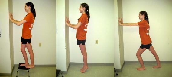
1.) Calf Stretch with Step 2.) Calf Strengthening Exercise 3.) Lunging Calf Stretch
Goals:
- First two weeks: ROM, decrease pain and swelling, protect ligaments against further injury
- Week 3 and onward: Restore normal ROM, strengthen ligaments and supporting muscles, training to improve endurance and balance [11]
- Most important long-term goal is to prevent re-injury! [12]
Patient Education:
- Surgeon/PT’s recommended weightbearing protocol
- Caution against vigorous physical activity until full weight bearing and dynamic balance has normalized. [7]
- Gait training with crutches or boot/brace [2]
- Fall risk [7]
Assistive Devices:
- Crutches- must be maintained until normal, pain free gait is obtained [6]
- Walking boot or stirrup brace for unstable injuries [2]
Modalities:
- RICE (rest, ice, compression, elevation) initially for 15 min 3x a day.[11]. However, Bleakley et al suggested that there is little evidence to support the use of RICE, although it is a widely accepted treatment
- Non-steroidal anti-inflammatory drugs and comfrey ointment have been shown to improve short-term recovery following acute ankle sprain. [13]
Therapeutic Exercise/ Neuromuscular Re-education:
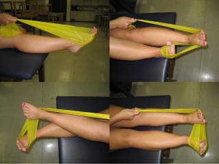
- First two weeks: AROM flexion, ankle alphabet, dorsiflexion/plantarflexion and inversion/eversion with theraband
- Weeks 3-4: Standing Stretch, seated dorsiflexion stretch with theraband, double heel raise progressing to single heel raise, and dorsiflexion stretching on step stool [11]
- Progressive weightbearing and treadmill gait training to promote normal gait pattern. ([6], [7])
- Neuromuscular Re-education: ankle proprioception, postural reflexes and balance
Ex: Single-leg stance, disk or balance pad training, aquatic therapy
- Progress to jogging, cycling, agility, jumping, and sport-specific drills. [7]
- Modify exercises to avoid hyperdorsiflexion (stresses mortise joint), subtalar eversion, and loaded external rotation. [6]
Manual Therapy:
- Passive accessory movement of the talocrural and subtalar joints and passive stretching may help stiffness. [7]
- Green et al: those subjects who used RICE with manual therapy were more likely to reach this normal ROM within the first 2 weeks of the ankle sprain than those who received RICE alone. [14]
- Collins et al: Subjects showed immediate ROM gains when Mulligan’s movement with mobilization was applied in the sub-acute sprains after ankle sprains and in patients with recurrent sprains. [15]
- Landrum et al: Reported that one 30-second A-P talocrural joint mob immediately increased ankle dorsiflexion ROM after prolonged mobilization. [16]
**The recovery for Syndesmotic Ankle Sprain is often twice that of a typical ankle sprain!**
1.) Theraband Plantarflexion 2.) Theraband Dorsiflexion
3.) Theraband Inversion 4.) Theraband Eversion
Key Research[edit | edit source]
Clanton T. Syndesmotic ankle sprains in athletes. International SportMed Journal. 2003;4 (4):1-10.
Dressendorfer R. Clinical Review: Syndesmotic Ankle Sprain. CINAHL. 2009.
Bleakley CM, McDonough SM, MacAuley DC. Some conservative strategies are effective when added to controlled mobilization with external support after acute ankle sprain: a systematic review. Aust J Physiother. 2008;54(1):7-20.
Resources
[edit | edit source]
Clinical Bottom Line[edit | edit source]
add text here
Recent Related Research (from Pubmed)[edit | edit source]
see tutorial on Adding PubMed Feed
Failed to load RSS feed from http://eutils.ncbi.nlm.nih.gov/entrez/eutils/erss.cgi?rss_guid=1VcNN1T1QuuXznqrgsIapdOm02wv-M2MCXquWkXfxO9Milxsyd|charset=UTF-8|short|max=10: Error parsing XML for RSS
References[edit | edit source]
see adding references tutorial.
- ↑ 1.0 1.1 Norkus S, Floyd RT, The Anatomy and Mechanisms of Synedesmotic Ankle Sprains. J Athletic Training. 2001;36:68-73
- ↑ 2.0 2.1 2.2 2.3 2.4 2.5 2.6 5. Clanton T. Syndesmotic ankle sprains in athletes. International SportMed Journal. 2003;4 (4):1-10.
6 Cite error: Invalid
<ref>tag; name "Clanton" defined multiple times with different content - ↑ Nussbaum E, Hosea T, Sieler S, Incremona B, Kessler D. Prospective Evaulation of Syndesmotic Ankle Sprains Without Diastasis. The American Journal of Sports Medicine. 2001;29 (1): 31-35.
- ↑ Lin CF, Gross ML, Weinhold P. Ankle syndesmosis injuries: anatomy, biomechanics, mechanism of injury, and clinical guidelines for diagnosis and intervention. JOSPT. 2006; 36(6):372-384. 3. Fincher L. Early Recognition of Syndesmotic Ankle Sprain. Athletic Therapy Today. 1999; 42-42.
- ↑ 5.0 5.1 Norwig JA. Injuring Management Update: Recognizing and Rehabilitating the High Ankle Sprain. Professional Jrnl of Athletic Trng &amp;amp;amp;amp;amp;amp; Ther. 1998, July; 12-13.
- ↑ 6.0 6.1 6.2 6.3 Fincher L. Early Recognition of Syndesmotic Ankle Sprain. Athletic Therapy Today. 1999; 42-42.
- ↑ 7.0 7.1 7.2 7.3 7.4 7.5 7.6 Dressendorfer R. Clinical Review: Syndesmotic Ankle Sprain. CINAHL. 2009.
- ↑ 8.0 8.1 Starkley C, Ryan J. Orthopedic &amp;amp;amp;amp;amp;amp;amp; Athletic Injury Evaluation Handbook. Phyladelphia, PA: Davis Company; 2003
- ↑ 9.0 9.1 Alonso A, Khoury L, Adams R. Clinical tests for Ankle Syndesmosis Injury: reliability and prediction of return to function. JOSPT. 1998; 27(4):276-84
- ↑ Magee D. Orthopedic Physical Assessment. 4th Edition. St. Louis, Missourie: Saunders Elsevier; 2006
- ↑ 11.0 11.1 11.2 Recovering from an ankle sprain. Harvard Women’s Health Watch. 2007 Feb; 14(6):4-6.
- ↑ Bleakley CM, McDonough SM, MacAuley DC. Some conservative strategies are effective when added to controlled mobilization with external support after acute ankle sprain: a systematic review. Aust J Physiother. 2008;54(1):7-20.
- ↑ Cite error: Invalid
<ref>tag; no text was provided for refs namedDolan - ↑ Green T, Refshauge K, Crosbie J, Adams A (2001) A randomized controlled trial of a passive accessory joint mobilization on acute ankle inversion sprains. Physical Therapy 81: 984-994
- ↑ Collins N, Teys P, Vicenzino B (2004) The initial effects of a Mulligan’s movilisation with movement technique on dorsiflexion and pain in subacute ankle sprains. Manual Therapy 9: 77-82.
- ↑ Landrum EL, Kelln BM,, Parente WR, Ingersoll CD, Hertel J. Immediate effects of anterior-to-posterior talocrural joint mobilization after prolonged ankle immobilization: a preliminary study. J Man Manip Ther 2008;16(2):100-105


