Search results
- <div class="primal-licensebox"> [http://www.primalpictures.com/ Primal Pictures] owns the copyright to this image.336 bytes (47 words) - 10:51, 19 April 2013
- [https://www.primalpictures.com/adoptions/physio-pedia/ Primal Pictures] owns the copyright to this image. Primal Pictures have given Physiopedia special permission to use this image in Physiopedia375 bytes (51 words) - 16:32, 20 April 2021
File:Cervical plexus phrenic nerve Primal.png [https://www.primalpictures.com/adoptions/physio-pedia/ Visit Primal Pictures] The copyright for this image is owned by Primal Pictures. They have given permission to use this image exclusively in Physiopedia. P(660 × 660 (358 KB)) - 18:43, 14 June 2021
File:Healthy alveoli Primal.png Visit Primal Pictures The copyright for this image is owned by Primal Pictures. They have given permission to use this image exclusively in Physiopedia. P(990 × 990 (1.04 MB)) - 23:58, 6 December 2020
File:Pneumonia Primal.png Visit Primal Pictures The copyright for this image is owned by Primal Pictures. They have given permission to use this image exclusively in Physiopedia. P(990 × 990 (1.05 MB)) - 00:02, 7 December 2020
File:Elbow joint Primal.png Visit Primal Pictures The copyright for this image is owned by Primal Pictures. They have given permission to use this image exclusively in Physiopedia. P(660 × 660 (235 KB)) - 00:11, 7 December 2020
File:Causes of pneumonia.png ...tm_medium=Banner_Physiopedia&utm_campaign=Pneumonia#products| Visit Primal Pictures] The copyright for this image is owned by Primal Pictures. They have given permission to use this image exclusively in Physiopedia. P(454 × 259 (80 KB)) - 19:30, 21 June 2020
File:Symptoms of pneumonia.png ...tm_medium=Banner_Physiopedia&utm_campaign=Pneumonia#products| Visit Primal Pictures] The copyright for this image is owned by Primal Pictures. They have given permission to use this image exclusively in Physiopedia. P(708 × 412 (176 KB)) - 19:35, 21 June 2020
File:Treatment of pneumonia.png ...tm_medium=Banner_Physiopedia&utm_campaign=Pneumonia#products| Visit Primal Pictures] The copyright for this image is owned by Primal Pictures. They have given permission to use this image exclusively in Physiopedia. P(708 × 412 (171 KB)) - 20:02, 21 June 2020
File:Lung tissue.png ...tm_medium=Banner_Physiopedia&utm_campaign=Pneumonia#products| Visit Primal Pictures] The copyright for this image is owned by Primal Pictures. They have given permission to use this image exclusively in Physiopedia. P(660 × 660 (276 KB)) - 17:59, 21 June 2020
File:What is pneumonia.png ...tm_medium=Banner_Physiopedia&utm_campaign=Pneumonia#products| Visit Primal Pictures] The copyright for this image is owned by Primal Pictures. They have given permission to use this image exclusively in Physiopedia. P(637 × 378 (141 KB)) - 18:09, 21 June 2020
File:Healthy alveoli and bronchiole.png ...tm_medium=Banner_Physiopedia&utm_campaign=Pneumonia#products| Visit Primal Pictures] The copyright for this image is owned by Primal Pictures. They have given permission to use this image exclusively in Physiopedia. P(990 × 990 (610 KB)) - 19:00, 21 June 2020
File:Respiratory membrane.png ...tm_medium=Banner_Physiopedia&utm_campaign=Pneumonia#products| Visit Primal Pictures] The copyright for this image is owned by Primal Pictures. They have given permission to use this image exclusively in Physiopedia. P(660 × 660 (659 KB)) - 19:20, 21 June 2020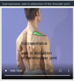
File:Supraspinatus aids in abduction.png ...Physiopedia&utm_campaign=Supraspinatus_tendinopathy#products| Visit Primal Pictures] The copyright for this image is owned by Primal Pictures. They have given permission to use this image exclusively in Physiopedia. P(261 × 286 (85 KB)) - 01:35, 21 June 2020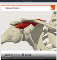
File:Rotator cuff tear.png ...Physiopedia&utm_campaign=Supraspinatus_tendinopathy#products| Visit Primal Pictures] The copyright for this image is owned by Primal Pictures. They have given permission to use this image exclusively in Physiopedia. P(561 × 587 (190 KB)) - 01:50, 21 June 2020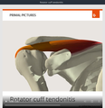
File:Rotator cuff tendonitis.png ...Physiopedia&utm_campaign=Supraspinatus_tendinopathy#products| Visit Primal Pictures] The copyright for this image is owned by Primal Pictures. They have given permission to use this image exclusively in Physiopedia. P(570 × 589 (168 KB)) - 01:57, 21 June 2020
File:Illustration of SLAP II lesion.png ...Physiopedia&utm_campaign=Supraspinatus_tendinopathy#products| Visit Primal Pictures] The copyright for this image is owned by Primal Pictures. They have given permission to use this image exclusively in Physiopedia. P(990 × 990 (516 KB)) - 02:13, 21 June 2020
File:Illustration of sublabral foramen.png ...Physiopedia&utm_campaign=Supraspinatus_tendinopathy#products| Visit Primal Pictures] The copyright for this image is owned by Primal Pictures. They have given permission to use this image exclusively in Physiopedia. P(990 × 990 (510 KB)) - 02:20, 21 June 2020
File:Tear of supraspinatus tendon.png ...Physiopedia&utm_campaign=Supraspinatus_tendinopathy#products| Visit Primal Pictures] The copyright for this image is owned by Primal Pictures. They have given permission to use this image exclusively in Physiopedia. P(868 × 568 (432 KB)) - 02:24, 21 June 2020
File:Calcification of rotator cuff.png ...Physiopedia&utm_campaign=Supraspinatus_tendinopathy#products| Visit Primal Pictures] The copyright for this image is owned by Primal Pictures. They have given permission to use this image exclusively in Physiopedia. P(704 × 575 (450 KB)) - 02:28, 21 June 2020
File:Muscles scapular region anterior aspect.png ...Physiopedia&utm_campaign=Supraspinatus_tendinopathy#products| Visit Primal Pictures] The copyright for this image is owned by Primal Pictures. They have given permission to use this image exclusively in Physiopedia. P(660 × 660 (485 KB)) - 00:54, 21 June 2020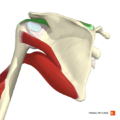
File:Supraspinatus muscle.png ...Physiopedia&utm_campaign=Supraspinatus_tendinopathy#products| Visit Primal Pictures] The copyright for this image is owned by Primal Pictures. They have given permission to use this image exclusively in Physiopedia. P(660 × 660 (257 KB)) - 01:04, 21 June 2020
File:Primalpictures logo.png Logo for Primal Pictures {{Primal AnatomyTV}}(1,128 × 362 (261 KB)) - 16:26, 4 December 2020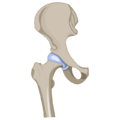
File:HipJoint.png The copyright for this image is owned by Primal Pictures. They have given permission to use this image exclusively in Physiopedia. P {{primal}}(640 × 640 (51 KB)) - 15:35, 2 March 2022
File:Flexor Carpi Radialis.png The copyright for this image is owned by Primal Pictures. They have given permission to use this image exclusively in Physiopedia. P {{primal}}(600 × 600 (116 KB)) - 13:07, 24 February 2022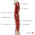
File:Deep extensor muscles of the forearm Primal.png The copyright for this image is owned by Primal Pictures. They have given permission to use this image exclusively in Physiopedia. P {{Primal AnatomyTV}}(660 × 660 (208 KB)) - 00:24, 7 December 2020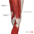
File:Intermediate muscles of the knee posterolateral aspect Primal.png The copyright for this image is owned by Primal Pictures. They have given permission to use this image exclusively in Physiopedia. P {{Primal AnatomyTV}}(660 × 660 (342 KB)) - 21:39, 7 December 2020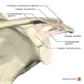
File:Acromioclavicular separation type 1 Primal.png The copyright for this image is owned by Primal Pictures. They have given permission to use this image exclusively in Physiopedia. P {{Primal AnatomyTV}}(660 × 660 (274 KB)) - 01:08, 8 December 2020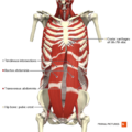
File:Anterior abdominal wall deep muscles Primal.png The copyright for this image is owned by Primal Pictures. They have given permission to use this image exclusively in Physiopedia. P {{Primal AnatomyTV}}(660 × 660 (412 KB)) - 21:47, 8 December 2020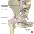
File:Ligaments of the hip joint anterior aspect Primal.png The copyright for this image is owned by Primal Pictures. They have given permission to use this image exclusively in Physiopedia. P {{Primal AnatomyTV}}(660 × 660 (361 KB)) - 20:29, 7 December 2020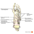
File:Ligaments of the foot dorsal aspect Primal.png The copyright for this image is owned by Primal Pictures. They have given permission to use this image exclusively in Physiopedia. P {{Primal AnatomyTV}}(660 × 660 (230 KB)) - 00:20, 8 December 2020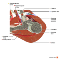
File:Axial section of the shoulder joint Primal.png The copyright for this image is owned by Primal Pictures. They have given permission to use this image exclusively in Physiopedia. P {{Primal AnatomyTV}}(990 × 990 (707 KB)) - 01:15, 8 December 2020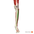
File:Tibialis anterior Primal.png The copyright for this image is owned by Primal Pictures. They have given permission to use this image exclusively in Physiopedia. P {{Primal AnatomyTV}}(660 × 660 (118 KB)) - 15:16, 10 January 2021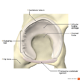
File:Radial fibrillated labral tear Primal.png The copyright for this image is owned by Primal Pictures. They have given permission to use this image exclusively in Physiopedia. P {{Primal AnatomyTV}}(990 × 990 (539 KB)) - 20:56, 7 December 2020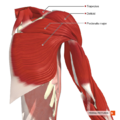
File:Muscles connecting the upper limb to the trunk anterior aspect Primal.png The copyright for this image is owned by Primal Pictures. They have given permission to use this image exclusively in Physiopedia. P {{Primal AnatomyTV}}(660 × 660 (485 KB)) - 00:58, 8 December 2020
File:Posterior disc hernia sagittal view Primal.png The copyright for this image is owned by Primal Pictures. They have given permission to use this image exclusively in Physiopedia. P {{Primal AnatomyTV}}(990 × 990 (660 KB)) - 15:21, 8 December 2020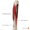
File:Deep flexor muscles of the forearm Primal.png The copyright for this image is owned by Primal Pictures. They have given permission to use this image exclusively in Physiopedia. P {{Primal AnatomyTV}}(660 × 660 (189 KB)) - 00:25, 7 December 2020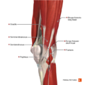
File:Intermediate muscles of the knee posteromedial aspect Primal.png The copyright for this image is owned by Primal Pictures. They have given permission to use this image exclusively in Physiopedia. P {{Primal AnatomyTV}}(660 × 660 (355 KB)) - 21:40, 7 December 2020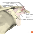
File:Acromioclavicular separation type 2 Primal.png The copyright for this image is owned by Primal Pictures. They have given permission to use this image exclusively in Physiopedia. P {{Primal AnatomyTV}}(660 × 660 (287 KB)) - 01:09, 8 December 2020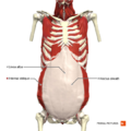
File:Anterior abdominal wall intermediate muscles Primal.png The copyright for this image is owned by Primal Pictures. They have given permission to use this image exclusively in Physiopedia. P {{Primal AnatomyTV}}(660 × 660 (345 KB)) - 21:48, 8 December 2020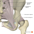
File:Ligaments of the hip joint posterior aspect Primal.png The copyright for this image is owned by Primal Pictures. They have given permission to use this image exclusively in Physiopedia. P {{Primal AnatomyTV}}(660 × 660 (395 KB)) - 20:40, 7 December 2020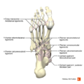
File:Ligaments of the foot plantar aspect Primal.png The copyright for this image is owned by Primal Pictures. They have given permission to use this image exclusively in Physiopedia. P {{Primal AnatomyTV}}(660 × 660 (234 KB)) - 00:22, 8 December 2020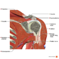
File:Coronal section of the tendon of long head of biceps Primal.png The copyright for this image is owned by Primal Pictures. They have given permission to use this image exclusively in Physiopedia. P {{Primal AnatomyTV}}(990 × 990 (900 KB)) - 01:17, 8 December 2020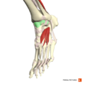
File:Capsule of ankle joint Primal.png The copyright for this image is owned by Primal Pictures. They have given permission to use this image exclusively in Physiopedia. P {{Primal AnatomyTV}}(660 × 660 (206 KB)) - 15:18, 10 January 2021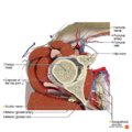
File:Axial section of the hip joint Primal.png The copyright for this image is owned by Primal Pictures. They have given permission to use this image exclusively in Physiopedia. P {{Primal AnatomyTV}}(990 × 990 (881 KB)) - 20:57, 7 December 2020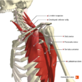
File:Muscles connecting the upper limb to the trunk deep muscles Primal.png The copyright for this image is owned by Primal Pictures. They have given permission to use this image exclusively in Physiopedia. P {{Primal AnatomyTV}}(660 × 660 (464 KB)) - 00:59, 8 December 2020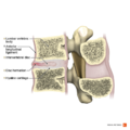
File:Anterior disc hernia sagittal view Primal.png The copyright for this image is owned by Primal Pictures. They have given permission to use this image exclusively in Physiopedia. P {{Primal AnatomyTV}}(990 × 990 (660 KB)) - 15:23, 8 December 2020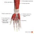
File:Muscles of the hand anterior aspect Primal.png The copyright for this image is owned by Primal Pictures. They have given permission to use this image exclusively in Physiopedia. P {{Primal AnatomyTV}}(660 × 660 (265 KB)) - 00:26, 7 December 2020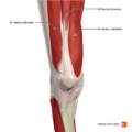
File:Muscles of the knee anterior aspect Primal.png The copyright for this image is owned by Primal Pictures. They have given permission to use this image exclusively in Physiopedia. P {{Primal AnatomyTV}}(660 × 660 (289 KB)) - 21:41, 7 December 2020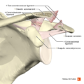
File:Acromioclavicular separation type 3 Primal.png The copyright for this image is owned by Primal Pictures. They have given permission to use this image exclusively in Physiopedia. P {{Primal AnatomyTV}}(660 × 660 (300 KB)) - 01:10, 8 December 2020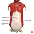
File:Anterior abdominal wall superficial muscles Primal.png The copyright for this image is owned by Primal Pictures. They have given permission to use this image exclusively in Physiopedia. P {{Primal AnatomyTV}}(660 × 660 (350 KB)) - 21:49, 8 December 2020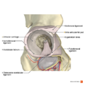
File:Acetabulum Primal.png The copyright for this image is owned by Primal Pictures. They have given permission to use this image exclusively in Physiopedia. P {{Primal AnatomyTV}}(990 × 990 (588 KB)) - 20:42, 7 December 2020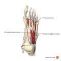
File:Muscles of the foot dorsal aspect Primal.png The copyright for this image is owned by Primal Pictures. They have given permission to use this image exclusively in Physiopedia. P {{Primal AnatomyTV}}(660 × 660 (223 KB)) - 00:22, 8 December 2020
File:Sagittal section of the rotator cuff muscles Primal.png The copyright for this image is owned by Primal Pictures. They have given permission to use this image exclusively in Physiopedia. P {{Primal AnatomyTV}}(990 × 990 (905 KB)) - 01:17, 8 December 2020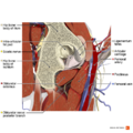
File:Sagittal section of the structures of the hip 1 Primal.png The copyright for this image is owned by Primal Pictures. They have given permission to use this image exclusively in Physiopedia. P {{Primal AnatomyTV}}(990 × 990 (1 MB)) - 20:58, 7 December 2020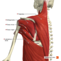
File:Muscles connecting the upper limb to the trunk posterior aspect Primal.png The copyright for this image is owned by Primal Pictures. They have given permission to use this image exclusively in Physiopedia. P {{Primal AnatomyTV}}(660 × 660 (487 KB)) - 00:59, 8 December 2020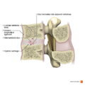
File:Intervertebral disc hernia into adjacent bodies sagittal view Primal.png The copyright for this image is owned by Primal Pictures. They have given permission to use this image exclusively in Physiopedia. P {{Primal AnatomyTV}}(990 × 990 (603 KB)) - 15:24, 8 December 2020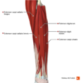
File:Superficial and intermediate extensor muscles of the forearm Primal.png The copyright for this image is owned by Primal Pictures. They have given permission to use this image exclusively in Physiopedia. P {{Primal AnatomyTV}}(660 × 660 (236 KB)) - 00:27, 7 December 2020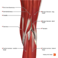
File:Superficial muscles of the knee posterior aspect Primal.png The copyright for this image is owned by Primal Pictures. They have given permission to use this image exclusively in Physiopedia. P {{Primal AnatomyTV}}(660 × 660 (372 KB)) - 21:42, 7 December 2020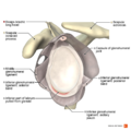
File:Illustration of Bankart lesion Primal.png The copyright for this image is owned by Primal Pictures. They have given permission to use this image exclusively in Physiopedia. P {{Primal AnatomyTV}}(990 × 990 (500 KB)) - 01:10, 8 December 2020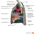
File:Right lung medial surface Primal.png The copyright for this image is owned by Primal Pictures. They have given permission to use this image exclusively in Physiopedia. P {{Primal AnatomyTV}}(660 × 660 (324 KB)) - 21:54, 8 December 2020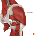
File:Intermediate muscles of the gluteal region Primal.png The copyright for this image is owned by Primal Pictures. They have given permission to use this image exclusively in Physiopedia. P {{Primal AnatomyTV}}(660 × 660 (507 KB)) - 20:44, 7 December 2020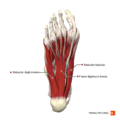
File:Plantar muscles of the foot first layer Primal.png The copyright for this image is owned by Primal Pictures. They have given permission to use this image exclusively in Physiopedia. P {{Primal AnatomyTV}}(660 × 660 (239 KB)) - 00:23, 8 December 2020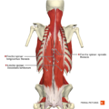
File:Muscles of the back erector spinae group Primal.png The copyright for this image is owned by Primal Pictures. They have given permission to use this image exclusively in Physiopedia. P {{Primal AnatomyTV}}(660 × 660 (354 KB)) - 15:12, 8 December 2020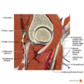
File:Sagittal section of the structures of the hip 2 Primal.png The copyright for this image is owned by Primal Pictures. They have given permission to use this image exclusively in Physiopedia. P {{Primal AnatomyTV}}(990 × 990 (1.04 MB)) - 21:02, 7 December 2020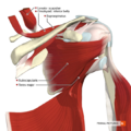
File:Muscles of the scapular region anterior aspect Primal.png The copyright for this image is owned by Primal Pictures. They have given permission to use this image exclusively in Physiopedia. P {{Primal AnatomyTV}}(660 × 660 (485 KB)) - 01:00, 8 December 2020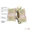
File:Intervertebral disc hernia into anterior body sagittal view Primal.png The copyright for this image is owned by Primal Pictures. They have given permission to use this image exclusively in Physiopedia. P {{Primal AnatomyTV}}(990 × 990 (623 KB)) - 15:26, 8 December 2020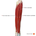
File:Superficial flexor muscles of the forearm Primal.png The copyright for this image is owned by Primal Pictures. They have given permission to use this image exclusively in Physiopedia. P {{Primal AnatomyTV}}(660 × 660 (194 KB)) - 00:28, 7 December 2020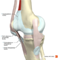
File:Patella bursae Primal.png The copyright for this image is owned by Primal Pictures. They have given permission to use this image exclusively in Physiopedia. P {{Primal AnatomyTV}}(660 × 660 (306 KB)) - 21:43, 7 December 2020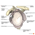
File:Illustration of Buford complex Primal.png The copyright for this image is owned by Primal Pictures. They have given permission to use this image exclusively in Physiopedia. P {{Primal AnatomyTV}}(990 × 990 (504 KB)) - 01:11, 8 December 2020
File:Left lung medial surface Primal.png The copyright for this image is owned by Primal Pictures. They have given permission to use this image exclusively in Physiopedia. P {{Primal AnatomyTV}}(660 × 660 (340 KB)) - 21:55, 8 December 2020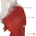
File:Superficial muscles of the gluteal region Primal.png The copyright for this image is owned by Primal Pictures. They have given permission to use this image exclusively in Physiopedia. P {{Primal AnatomyTV}}(660 × 660 (474 KB)) - 20:46, 7 December 2020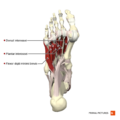
File:Plantar muscles of the foot fourth layer Primal.png The copyright for this image is owned by Primal Pictures. They have given permission to use this image exclusively in Physiopedia. P {{Primal AnatomyTV}}(660 × 660 (205 KB)) - 00:24, 8 December 2020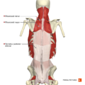
File:Muscles of the back intermediate layer Primal.png The copyright for this image is owned by Primal Pictures. They have given permission to use this image exclusively in Physiopedia. P {{Primal AnatomyTV}}(660 × 660 (300 KB)) - 15:14, 8 December 2020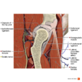
File:Sagittal section of the structures of the hip 3 Primal.png The copyright for this image is owned by Primal Pictures. They have given permission to use this image exclusively in Physiopedia. P {{Primal AnatomyTV}}(990 × 990 (1.01 MB)) - 21:03, 7 December 2020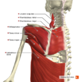
File:Muscles of the scapular region posterior aspect Primal.png The copyright for this image is owned by Primal Pictures. They have given permission to use this image exclusively in Physiopedia. P {{Primal AnatomyTV}}(660 × 660 (480 KB)) - 01:01, 8 December 2020
File:Cross-section of a functional spinal unit Primal.png The copyright for this image is owned by Primal Pictures. They have given permission to use this image exclusively in Physiopedia. P {{Primal AnatomyTV}}(990 × 990 (535 KB)) - 15:37, 8 December 2020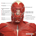
File:Superficial muscles of the head and neck anterior aspect Primal.png The copyright for this image is owned by Primal Pictures. They have given permission to use this image exclusively in Physiopedia. P {{Primal AnatomyTV}}(660 × 660 (582 KB)) - 20:00, 7 December 2020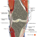
File:Coronal section of the knee joint 1 Primal.png The copyright for this image is owned by Primal Pictures. They have given permission to use this image exclusively in Physiopedia. P {{Primal AnatomyTV}}(660 × 660 (658 KB)) - 21:44, 7 December 2020
File:Illustration of SLAP II lesion Primal.png The copyright for this image is owned by Primal Pictures. They have given permission to use this image exclusively in Physiopedia. P {{Primal AnatomyTV}}(990 × 990 (516 KB)) - 01:11, 8 December 2020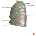
File:Left lung lateral surface Primal.png The copyright for this image is owned by Primal Pictures. They have given permission to use this image exclusively in Physiopedia. P {{Primal AnatomyTV}}(660 × 660 (310 KB)) - 21:56, 8 December 2020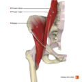
File:Muscles of the iliac region Primal.png The copyright for this image is owned by Primal Pictures. They have given permission to use this image exclusively in Physiopedia. P {{Primal AnatomyTV}}(660 × 660 (281 KB)) - 20:47, 7 December 2020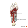
File:Plantar muscles of the foot second layer Primal.png The copyright for this image is owned by Primal Pictures. They have given permission to use this image exclusively in Physiopedia. P {{Primal AnatomyTV}}(660 × 660 (223 KB)) - 00:24, 8 December 2020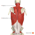
File:Muscles of the back superficial layer Primal.png The copyright for this image is owned by Primal Pictures. They have given permission to use this image exclusively in Physiopedia. P {{Primal AnatomyTV}}(660 × 660 (317 KB)) - 15:15, 8 December 2020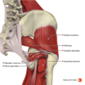
File:Deep muscles of the gluteal region Primal.png The copyright for this image is owned by Primal Pictures. They have given permission to use this image exclusively in Physiopedia. P {{Primal AnatomyTV}}(660 × 660 (445 KB)) - 21:20, 7 December 2020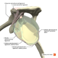
File:Ligaments of the shoulder joint sagittal section Primal.png The copyright for this image is owned by Primal Pictures. They have given permission to use this image exclusively in Physiopedia. P {{Primal AnatomyTV}}(660 × 660 (275 KB)) - 01:05, 8 December 2020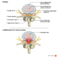
File:Cauda equina syndrome Primal.png The copyright for this image is owned by Primal Pictures. They have given permission to use this image exclusively in Physiopedia. P {{Primal AnatomyTV}}(990 × 990 (470 KB)) - 15:37, 8 December 2020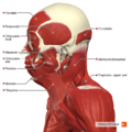
File:Superficial muscles of the head and neck lateral aspect Primal.png The copyright for this image is owned by Primal Pictures. They have given permission to use this image exclusively in Physiopedia. P {{Primal AnatomyTV}}(660 × 660 (472 KB)) - 20:01, 7 December 2020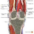
File:Coronal section of the knee joint 2 Primal.png The copyright for this image is owned by Primal Pictures. They have given permission to use this image exclusively in Physiopedia. P {{Primal AnatomyTV}}(660 × 660 (629 KB)) - 21:45, 7 December 2020
File:Illustration of glenohumeral ligaments lateral view Primal.png The copyright for this image is owned by Primal Pictures. They have given permission to use this image exclusively in Physiopedia. P {{Primal AnatomyTV}}(990 × 990 (494 KB)) - 01:12, 8 December 2020
File:Right lung lateral surface Primal.png The copyright for this image is owned by Primal Pictures. They have given permission to use this image exclusively in Physiopedia. P {{Primal AnatomyTV}}(660 × 660 (280 KB)) - 21:57, 8 December 2020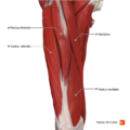
File:Muscles of the thigh anterior compartment Primal.png The copyright for this image is owned by Primal Pictures. They have given permission to use this image exclusively in Physiopedia. P {{Primal AnatomyTV}}(660 × 660 (326 KB)) - 20:48, 7 December 2020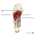
File:Plantar muscles of the foot third layer Primal.png The copyright for this image is owned by Primal Pictures. They have given permission to use this image exclusively in Physiopedia. P {{Primal AnatomyTV}}(660 × 660 (218 KB)) - 00:25, 8 December 2020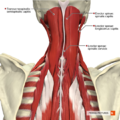
File:Muscles of the cervical region intermediate muscles Primal.png The copyright for this image is owned by Primal Pictures. They have given permission to use this image exclusively in Physiopedia. P {{Primal AnatomyTV}}(660 × 660 (619 KB)) - 15:16, 8 December 2020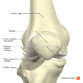
File:Knee joint anterior aspect Primal.png The copyright for this image is owned by Primal Pictures. They have given permission to use this image exclusively in Physiopedia. P {{Primal AnatomyTV}}(660 × 660 (279 KB)) - 21:36, 7 December 2020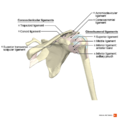
File:Ligaments of the shoulder anterior aspect Primal.png The copyright for this image is owned by Primal Pictures. They have given permission to use this image exclusively in Physiopedia. P {{Primal AnatomyTV}}(990 × 990 (340 KB)) - 01:05, 8 December 2020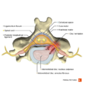
File:Posterolateral disc hernia axial view Primal.png The copyright for this image is owned by Primal Pictures. They have given permission to use this image exclusively in Physiopedia. P {{Primal AnatomyTV}}(660 × 660 (287 KB)) - 15:38, 8 December 2020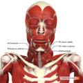
File:Intermediate muscles of the head and neck anterior aspect Primal.png The copyright for this image is owned by Primal Pictures. They have given permission to use this image exclusively in Physiopedia. P {{Primal AnatomyTV}}(660 × 660 (557 KB)) - 20:05, 7 December 2020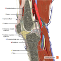
File:Sagittal section of the knee joint Primal.png The copyright for this image is owned by Primal Pictures. They have given permission to use this image exclusively in Physiopedia. P {{Primal AnatomyTV}}(660 × 660 (607 KB)) - 21:46, 7 December 2020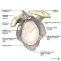
File:Illustration of sublabral foramen Primal.png The copyright for this image is owned by Primal Pictures. They have given permission to use this image exclusively in Physiopedia. P {{Primal AnatomyTV}}(990 × 990 (510 KB)) - 01:13, 8 December 2020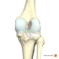
File:Posterior menisco-meniscal ligament Primal.png The copyright for this image is owned by Primal Pictures. They have given permission to use this image exclusively in Physiopedia. P {{Primal AnatomyTV}}(660 × 660 (173 KB)) - 15:13, 10 January 2021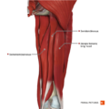
File:Muscles of the thigh posterior compartment Primal.png The copyright for this image is owned by Primal Pictures. They have given permission to use this image exclusively in Physiopedia. P {{Primal AnatomyTV}}(660 × 660 (315 KB)) - 20:49, 7 December 2020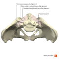
File:Sacro-iliac joint Primal.png The copyright for this image is owned by Primal Pictures. They have given permission to use this image exclusively in Physiopedia. P {{Primal AnatomyTV}}(660 × 660 (239 KB)) - 00:49, 8 December 2020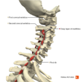
File:Muscles of the cervical region multifidus deep layer Primal.png The copyright for this image is owned by Primal Pictures. They have given permission to use this image exclusively in Physiopedia. P {{Primal AnatomyTV}}(660 × 660 (290 KB)) - 15:17, 8 December 2020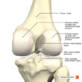
File:Knee joint posterior aspect Primal.png The copyright for this image is owned by Primal Pictures. They have given permission to use this image exclusively in Physiopedia. P {{Primal AnatomyTV}}(660 × 660 (289 KB)) - 21:37, 7 December 2020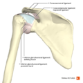
File:Ligaments of the shoulder posterior aspect Primal.png The copyright for this image is owned by Primal Pictures. They have given permission to use this image exclusively in Physiopedia. P {{Primal AnatomyTV}}(660 × 660 (256 KB)) - 01:06, 8 December 2020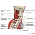
File:Sagittal section of the cervical spine Primal.png The copyright for this image is owned by Primal Pictures. They have given permission to use this image exclusively in Physiopedia. P {{Primal AnatomyTV}}(990 × 990 (792 KB)) - 15:39, 8 December 2020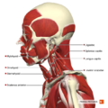
File:Intermediate muscles of the head and neck lateral aspect Primal.png The copyright for this image is owned by Primal Pictures. They have given permission to use this image exclusively in Physiopedia. P {{Primal AnatomyTV}}(660 × 660 (437 KB)) - 20:06, 7 December 2020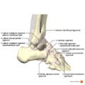
File:Ligaments of the ankle lateral aspect Primal.png The copyright for this image is owned by Primal Pictures. They have given permission to use this image exclusively in Physiopedia. P {{Primal AnatomyTV}}(990 × 990 (433 KB)) - 00:16, 8 December 2020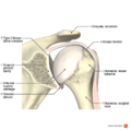
File:Illustration of type I biceps labral complex Primal.png The copyright for this image is owned by Primal Pictures. They have given permission to use this image exclusively in Physiopedia. P {{Primal AnatomyTV}}(990 × 990 (498 KB)) - 01:13, 8 December 2020
File:Extensor retinaculum of hand Primal.png The copyright for this image is owned by Primal Pictures. They have given permission to use this image exclusively in Physiopedia. P {{Primal AnatomyTV}}(660 × 660 (193 KB)) - 15:14, 10 January 2021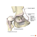
File:Hip joint Primal.png The copyright for this image is owned by Primal Pictures. They have given permission to use this image exclusively in Physiopedia. P {{Primal AnatomyTV}}(990 × 990 (443 KB)) - 20:52, 7 December 2020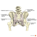
File:Ligaments of the pelvis posterior aspect Primal.png The copyright for this image is owned by Primal Pictures. They have given permission to use this image exclusively in Physiopedia. P {{Primal AnatomyTV}}(990 × 990 (537 KB)) - 00:50, 8 December 2020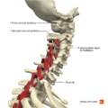
File:Muscles of the cervical region multifidus intermediate layer Primal.png The copyright for this image is owned by Primal Pictures. They have given permission to use this image exclusively in Physiopedia. P {{Primal AnatomyTV}}(660 × 660 (307 KB)) - 15:18, 8 December 2020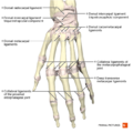
File:Ligaments of the hand dorsal aspect Primal.png The copyright for this image is owned by Primal Pictures. They have given permission to use this image exclusively in Physiopedia. P {{Primal AnatomyTV}}(660 × 660 (297 KB)) - 00:20, 7 December 2020
File:Ligaments of the knee joint superior aspect Primal.png The copyright for this image is owned by Primal Pictures. They have given permission to use this image exclusively in Physiopedia. P {{Primal AnatomyTV}}(990 × 990 (409 KB)) - 21:37, 7 December 2020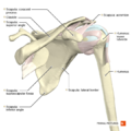
File:Shoulder anterior aspect Primal.png The copyright for this image is owned by Primal Pictures. They have given permission to use this image exclusively in Physiopedia. P {{Primal AnatomyTV}}(660 × 660 (266 KB)) - 01:07, 8 December 2020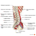
File:Sagittal section of the lumbar spine Primal.png The copyright for this image is owned by Primal Pictures. They have given permission to use this image exclusively in Physiopedia. P {{Primal AnatomyTV}}(990 × 990 (683 KB)) - 15:41, 8 December 2020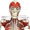
File:Deep muscles of the head and neck anterior aspect Primal.png The copyright for this image is owned by Primal Pictures. They have given permission to use this image exclusively in Physiopedia. P {{Primal AnatomyTV}}(660 × 660 (524 KB)) - 20:07, 7 December 2020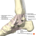
File:Ligaments of the ankle medial aspect Primal.png The copyright for this image is owned by Primal Pictures. They have given permission to use this image exclusively in Physiopedia. P {{Primal AnatomyTV}}(660 × 660 (335 KB)) - 00:18, 8 December 2020
File:Illustration of type II biceps labral complex Primal.png The copyright for this image is owned by Primal Pictures. They have given permission to use this image exclusively in Physiopedia. P {{Primal AnatomyTV}}(990 × 990 (497 KB)) - 01:14, 8 December 2020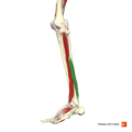
File:Peroneus brevis Primal.png The copyright for this image is owned by Primal Pictures. They have given permission to use this image exclusively in Physiopedia. P {{Primal AnatomyTV}}(660 × 660 (113 KB)) - 15:15, 10 January 2021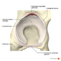
File:Peripheral longitudinal labral tear Primal.png The copyright for this image is owned by Primal Pictures. They have given permission to use this image exclusively in Physiopedia. P {{Primal AnatomyTV}}(990 × 990 (552 KB)) - 20:53, 7 December 2020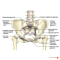
File:Ligaments of the pelvis anterior aspect Primal.png The copyright for this image is owned by Primal Pictures. They have given permission to use this image exclusively in Physiopedia. P {{Primal AnatomyTV}}(990 × 990 (557 KB)) - 00:51, 8 December 2020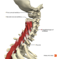
File:Muscles of the cervical region multifidus superficial layer Primal.png The copyright for this image is owned by Primal Pictures. They have given permission to use this image exclusively in Physiopedia. P {{Primal AnatomyTV}}(660 × 660 (314 KB)) - 15:19, 8 December 2020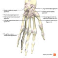
File:Ligaments of the hand palmar aspect Primal.png The copyright for this image is owned by Primal Pictures. They have given permission to use this image exclusively in Physiopedia. P {{Primal AnatomyTV}}(660 × 660 (288 KB)) - 00:22, 7 December 2020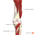
File:Deep muscles of the knee posterior aspect Primal.png The copyright for this image is owned by Primal Pictures. They have given permission to use this image exclusively in Physiopedia. P {{Primal AnatomyTV}}(660 × 660 (250 KB)) - 21:38, 7 December 2020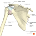
File:Shoulder posterior aspect Primal.png The copyright for this image is owned by Primal Pictures. They have given permission to use this image exclusively in Physiopedia. P {{Primal AnatomyTV}}(660 × 660 (280 KB)) - 01:07, 8 December 2020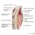
File:Sagittal section of the thoracic spine Primal.png The copyright for this image is owned by Primal Pictures. They have given permission to use this image exclusively in Physiopedia. P {{Primal AnatomyTV}}(990 × 990 (709 KB)) - 15:41, 8 December 2020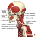
File:Deep muscles of the head and neck lateral aspect Primal.png The copyright for this image is owned by Primal Pictures. They have given permission to use this image exclusively in Physiopedia. P {{Primal AnatomyTV}}(660 × 660 (418 KB)) - 20:09, 7 December 2020
File:Ligaments of the ankle posterior aspect Primal.png The copyright for this image is owned by Primal Pictures. They have given permission to use this image exclusively in Physiopedia. P {{Primal AnatomyTV}}(660 × 660 (286 KB)) - 00:18, 8 December 2020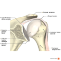
File:Illustration of type III biceps labral complex Primal.png The copyright for this image is owned by Primal Pictures. They have given permission to use this image exclusively in Physiopedia. P {{Primal AnatomyTV}}(990 × 990 (510 KB)) - 01:15, 8 December 2020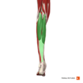
File:Gastrocnemius Primal.png The copyright for this image is owned by Primal Pictures. They have given permission to use this image exclusively in Physiopedia. P {{Primal AnatomyTV}}(660 × 660 (112 KB)) - 15:16, 10 January 2021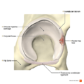
File:Radial flap labral tear Primal.png The copyright for this image is owned by Primal Pictures. They have given permission to use this image exclusively in Physiopedia. P {{Primal AnatomyTV}}(990 × 990 (542 KB)) - 20:54, 7 December 2020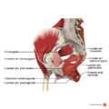
File:Muscles of the pelvic diaphragm Primal.png The copyright for this image is owned by Primal Pictures. They have given permission to use this image exclusively in Physiopedia. P {{Primal AnatomyTV}}(990 × 990 (574 KB)) - 00:51, 8 December 2020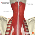
File:Muscles of the cervical region superficial muscles Primal.png The copyright for this image is owned by Primal Pictures. They have given permission to use this image exclusively in Physiopedia. P {{Primal AnatomyTV}}(660 × 660 (604 KB)) - 15:20, 8 December 2020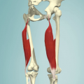
File:Biceps fem 1.png 3D anatomy images. Copyright of Primal Pictures Ltd. www.primalpictures.com(385 × 385 (149 KB)) - 01:32, 8 May 2015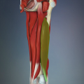
File:Biceps fem 2.png 3D anatomy images. Copyright of Primal Pictures Ltd. www.primalpictures.com(385 × 385 (159 KB)) - 01:33, 8 May 2015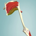
File:Teres maj 1.png 3D anatomy images. Copyright of Primal Pictures Ltd. www.primalpictures.com(500 × 500 (233 KB)) - 18:46, 9 May 2015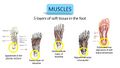
File:Layers of the foot.jpg Ligaments of the plantar surface image - Primal pictures(1,280 × 720 (128 KB)) - 15:16, 21 September 2021- **primal|Primal Pictures Image Bank **Primal AnatomyTV|Primal Pictures Anatomy TV2 KB (233 words) - 09:41, 1 February 2024
- Primal Pictures 3D Human Anatomy Software - The Most Complete, Detailed and Accurate 3D Mod Since 1991, Primal Pictures has been providing cutting-edge anatomy and physiology solutions to student2 KB (338 words) - 18:12, 23 July 2013
- ...tures in articles that you are creating or updating in Physiopedia. Primal pictures retain the copyright on these images and request that they are not used out To use the pictures:21 KB (3,064 words) - 18:47, 14 June 2021
- ...hand_anterior_aspect_Primal.png|thumb|right|200px|Image courtesy of Primal Pictures]]3 KB (393 words) - 16:40, 28 February 2021
- **[[Template:Primal AnatomyTV|Primal Pictures Anatomy TV]]7 KB (788 words) - 14:16, 6 May 2024
- *[http://www.physio-pedia.com/Primal_Pictures_Image_Bank the Primal Picture image bank] #*Have permission from the source of the image e.g. the Primal Picture image bank (http://www.physio-pedia.com/Primal_Pictures_Image_Bank)10 KB (1,577 words) - 00:54, 25 November 2019
- ...ead' (deep). It is part of the [[hamstrings]].<ref name="anatomytv">Primal Pictures. Available from:http://www.anatomy.tv/interactivehip (accessed 8 May 2015)< ...png|center|300x300px|Biceps_fem_1]]<ref name="primalonlinelearning">Primal Pictures. Available from: https://www.anatomy.tv/#bicepsfemoris (accessed 8 May 201510 KB (1,461 words) - 01:58, 23 March 2024
- ...r of the [[patella]].<ref name=":0">Anatomy.tv | 3D Human Anatomy | Primal Pictures [Internet]. Anatomy.tv. 2018 [cited 16 March 2018]. Available from: <nowiki5 KB (759 words) - 05:17, 1 April 2022
- * [[Partner Images|Primal Pictures]] - Anatomy images11 KB (1,855 words) - 11:20, 22 May 2023
- [[Image:Lumbopelvic.png|thumb|right|300px|Courtesy of Primal Pictures Ltd. www.primalpictures.com]]10 KB (1,336 words) - 03:10, 18 April 2020
- ...Sciences; 2012.</ref><ref name=":1">Anatomy.tv | 3D Human Anatomy | Primal Pictures [Internet]. Anatomy.tv. 2018 [cited 30 April 2018]. Available from: <nowiki ...ol. 2014 Jun;43(6):781-91</ref>:[[File:Coronal section of the knee joint 2 Primal.png|thumb|Distal semimembranosus complex: insertions|alt=]]12 KB (1,726 words) - 11:04, 22 January 2024
- ...exus|sacral plexus.]]<ref name=":0">Anatomy.tv | 3D Human Anatomy | Primal Pictures [Internet]. Anatomy.tv. 2018 [cited 1 May 2018]. Available from: <nowiki>ht13 KB (1,905 words) - 03:53, 30 January 2024
- [[File:Ligaments of the hip joint anterior aspect Primal.png|right|frameless|400x400px]] ...mal.png|thumb|300x300px|Acetabular Labrum copyright and courtesy of Primal Pictures Ltd]]15 KB (2,281 words) - 12:13, 17 December 2023
- ...igaments of the hip joint anterior aspect Primal.png|thumb|center|© Primal Pictures]]</div> ...Image:Peripheral_longitudinal_labral_tear_Primal.png|thumb|center|© Primal Pictures]]</div>32 KB (4,647 words) - 13:44, 18 March 2024
- ...cles Primal.png|thumb|Figure 1. Muscles of the cervical region from Primal Pictures.]]18 KB (2,531 words) - 02:06, 18 October 2023
- ...low pectoralis minor <ref name=":9">Anatomy.tv | 3D Human Anatomy | Primal Pictures [Internet]. Anatomy.tv. 2018 [cited 22 July 2019]. Available from: <nowiki>20 KB (2,786 words) - 00:15, 24 March 2024
- ...les_of_the_back_erector_spinae_group_Primal.png|thumb|right|250px|© Primal Pictures]]23 KB (3,461 words) - 11:16, 28 August 2023
- ...right|200px|Anatomy of the Pelvic Floor - copyright and courtesy of Primal Pictures Ltd. www.primalpictures.com]]25 KB (3,632 words) - 10:49, 24 May 2022
- ...the_scapular_region_posterior_aspect_Primal.png|thumb|right|250px|© Primal Pictures]]Movements of the [[scapula]] can be broken up into 3 motions and 2 transla24 KB (3,413 words) - 04:05, 26 January 2024
- ..._of_the_head_and_neck_lateral_aspect_Primal.png|thumb|right|250px|© Primal Pictures]] Temporomandibular Disorder (TMD) is a broad term that encompasses disorde26 KB (3,849 words) - 15:15, 7 May 2024
- ...tensor retinaculum of hand Primal.png|thumb|right|300px|Courtesy of Primal Pictures]]31 KB (4,793 words) - 09:57, 12 December 2022
- ...medium=Physiopedia&utm_campaign=Supraspinatus_tendinopathy#products Primal Pictures Anatomy TV]</ref> </div>29 KB (4,126 words) - 16:59, 2 August 2023


