Sacral Insufficiency Fractures: Difference between revisions
Andrea Nees (talk | contribs) No edit summary |
No edit summary |
||
| Line 5: | Line 5: | ||
*Sacral insufficiency fractures | *Sacral insufficiency fractures | ||
*SIF | *SIF | ||
*Physical therapy OR Physiotherapy OR | *Physical therapy OR Physiotherapy OR Exercise OR Treatment | ||
*Mobilization | *Mobilization | ||
*Hydrotherapy OR Aquatherapy | *Hydrotherapy OR Aquatherapy | ||
| Line 17: | Line 17: | ||
*Google | *Google | ||
<br> | <br> | ||
= Definition/Description = | = Definition/Description = | ||
Sacral insufficiency fractures (SIFs) are a cause of low back pain.<sup>(10)</sup><br>These kind of fractures are a subtype of stress fractures. They result from normal stress applied to a bone with reduced elasticity.<sup>(1; Levels of evidence: C, 5; Levels of evidence: C)</sup> | |||
An underlying condition like [[Osteoporosis|osteoporosis]] or other metabolic bone diseases are often a cause of SIFs. This is why SIFs are more common with elder women.<sup>(1, 5, 9; Levels of evidence: C)</sup><br>Bone insufficiency fractures are described first by Lourie in 1982.<sup>(5, 9)</sup><br><br> | |||
= Clinically Relevant Anatomy = | |||
[[Image:Sacrum_and_Coccyx.jpg|right|200px]] | |||
The sacrum is a triangular bone formed by 5 vertebral segments. It articulates superiorly with the fifth lumbar vertebra and inferiorly with the coccyx. The lateral surface of the upper part of the lateral masses (auricular surface) articulates with the ilium. <sup>(7; Levels of evidence: C)</sup> | The sacrum is a triangular bone formed by 5 vertebral segments. It articulates superiorly with the fifth lumbar vertebra and inferiorly with the coccyx. The lateral surface of the upper part of the lateral masses (auricular surface) articulates with the ilium. <sup>(7; Levels of evidence: C)</sup> | ||
| Line 33: | Line 33: | ||
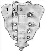
Denis et al classified traumatic sacral fractures by dividing the sacrum into 3 zones (Fig.1). These traumatic fractures are not directly related to SIFs, but the classification system of Denis is very useful for the description of the fractures.<sup>(7)</sup> | Denis et al classified traumatic sacral fractures by dividing the sacrum into 3 zones (Fig.1). These traumatic fractures are not directly related to SIFs, but the classification system of Denis is very useful for the description of the fractures.<sup>(7)</sup> | ||
Zone 1: involves the sacral ala (lateral to the sacral foramina). This is the most common site of SIFs.<br>Zone 2: involves the sacral (neural) foramina (but the fracture does not enter the central sacral canal). The fractures in this site are associated with unilateral lumbosacral radiculopathies.<br>Zone 3: involves the sacral bodies and the transverse central canal. <sup>(1, 7) <br></sup> | Zone 1: involves the sacral ala (lateral to the sacral foramina). This is the most common site of SIFs.<br>Zone 2: involves the sacral (neural) foramina (but the fracture does not enter the central sacral canal). The fractures in this site are associated with unilateral lumbosacral radiculopathies.<br>Zone 3: involves the sacral bodies and the transverse central canal. <sup>(1, 7) <br></sup> | ||
[[Image: | [[Image:Denis classification.jpg|right|Fig]]<br> Fig. 1: Denis classification<sup>(8)</sup> | ||
= Epidemiology /Etiology <br> = | = Epidemiology /Etiology <br> = | ||
| Line 58: | Line 58: | ||
Patients with SIFs are mostly time older than 55 years old. The mean age is between 70 and 75 years old.<br>The precise incidence of SIFs is unknown but some studies reported a prevalence of 1% - 5% in at-risk patient populations.<br>Two-third of patients were a-traumatic. <sup>(1)</sup> | Patients with SIFs are mostly time older than 55 years old. The mean age is between 70 and 75 years old.<br>The precise incidence of SIFs is unknown but some studies reported a prevalence of 1% - 5% in at-risk patient populations.<br>Two-third of patients were a-traumatic. <sup>(1)</sup> | ||
SFI can occur in a younger population. For example: pregnant women. This can be related to pregnancy-associated osteoporosis<sup>.(4)</sup><sup></sup> | SFI can occur in a younger population. For example: pregnant women. This can be related to pregnancy-associated osteoporosis<sup>.(4)</sup><sup></sup> | ||
= Characteristics/Clinical Presentation = | = Characteristics/Clinical Presentation = | ||
| Line 69: | Line 69: | ||
*Nerve damage (unusual) <sup>(1, 5)</sup> | *Nerve damage (unusual) <sup>(1, 5)</sup> | ||
<br> | <br> | ||
= Differential Diagnosis = | = Differential Diagnosis = | ||
| Line 78: | Line 78: | ||
*Radiculopathy<sup>(1)</sup> | *Radiculopathy<sup>(1)</sup> | ||
*Disc disease | *Disc disease | ||
*[[ | *[[Spinal Stenosis|Spinal stenosis]] | ||
*[[ | *[[Cauda Equina Syndrome|Cauda equina syndrome]] | ||
<br> | <br> | ||
<br> | <br> | ||
= Diagnostic Procedures = | = Diagnostic Procedures = | ||
| Line 89: | Line 89: | ||
SIF can be diagnosed with radiology. Bone scintigraphy is the most sensitive study to detect SIFs. Other radiographic procedures that can help to diagnose SIFs are magnetic resonance imaging (MRI) and computed tomography (CT) scans. <sup>(1, 5, 8; Levels of evidence: B)</sup> | SIF can be diagnosed with radiology. Bone scintigraphy is the most sensitive study to detect SIFs. Other radiographic procedures that can help to diagnose SIFs are magnetic resonance imaging (MRI) and computed tomography (CT) scans. <sup>(1, 5, 8; Levels of evidence: B)</sup> | ||
<br> | <br> | ||
= Examination = | = Examination = | ||
| Line 98: | Line 98: | ||
*SI-joint tests are often positive (this test is not specific for SFI) | *SI-joint tests are often positive (this test is not specific for SFI) | ||
*Gait is slow and antalgic | *Gait is slow and antalgic | ||
*[[ | *[[Trendelenburg Test|Trendelenburg test]] is normal | ||
*Sciatic nerve tension tests (Lasegue and [[ | *Sciatic nerve tension tests (Lasegue and [[Straight Leg Raise Test|Straight Leg Raise]] (SLR)) are normal | ||
<br> | <br> | ||
= Physical Therapy Management = | = Physical Therapy Management = | ||
| Line 107: | Line 107: | ||
<br> | <br> | ||
Early rehabilitation and moderate weight-bearing exercises, within the boundaries of pain tolerance, has been suggested. The earlier rehabilitation will stimulate the bone formation by the osteoblasts and improve muscle tension. <br>Mobilization is recommended because long periods of immobilization has a lot of complications (deep vein thrombosis, pulmonary embolus, loss of muscle strength, etc.). In the earlier stages of fracture healing is assisted mobilization whit external devices (e.g. walking frames or hydrotherapy) better tolerated by many patients<sup>(7)</sup>. | Early rehabilitation and moderate weight-bearing exercises, within the boundaries of pain tolerance, has been suggested. The earlier rehabilitation will stimulate the bone formation by the osteoblasts and improve muscle tension. <br>Mobilization is recommended because long periods of immobilization has a lot of complications (deep vein thrombosis, pulmonary embolus, loss of muscle strength, etc.). In the earlier stages of fracture healing is assisted mobilization whit external devices (e.g. walking frames or hydrotherapy) better tolerated by many patients<sup>(7)</sup>. | ||
<br> | <br> | ||
= Resources = | = Resources = | ||
| Line 115: | Line 115: | ||
http://www.artrose-blog.nl/rugaandoeningen/insufficientiefracturen-van-het-sacrum | http://www.artrose-blog.nl/rugaandoeningen/insufficientiefracturen-van-het-sacrum | ||
<br> | <br> | ||
= References = | = References = | ||
1. LYDERS E.M., WHITLOW C.T., BAKER M.D., MORRIS P.P., Imaging and treatment of sacral insufficiency fractures (review), Am. J. Neuroradiology 31:201-10, Februari 2010.<br>(Levels of evidence: C)<br>2. KOS C.B., TACONIS W.K., VAN DER EIJKEN J.W., Insufficiëntiefracturen van het sacrum, Ned. Tijdschr. Geneeskd 1999, 13 februari; 143(7)<br>(Levels of evidence: C)<br>3. THEIN R., BURSTEIN G., SHABSHIN N., Labor-related sacral stress fracture presenting as lower limb radicular pain. Orthopedics June 2009; 32(6):447<br>(Levels of evidence: B)<br>4. KARATAS M., BASARAN C., OZGUL E., TARHAN E., AGILDERE A.M., Postpartum sacral stress fracture, An unusual case of low-back and buttock pain. Am. J. Phys. Med. Rehabil. Vol 87, No. 5. 2008<br>(Levels of evidence: C)<br>5. WILD A., JAEGER M., HAAK H., MEHDIAN S.H., Sacral insufficiency fracture, an unsuspected cause of low-back pain in elderly women. Arch. Orthop. Trauma. Surg (2002) 122:58-60<br>(Levels of evidence: C)<br>6. DASGUPTA B., SHAH N., BROWN H., GORDON T.E., TANQUERAY A.B., MELLOR J.A., Sacral insufficiency fractures: an unsuspected cause of low back pain. British Journal of Rheumatology 1998; 37: 789-793<br>(Levels of evidence: B)<br>7. TSIRIDIS E., UPADHYAY N., GIANNOUDIS P.V., Sacral insufficiency fractures: current concepts of management. Osteoporos. Int. (2006) 17: 1716-1725<br>(Levels of evidence: C)<br>8. GOTIS-GRAHAM I., McGUIGAN L., DIAMOND T., PORTEK I., QUINN R., STURGESS A., TULLOCH R., Sacral insufficiency fractures in the elderly. J. Bone Joint Surg. 1994; 76-B: 882-6<br>(Levels of evidence: B)<br>9. YONG-LEE J., BONG-JIN H., KIM J.T., CHUNG D.S., Sacral insufficiency fracture, most overlooked cause of lumbosacral pain. J. Kor. Neurosurg. Soc. September 2008, 44 (3): 166-169<br>(Levels of evidence: C)<br>10. MEEUSEN, R. Praktijkgids rug- en nekletsels. Deel 1. Dienst uitgaven Vrije Universiteit Brussel. 218 blz.<br> | 1. LYDERS E.M., WHITLOW C.T., BAKER M.D., MORRIS P.P., Imaging and treatment of sacral insufficiency fractures (review), Am. J. Neuroradiology 31:201-10, Februari 2010.<br>(Levels of evidence: C)<br>2. KOS C.B., TACONIS W.K., VAN DER EIJKEN J.W., Insufficiëntiefracturen van het sacrum, Ned. Tijdschr. Geneeskd 1999, 13 februari; 143(7)<br>(Levels of evidence: C)<br>3. THEIN R., BURSTEIN G., SHABSHIN N., Labor-related sacral stress fracture presenting as lower limb radicular pain. Orthopedics June 2009; 32(6):447<br>(Levels of evidence: B)<br>4. KARATAS M., BASARAN C., OZGUL E., TARHAN E., AGILDERE A.M., Postpartum sacral stress fracture, An unusual case of low-back and buttock pain. Am. J. Phys. Med. Rehabil. Vol 87, No. 5. 2008<br>(Levels of evidence: C)<br>5. WILD A., JAEGER M., HAAK H., MEHDIAN S.H., Sacral insufficiency fracture, an unsuspected cause of low-back pain in elderly women. Arch. Orthop. Trauma. Surg (2002) 122:58-60<br>(Levels of evidence: C)<br>6. DASGUPTA B., SHAH N., BROWN H., GORDON T.E., TANQUERAY A.B., MELLOR J.A., Sacral insufficiency fractures: an unsuspected cause of low back pain. British Journal of Rheumatology 1998; 37: 789-793<br>(Levels of evidence: B)<br>7. TSIRIDIS E., UPADHYAY N., GIANNOUDIS P.V., Sacral insufficiency fractures: current concepts of management. Osteoporos. Int. (2006) 17: 1716-1725<br>(Levels of evidence: C)<br>8. GOTIS-GRAHAM I., McGUIGAN L., DIAMOND T., PORTEK I., QUINN R., STURGESS A., TULLOCH R., Sacral insufficiency fractures in the elderly. J. Bone Joint Surg. 1994; 76-B: 882-6<br>(Levels of evidence: B)<br>9. YONG-LEE J., BONG-JIN H., KIM J.T., CHUNG D.S., Sacral insufficiency fracture, most overlooked cause of lumbosacral pain. J. Kor. Neurosurg. Soc. September 2008, 44 (3): 166-169<br>(Levels of evidence: C)<br>10. MEEUSEN, R. Praktijkgids rug- en nekletsels. Deel 1. Dienst uitgaven Vrije Universiteit Brussel. 218 blz.<br> | ||
Revision as of 23:50, 19 April 2014
Search Strategy[edit | edit source]
Keywords:
- Sacral insufficiency fractures
- SIF
- Physical therapy OR Physiotherapy OR Exercise OR Treatment
- Mobilization
- Hydrotherapy OR Aquatherapy
Databases:
- Pubmed
- Web of Knowledge
- PEDro
- Google scholar
Definition/Description[edit | edit source]
Sacral insufficiency fractures (SIFs) are a cause of low back pain.(10)
These kind of fractures are a subtype of stress fractures. They result from normal stress applied to a bone with reduced elasticity.(1; Levels of evidence: C, 5; Levels of evidence: C)
An underlying condition like osteoporosis or other metabolic bone diseases are often a cause of SIFs. This is why SIFs are more common with elder women.(1, 5, 9; Levels of evidence: C)
Bone insufficiency fractures are described first by Lourie in 1982.(5, 9)
Clinically Relevant Anatomy[edit | edit source]
The sacrum is a triangular bone formed by 5 vertebral segments. It articulates superiorly with the fifth lumbar vertebra and inferiorly with the coccyx. The lateral surface of the upper part of the lateral masses (auricular surface) articulates with the ilium. (7; Levels of evidence: C)
Denis et al classified traumatic sacral fractures by dividing the sacrum into 3 zones (Fig.1). These traumatic fractures are not directly related to SIFs, but the classification system of Denis is very useful for the description of the fractures.(7)
Zone 1: involves the sacral ala (lateral to the sacral foramina). This is the most common site of SIFs.
Zone 2: involves the sacral (neural) foramina (but the fracture does not enter the central sacral canal). The fractures in this site are associated with unilateral lumbosacral radiculopathies.
Zone 3: involves the sacral bodies and the transverse central canal. (1, 7)
Fig. 1: Denis classification(8)
Epidemiology /Etiology
[edit | edit source]
The most important cause of SIF is osteoporosis(1, 5, 6, 9). Other risk factors are:
- Pelvic radiation
- Steroid-induced osteopenia(9)
- Rheumatoid arthritis(6; Levels of evidence: B)
- Multiple myeloma
- Paget disease(9)
- Renal osteodystrophy
- Hyperparathyroidism(1, 4, 5, 9)
- Corticosteroid medication(1, 5)
- Metastatic disease(1, 5)
- Marrow replacement processes(1, 5)
- Fibrous dysplasia(4; Levels of evidence: C)
- Osteogenesis imperfect(4)
- Osteopetrosis(4)
- Osteomalacia(4)
Patients with SIFs are mostly time older than 55 years old. The mean age is between 70 and 75 years old.
The precise incidence of SIFs is unknown but some studies reported a prevalence of 1% - 5% in at-risk patient populations.
Two-third of patients were a-traumatic. (1)
SFI can occur in a younger population. For example: pregnant women. This can be related to pregnancy-associated osteoporosis.(4)
Characteristics/Clinical Presentation[edit | edit source]
Patients with SIFs may have:
- Tenderness of palpation (lower back and sacral region) (1, 2, 5, 6)
- Pain (at the buttock, back, hip, pelvic or groin)(3; Levels of evidence: B)
- Problems with walking (slowly and painfull)
- Nerve damage (unusual) (1, 5)
Differential Diagnosis[edit | edit source]
Sacral insufficiency fractures are difficult to diagnose because the signs and symptoms are vague and non-specific.
SIFs can be confused with:
- Metastatic disease(5)
- Radiculopathy(1)
- Disc disease
- Spinal stenosis
- Cauda equina syndrome
Diagnostic Procedures[edit | edit source]
SIF can be diagnosed with radiology. Bone scintigraphy is the most sensitive study to detect SIFs. Other radiographic procedures that can help to diagnose SIFs are magnetic resonance imaging (MRI) and computed tomography (CT) scans. (1, 5, 8; Levels of evidence: B)
Examination[edit | edit source]
The physical examination shows(7):
- Sacral tenderness (on lateral compression)
- SI-joint tests are often positive (this test is not specific for SFI)
- Gait is slow and antalgic
- Trendelenburg test is normal
- Sciatic nerve tension tests (Lasegue and Straight Leg Raise (SLR)) are normal
Physical Therapy Management[edit | edit source]
Early rehabilitation and moderate weight-bearing exercises, within the boundaries of pain tolerance, has been suggested. The earlier rehabilitation will stimulate the bone formation by the osteoblasts and improve muscle tension.
Mobilization is recommended because long periods of immobilization has a lot of complications (deep vein thrombosis, pulmonary embolus, loss of muscle strength, etc.). In the earlier stages of fracture healing is assisted mobilization whit external devices (e.g. walking frames or hydrotherapy) better tolerated by many patients(7).
Resources[edit | edit source]
http://www.artrose-blog.nl/rugaandoeningen/insufficientiefracturen-van-het-sacrum
References[edit | edit source]
1. LYDERS E.M., WHITLOW C.T., BAKER M.D., MORRIS P.P., Imaging and treatment of sacral insufficiency fractures (review), Am. J. Neuroradiology 31:201-10, Februari 2010.
(Levels of evidence: C)
2. KOS C.B., TACONIS W.K., VAN DER EIJKEN J.W., Insufficiëntiefracturen van het sacrum, Ned. Tijdschr. Geneeskd 1999, 13 februari; 143(7)
(Levels of evidence: C)
3. THEIN R., BURSTEIN G., SHABSHIN N., Labor-related sacral stress fracture presenting as lower limb radicular pain. Orthopedics June 2009; 32(6):447
(Levels of evidence: B)
4. KARATAS M., BASARAN C., OZGUL E., TARHAN E., AGILDERE A.M., Postpartum sacral stress fracture, An unusual case of low-back and buttock pain. Am. J. Phys. Med. Rehabil. Vol 87, No. 5. 2008
(Levels of evidence: C)
5. WILD A., JAEGER M., HAAK H., MEHDIAN S.H., Sacral insufficiency fracture, an unsuspected cause of low-back pain in elderly women. Arch. Orthop. Trauma. Surg (2002) 122:58-60
(Levels of evidence: C)
6. DASGUPTA B., SHAH N., BROWN H., GORDON T.E., TANQUERAY A.B., MELLOR J.A., Sacral insufficiency fractures: an unsuspected cause of low back pain. British Journal of Rheumatology 1998; 37: 789-793
(Levels of evidence: B)
7. TSIRIDIS E., UPADHYAY N., GIANNOUDIS P.V., Sacral insufficiency fractures: current concepts of management. Osteoporos. Int. (2006) 17: 1716-1725
(Levels of evidence: C)
8. GOTIS-GRAHAM I., McGUIGAN L., DIAMOND T., PORTEK I., QUINN R., STURGESS A., TULLOCH R., Sacral insufficiency fractures in the elderly. J. Bone Joint Surg. 1994; 76-B: 882-6
(Levels of evidence: B)
9. YONG-LEE J., BONG-JIN H., KIM J.T., CHUNG D.S., Sacral insufficiency fracture, most overlooked cause of lumbosacral pain. J. Kor. Neurosurg. Soc. September 2008, 44 (3): 166-169
(Levels of evidence: C)
10. MEEUSEN, R. Praktijkgids rug- en nekletsels. Deel 1. Dienst uitgaven Vrije Universiteit Brussel. 218 blz.


