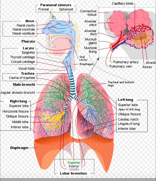
Respiratory System
Introduction[edit | edit source]
The lungs and respiratory system allow us to breathe. They bring oxygen into our bodies (called inspiration, or inhalation) and send carbon dioxide out (called expiration, or exhalation)[1].
- The respiratory system of animals is crucial for the life as it allows the exchange of gases between an organism and the environment.[2]
- This exchange of oxygen and carbon dioxide is called respiration.
Parts of the Respiratory System[edit | edit source]
The human respiratory system comprises of
- Nose and Mouth: Air enters the respiratory system through the nose or the mouth. If it goes in the nostrils, the air is warmed and humidified. Cilia (tiny hairs) protect the nasal passageways and other parts of the respiratory tract, filtering out dust and other particles that enter the nose through the breathed air.
- Pharynx (throat): The nasal cavity and the mouth openings meet at the pharynx at the back of the nose and mouth. The pharynx is part of the digestive system as well as the respiratory system because it carries both food and air. At the bottom of the pharynx, this pathway divides in two, the esophagus, which leads to the stomach and the other for air. The epiglottis, a small flap of tissue, covers the air-only passage when we swallow, keeping food and liquid from going into the lungs.
- The Larynx (voice box): Top part of Trachea. This short tube contains a pair of vocal cords, which vibrate to make sounds.
- Trachea: The walls of the trachea are strengthened by stiff rings of cartilage to keep it open. The trachea is also lined with cilia, which sweep fluids and foreign particles out of the airway so that they stay out of the lungs.
- Bronchi: At its bottom end, the trachea divides into left and right air tubes called bronchi, which connect to the lungs. Within the lungs, the bronchi branch into smaller bronchi and even smaller tubes called bronchioles.
- Lungs: The lungs are the functional units of respiration and are key to survival. They contain 1500 miles of airways, 300-500 million alveoli and have a combined surface area of 70 square metres (half a tennis court). Each lung weighs approximately 1.1 kg. They are affected by a wide range of pathology that results in a diverse range of illnesses.[3]
- Alveoli: Bronchioles end in tiny air sacs called alveoli, where the exchange of oxygen and carbon dioxide actually takes place. Each person has hundreds of millions of alveoli in their lungs.
This network of alveoli, bronchioles, bronchi and trachea is known as the tracheobronchial tree.[1]
The chest cavity, or thorax, houses the bronchial tree, lungs, heart, and other structures.
- The top and sides of the thorax are formed by the ribs and attached muscles, and the bottom is formed by the diaphragm.
- The chest walls form a protective cage around the lungs and other contents of the chest cavity[1].
Tracheobronchial tree[edit | edit source]
The tracheobronchial tree is a branching structure of tubes of an ever-decreasing diameter that start at the larynx and end in the alveoli. It can broadly be divided into conduction and respiratory zones.
- Conduction zone
Gas is warmed and humidified as it is conducted from the oropharynx to the functional portion of the lung where gas exchange occurs. The conduction zone is composed of the trachea, bronchi, bronchioles and terminal bronchioles. Their function is to optimise gas delivery to the functional portion of the lung. Their walls contain cilia to remove particulates from the inspired gas and cartilage to ensure that they do not collapse in expiration.
2. Respiratory zone
The respiratory zone is an extension of the tracheobronchial tree at the level of the terminal bronchioles. It is composed of respiratory bronchioles, alveolar ducts and alveoli, and is the location of gas transfer within the lung[3].
Alveoli[edit | edit source]
The alveoli (singular: alveolus) are tiny hollow air sacs that comprise the basic unit of respiration.
Alveoli are found within the lung parenchyma and are found at the terminal ends of the respiratory tree, clustered around alveolar sacs and alveolar ducts. Each alveolus is approximately 0.2 mm in diameter. There are around 300 million to 1 billion alveoli in the human lungs, covering an area of 70 square metres.
- The alveolar walls are comprised of collagen and elastic fibres which facilitate expansion during inspiration and return to the original shape during expiration. There are numerous capillaries within the alveolar walls where gas exchange occurs. Pores of Kohn are also located within the walls.
- Alveoli contain two major types of epithelial cells. The most abundant, type 1 pneumocytes (95%) are squamous cells in which gas exchange occurs. The remaining 5%, type 2 pneumocytes, are granular cells which secrete surfactant. Surfactant is a lipoprotein with a high phospholipid content which reduces surface tension. This increases pulmonary compliance, prevents atelectasis and aids recruitment of collapsed airways.
- Alveolar macrophages are also located in the alveoli. They protect the alveoli from foreign material by engulfing it, including bacteria, dust and carbon particles[4].
References[edit | edit source]
- ↑ 1.0 1.1 1.2 Teenhealth Lungs and Respiratory System Available from:https://kidshealth.org/en/teens/lungs.html (last accessed 1.10.2020)
- ↑ AU essays Evolution of Respiratory Systems in Animals Available from:https://www.auessays.com/essays/biology/evolution-respiratory-systems-1447.php (last accessed 1.10.2020)
- ↑ 3.0 3.1 Radiopedia The Lungs Available from:https://radiopaedia.org/articles/lung (last accessed 1.10.2020)
- ↑ Radiopedia Alveoli Available from:https://radiopaedia.org/articles/alveoli?lang=gb (last accessed 1.10.2020)


