Respiratory System: Difference between revisions
No edit summary |
No edit summary |
||
| Line 18: | Line 18: | ||
* Lungs: The lungs are the functional units of respiration and are key to survival. They contain 1500 miles of airways, 300-500 million alveoli and have a combined surface area of 70 square metres (half a tennis court). Each lung weighs approximately 1.1 kg. They are affected by a wide range of pathology that results in a diverse range of illnesses.<ref name=":1">Radiopedia [https://radiopaedia.org/articles/lung The Lungs] Available from:https://radiopaedia.org/articles/lung (last accessed 1.10.2020)</ref> | * Lungs: The lungs are the functional units of respiration and are key to survival. They contain 1500 miles of airways, 300-500 million alveoli and have a combined surface area of 70 square metres (half a tennis court). Each lung weighs approximately 1.1 kg. They are affected by a wide range of pathology that results in a diverse range of illnesses.<ref name=":1">Radiopedia [https://radiopaedia.org/articles/lung The Lungs] Available from:https://radiopaedia.org/articles/lung (last accessed 1.10.2020)</ref> | ||
* Alveoli: Bronchioles end in tiny air sacs called alveoli, where the exchange of oxygen and carbon dioxide actually takes place. Each person has hundreds of millions of alveoli in their lungs. | * Alveoli: Bronchioles end in tiny air sacs called alveoli, where the exchange of oxygen and carbon dioxide actually takes place. Each person has hundreds of millions of alveoli in their lungs. | ||
This network of alveoli, bronchioles, bronchi and trachea is known as the tracheobronchial tree.<ref name=":0" /> | This network of alveoli, bronchioles, bronchi and trachea is known as the tracheobronchial tree.<ref name=":0" /><ref name=":2" />. | ||
The chest cavity ([[Thoracic Anatomy|thorax]]) houses the bronchial tree, lungs, [[Anatomy of the Human Heart|heart]], and other structures. | The chest cavity ([[Thoracic Anatomy|thorax]]) houses the bronchial tree, lungs, [[Anatomy of the Human Heart|heart]], and other structures. | ||
* The top and sides of the thorax are formed by the [[ribs]] and attached muscles, and the bottom is formed by the [[Diaphragmatic Breathing Exercises|diaphragm]]. | * The top and sides of the thorax are formed by the [[ribs]] and attached muscles, and the bottom is formed by the [[Diaphragmatic Breathing Exercises|diaphragm]]. | ||
| Line 55: | Line 55: | ||
* Alveoli contain two major types of epithelial cells. The most abundant, type 1 pneumocytes (95%) are squamous cells in which gas exchange occurs. The remaining 5%, type 2 pneumocytes, are granular cells which secrete surfactant. Surfactant is a lipoprotein with a high phospholipid content which reduces surface tension. This increases pulmonary compliance, prevents atelectasis and aids recruitment of collapsed airways. | * Alveoli contain two major types of epithelial cells. The most abundant, type 1 pneumocytes (95%) are squamous cells in which gas exchange occurs. The remaining 5%, type 2 pneumocytes, are granular cells which secrete surfactant. Surfactant is a lipoprotein with a high phospholipid content which reduces surface tension. This increases pulmonary compliance, prevents atelectasis and aids recruitment of collapsed airways. | ||
* Alveolar macrophages are also located in the alveoli. They protect the alveoli from foreign material by engulfing it, including bacteria, dust and carbon particles<ref>Radiopedia [https://radiopaedia.org/articles/alveoli?lang=gb Alveoli] Available from:https://radiopaedia.org/articles/alveoli?lang=gb (last accessed 1.10.2020)</ref>. | * Alveolar macrophages are also located in the alveoli. They protect the alveoli from foreign material by engulfing it, including bacteria, dust and carbon particles<ref>Radiopedia [https://radiopaedia.org/articles/alveoli?lang=gb Alveoli] Available from:https://radiopaedia.org/articles/alveoli?lang=gb (last accessed 1.10.2020)</ref>. | ||
Gas exchange occurs in the lungs between alveolar air and blood of the pulmonary capillaries. For effective gas exchange to occur, alveoli must be ventilated and perfused. Ventilation (V) refers to the flow of air into and out of the alveoli, while perfusion (Q) refers to the flow of blood to alveolar capillaries. | |||
* Individual alveoli have variable degrees of ventilation and perfusion in different regions of the heart. | |||
* Collective changes in ventilation and perfusion in the lungs are measured clinically using the ratio of ventilation to perfusion (V/Q). | |||
* Changes in the V/Q ratio can affect gas exchange and can contribute to hypoxemia<ref>Powers KA, Dhamoon AS. [https://www.ncbi.nlm.nih.gov/books/NBK539907/ Physiology, Pulmonary, Ventilation and Perfusion].2019 Available from:https://www.ncbi.nlm.nih.gov/books/NBK539907/ (last accessed 2.10.2020)</ref>. | |||
== References == | == References == | ||
Revision as of 06:52, 2 October 2020
Introduction[edit | edit source]
The lungs and respiratory system allow us to breathe.
They bring oxygen into our bodies (called inspiration, or inhalation) and send carbon dioxide out (called expiration, or exhalation)[1].
- The respiratory system of animals is crucial for the life as it allows the exchange of gases between an organism and the environment.[2]
- This exchange of oxygen and carbon dioxide is called respiration.
Parts of the Respiratory System[edit | edit source]
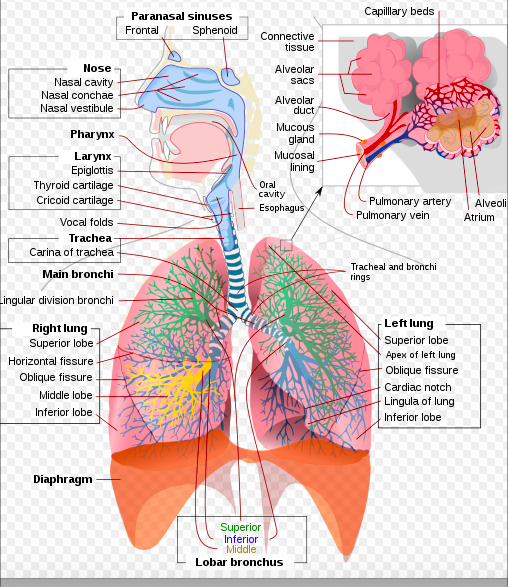
The human respiratory system comprises of
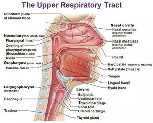
- Nose and Mouth: Air enters the respiratory system through the nose or the mouth. If it goes in the nostrils, the air is warmed and humidified. Cilia (tiny hairs) protect the nasal passageways and other parts of the respiratory tract, filtering out dust and other particles that enter the nose through the breathed air.
- Pharynx (throat): The nasal cavity and the mouth openings meet at the pharynx at the back of the nose and mouth. The pharynx is part of the digestive system as well as the respiratory system because it carries both food and air. At the bottom of the pharynx, this pathway divides in two, the esophagus, which leads to the stomach and the other for air. The epiglottis, a small flap of tissue, covers the air-only passage when we swallow, keeping food and liquid from going into the lungs.
- The Larynx (voice box): Top part of Trachea. This short tube contains a pair of vocal cords, which vibrate to make sounds.
- Trachea: The walls of the trachea are strengthened by stiff rings of cartilage to keep it open. The trachea is also lined with cilia, which sweep fluids and foreign particles out of the airway so that they stay out of the lungs. (1.Endotracheal tube (intubation) - A procedure by which a flexible plastic tube is inserted through the mouth to go down into the trachea; the clinician inserts the tube with the help of a laryngoscope, purpose of endotracheal intubation is to permit air to pass freely to and from the lungs in order to ventilate the lungs, may be connected to ventilator machines to provide artificial positive pressure ventilation.2.Tracheotomy - A surgical operation which creates an opening into the trachea with a tube inserted to provide a passage for air; performed when the pharynx is obstructed by eg edema; cancer)[3].
- Carina - The angle made between the two primary bronchi when they diverge at the tracheal bifurcation; it is richly innervated with sensory nerve endings to respond to the arrival of any aspirated material by initiating a cough reflex; it may be visualised as a ridge within the bronchial tree when using a bronchoscope[3].
- Bronchi: At its bottom end, the trachea divides into left and right air tubes called bronchi, which connect to the lungs. Within the lungs, the bronchi branch into smaller bronchi and even smaller tubes called bronchioles.
- Lungs: The lungs are the functional units of respiration and are key to survival. They contain 1500 miles of airways, 300-500 million alveoli and have a combined surface area of 70 square metres (half a tennis court). Each lung weighs approximately 1.1 kg. They are affected by a wide range of pathology that results in a diverse range of illnesses.[4]
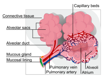
- Alveoli: Bronchioles end in tiny air sacs called alveoli, where the exchange of oxygen and carbon dioxide actually takes place. Each person has hundreds of millions of alveoli in their lungs.
This network of alveoli, bronchioles, bronchi and trachea is known as the tracheobronchial tree.[1][3].
The chest cavity (thorax) houses the bronchial tree, lungs, heart, and other structures.
- The top and sides of the thorax are formed by the ribs and attached muscles, and the bottom is formed by the diaphragm.
- The chest walls form a protective cage around the lungs and other contents of the chest cavity[1].
How the Respiratory System Works[edit | edit source]
The cells in our bodies need oxygen to stay alive. Carbon dioxide is a by-product of respiration. The lungs and respiratory system allow oxygen in the air to be taken into the body, while also letting the body get rid of carbon dioxide in the air breathed out.
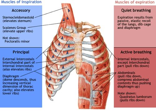
- When you breathe in, the diaphragm moves downward toward the abdomen, and the rib muscles pull the ribs upward and outward (making the chest cavity bigger and pulling air through the nose or mouth into the lungs). See muscles of Respiration.
- In exhalation, the diaphragm moves upward and the chest wall muscles relax, which causes the chest cavity to get smaller and push air out of the respiratory system through the nose or mouth.
- With each inhalation, air fills a large portion of the millions of alveoli. Oxygen diffuses from the alveoli to the blood through the capillaries lining the alveolar walls. Once in the bloodstream, oxygen gets picked up by the hemoglobin in red blood cells. This oxygen-rich blood then flows back to the heart, which pumps it through the arteries to oxygen needy tissues throughout the body.
- In the capillaries of the body tissues, oxygen is freed from the hemoglobin and moves into the cells.
- Carbon dioxide produced moves out of the cells into the capillaries, where most of it dissolves in the plasma of the blood.
- Blood rich in carbon dioxide then returns to the heart via the veins.
- From the heart, this blood is pumped to the lungs, where carbon dioxide passes into the alveoli to be exhaled[1].
Tracheobronchial tree[edit | edit source]
The tracheobronchial tree is a branching structure of tubes of an ever-decreasing diameter that start at the larynx and end in the alveoli. It can broadly be divided into conduction and respiratory zones.
- Gas is warmed and humidified as it is conducted from the oropharynx to the functional portion of the lung where gas exchange occurs.
- The conduction zone is composed of the trachea, bronchi, bronchioles and terminal bronchioles.
- Their function is to optimise gas delivery to the functional portion of the lung.
- Their walls contain cilia to remove particulates from the inspired gas and cartilage to ensure that they do not collapse in expiration.
2. Respiratory zone
- The respiratory zone is an extension of the tracheobronchial tree at the level of the terminal bronchioles.
- It is composed of respiratory bronchioles, alveolar ducts and alveoli, and is the location of gas transfer within the lung[4].
Alveoli[edit | edit source]
The alveoli (singular: alveolus) are tiny hollow air sacs that comprise the basic unit of respiration.
Alveoli are found within the lung parenchyma and are found at the terminal ends of the respiratory tree, clustered around alveolar sacs and alveolar ducts. Each alveolus is approximately 0.2 mm in diameter. There are around 300 million to 1 billion alveoli in the human lungs, covering an area of 70 square metres.
- The alveolar walls are comprised of collagen and elastic fibres which facilitate expansion during inspiration and return to the original shape during expiration. There are numerous capillaries within the alveolar walls where gas exchange occurs. Pores of Kohn are also located within the walls.
- Alveoli contain two major types of epithelial cells. The most abundant, type 1 pneumocytes (95%) are squamous cells in which gas exchange occurs. The remaining 5%, type 2 pneumocytes, are granular cells which secrete surfactant. Surfactant is a lipoprotein with a high phospholipid content which reduces surface tension. This increases pulmonary compliance, prevents atelectasis and aids recruitment of collapsed airways.
- Alveolar macrophages are also located in the alveoli. They protect the alveoli from foreign material by engulfing it, including bacteria, dust and carbon particles[5].
Gas exchange occurs in the lungs between alveolar air and blood of the pulmonary capillaries. For effective gas exchange to occur, alveoli must be ventilated and perfused. Ventilation (V) refers to the flow of air into and out of the alveoli, while perfusion (Q) refers to the flow of blood to alveolar capillaries.
- Individual alveoli have variable degrees of ventilation and perfusion in different regions of the heart.
- Collective changes in ventilation and perfusion in the lungs are measured clinically using the ratio of ventilation to perfusion (V/Q).
- Changes in the V/Q ratio can affect gas exchange and can contribute to hypoxemia[6].
References[edit | edit source]
- ↑ 1.0 1.1 1.2 1.3 Teenhealth Lungs and Respiratory System Available from:https://kidshealth.org/en/teens/lungs.html (last accessed 1.10.2020)
- ↑ AU essays Evolution of Respiratory Systems in Animals Available from:https://www.auessays.com/essays/biology/evolution-respiratory-systems-1447.php (last accessed 1.10.2020)
- ↑ 3.0 3.1 3.2 Respiratory systemhttp://apsubiology.org/anatomy/2020/2020_Exam_Reviews/Exam_3/CH22_Respiratory_Tree.htm (last accessed 1.10.2020)
- ↑ 4.0 4.1 Radiopedia The Lungs Available from:https://radiopaedia.org/articles/lung (last accessed 1.10.2020)
- ↑ Radiopedia Alveoli Available from:https://radiopaedia.org/articles/alveoli?lang=gb (last accessed 1.10.2020)
- ↑ Powers KA, Dhamoon AS. Physiology, Pulmonary, Ventilation and Perfusion.2019 Available from:https://www.ncbi.nlm.nih.gov/books/NBK539907/ (last accessed 2.10.2020)


