Reflexes: Difference between revisions
No edit summary |
(text) |
||
| Line 21: | Line 21: | ||
==== '''Deep Tendon (muscle stretch) Reflexes''' ==== | ==== '''Deep Tendon (muscle stretch) Reflexes''' ==== | ||
Evaluates afferent nerves, synaptic connections within the spinal cord, motor nerves, and descending motor pathways. Lower motor neuron lesions (eg affecting the anterior horn cell, spinal root or peripheral nerve) depress reflexes: upper motor neuron lesions increase the reflexes. | |||
Reflexes tested include the following: | Reflexes tested include the following: | ||
| Line 39: | Line 39: | ||
Jaw jerk (by the 5th cranial nerve) | Jaw jerk (by the 5th cranial nerve) | ||
Note any asymmetric increase or depression. Jendrassik maneuver can be used to augment hypoactive reflexes ie the patient locks the hands together and pulls vigorously apart as a tendon in the lower extremity is tapped or can push the knees together against each other, while the upper limb tendon is tested. | |||
==== '''Pathologic reflexes''' ==== | |||
Pathologic reflexes (eg, Babinski, rooting, grasp) are reversions to primitive responses and indicate loss of cortical inhibition. | |||
==== '''Other reflexes''' ==== | |||
Clonus (rhythmic, rapid alternation of muscle contraction and relaxation caused by sudden, passive tendon stretching) testing is done by rapid dorsiflexion of the foot at the ankle. Sustained clonus indicates an upper motor neuron disorder.<ref>MSD Manual. [https://www.msdmanuals.com/professional/neurologic-disorders/neurologic-examination/how-to-assess-reflexes How to access reflexes]. Available from: https://www.msdmanuals.com/professional/neurologic-disorders/neurologic-examination/how-to-assess-reflexes (last accessed 21.4.2019)</ref> | |||
<section> | |||
Evaluates afferent nerves, synaptic connections within the spinal cord, motor nerves, and descending motor pathways. Lower motor neuron lesions (eg, affecting the anterior horn cell, spinal root, or peripheral nerve) depress reflexes; upper motor neuron lesions (ie, non–basal ganglia disorders anywhere above the anterior horn cell) increase reflexes. | Evaluates afferent nerves, synaptic connections within the spinal cord, motor nerves, and descending motor pathways. Lower motor neuron lesions (eg, affecting the anterior horn cell, spinal root, or peripheral nerve) depress reflexes; upper motor neuron lesions (ie, non–basal ganglia disorders anywhere above the anterior horn cell) increase reflexes. | ||
Revision as of 07:18, 21 April 2019
Original Editor - Your name will be added here if you created the original content for this page.
Lead Editors
Spinal Reflex/The Reflex Arc[edit | edit source]
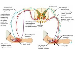
A reflex is an involuntary and nearly instantaneous movement in response to a stimulus. The reflex is an automatic response to a stimulus that does not receive or need conscious thought as it occurs through a reflex arc. Reflex arcs act on an impulse before that impulse reaches the brain.[1]
Relex arcs can be
- Monosynaptic ie contain only two neurons, a sensory and a motor neuron. Examples of monosynaptic reflex arcs in humans include the patellar reflex and the Achilles reflex.
- Polysynaptic ie multiple interneurons (also called relay neurons) that interface between the sensory and motor neurons in the reflex pathway.[2]
 Illustration of the reflex arc.
The video below illustrates the reflex arc
Reflex Testing[edit | edit source]
Deep Tendon (muscle stretch) Reflexes[edit | edit source]
Evaluates afferent nerves, synaptic connections within the spinal cord, motor nerves, and descending motor pathways. Lower motor neuron lesions (eg affecting the anterior horn cell, spinal root or peripheral nerve) depress reflexes: upper motor neuron lesions increase the reflexes.
Reflexes tested include the following:
Biceps (innervated by C5 and C6)
Radial brachialis (by C6)
Triceps (by C7)
Distal finger flexors (by C8)
Quadriceps knee jerk (by L4)
Ankle jerk (by S1)
Jaw jerk (by the 5th cranial nerve)
Note any asymmetric increase or depression. Jendrassik maneuver can be used to augment hypoactive reflexes ie the patient locks the hands together and pulls vigorously apart as a tendon in the lower extremity is tapped or can push the knees together against each other, while the upper limb tendon is tested.
Pathologic reflexes[edit | edit source]
Pathologic reflexes (eg, Babinski, rooting, grasp) are reversions to primitive responses and indicate loss of cortical inhibition.
Other reflexes[edit | edit source]
Clonus (rhythmic, rapid alternation of muscle contraction and relaxation caused by sudden, passive tendon stretching) testing is done by rapid dorsiflexion of the foot at the ankle. Sustained clonus indicates an upper motor neuron disorder.[4]
<section> Evaluates afferent nerves, synaptic connections within the spinal cord, motor nerves, and descending motor pathways. Lower motor neuron lesions (eg, affecting the anterior horn cell, spinal root, or peripheral nerve) depress reflexes; upper motor neuron lesions (ie, non–basal ganglia disorders anywhere above the anterior horn cell) increase reflexes.
Reflexes tested include the following:
- Biceps (innervated by C5 and C6)
- Radial brachialis (by C6)
- Triceps (by C7)
- Distal finger flexors (by C8)
- Quadriceps knee jerk (by L4)
- Ankle jerk (by S1)
- Jaw jerk (by the 5th cranial nerve)
Note any asymmetric increase or decrease. Jendrassik maneuver can be used to augment hypoactive reflexes ie the patient locks hands together and pulls vigorously apart as a tendon in the lower extremity is tapped or pushs the knees together against each other, while the upper limb tendon is tested.[5] </section><section></section>
Evidence[edit | edit source]
Provide the evidence for this technique here
Resources[edit | edit source]
add any relevant resources here
References[edit | edit source]
- ↑ Wikipedia. Reflex. Available from: https://en.wikipedia.org/wiki/Reflex (last accessed 21.4.2019)
- ↑ Lumen. Reflexes. https://courses.lumenlearning.com/boundless-ap/chapter/reflexes/ (last accessed 21.4.2019)
- ↑ Dr Matt and Dr Mikes Medical youtube. Spinal reflex Available from: https://www.youtube.com/watch?v=_qXS9KjyDC4&feature=youtu.be (last accessed 21.4.2019)
- ↑ MSD Manual. How to access reflexes. Available from: https://www.msdmanuals.com/professional/neurologic-disorders/neurologic-examination/how-to-assess-reflexes (last accessed 21.4.2019)
- ↑ MDA Maunaul. How to assess reflexes. Available from: https://www.msdmanuals.com/professional/neurologic-disorders/neurologic-examination/how-to-assess-reflexes (last accessed 21.4.2019)


