Pressure Ulcers
Original Editor - Jayati Mehta
Top Contributors - Jayati Mehta, Kim Jackson, Naomi O'Reilly, Lucinda hampton, Adam Vallely Farrell, Rucha Gadgil, Tony Lowe, Tarina van der Stockt, WikiSysop, Vidya Acharya, Lauren Lopez, Samuel Winter, Jess Bell and Robin Tacchetti
Introduction[edit | edit source]
Decubitus ulcers, also termed bedsores or pressure ulcers, are skin and soft tissue injuries that form as a result of constant or prolonged pressure exerted on the skin.
These ulcers
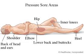
- Occur at bony areas of the body such as the ischium, greater trochanter, sacrum, heel, malleolus (lateral than medial), and occiput.
- Mostly occur in people with conditions that decrease their mobility making postural change difficult[1].
The terms decubitus ulcer (from Latin decumbere, “to lie down”), pressure sore, pressure ulcer and bedsores are often used interchangeably
Etiology[edit | edit source]
The development of decubitus ulcers is complex and multifactorial.
Loss of sensory perception, locally and general impaired loss of consciousness, along with decreased mobility, are the most important causes that aid in the formation of these ulcers (patients are not aware of discomfort hence do not relieve the pressure).
Both external and internal factors work simultaneously, forming these ulcers.
- External factors; pressure, friction, shear force, and moisture
- Internal factors; fever, malnutrition, anemia, and endothelial dysfunction speed up the process of these lesions.
The dysfunction of nervous regulatory mechanisms responsible for the regulation of local blood flow is somewhat culpable in the formation of these ulcers
- Prolonged pressure on tissues can cause capillary bed occlusion and, thus, low oxygen levels in the area.
- Over time, the ischemic tissue begins to accumulate toxic metabolites.
- Subsequently, tissue ulceration and necrosis occur.
Risk Factors Include:
- Neurologic disease
- Cardiovascular disease
- Prolonged anesthesia
- Dehydration
- Malnutrition
- Hypotension
- Surgical patients[1]
Epidemiology[edit | edit source]
Decubitus ulcers are a worldwide health care concern affecting tens of thousands of patients and costing over a billion dollars a year.[2]
- Their management costs billions of dollars per annum, burdening the already scarce health economy.
- Elderly patients are more prone to sacral decubitus ulcers
- Two-thirds of ulcers occur in patients who are over 70 years old
- Patients who are incontinent, paralyzed, or debilitated are more prone to getting them.
- Patients with normal sensory status, mobility, and mental status are less likely to form these ulcers because their normal physiologic feedback system leads to frequent physical positional shifts. .
- Data that shows 83% of hospitalized patients with ulcers developed them within five days of their hospitalization[1]
Pathophysiology[edit | edit source]
Many factors contribute to the development of pressure ulcers, but pressure leading to ischemia and necrosis is the final common pathway.
Pressure ulcers
- Result from constant pressure sufficient to impair local blood flow to soft tissue for an extended period.
- External pressure must be greater than the arterial capillary pressure (32 mm Hg) to impair inflow for an extended time
- Greater than the venous capillary closing pressure (8-12 mm Hg) to impede the return of flow for an extended time. [3]
- Tissues are capable of withstanding enormous pressures for brief periods, but prolonged exposure to pressures just slightly above capillary filling pressure initiates a downward spiral toward tissue necrosis and ulceration.
- The superficial dermis can tolerate ischemia for 2 to 8 hours before breakdown occurs.
- Deeper muscle, connective tissue, and fat tissues tolerate pressures for 2 hours or less (probably because of its increased need for oxygen and higher metabolic requirements).
- Often there is significant damage to underlying tissues while the epidermis and dermis remain intact.
- By the time ulceration is present through the skin level, significant damage of underlying muscle may already have occurred, making the overall shape of the ulcer an inverted cone.[4].
- Friction caused by skin rubbing against surfaces like clothing or bedding can also lead to the development of ulcers by contributing to breaks in the superficial layers of the skin.
- Moisture can cause ulcers and worsens existing ulcers via tissue breakdown and maceration[1].
Complications[edit | edit source]
Complications of pressure ulcers, some may be life-threatening, include:
- Cellulitis - Cellulitis is an infection of the skin and connected soft tissues. It can cause warmth, redness and swelling of the affected area. People with nerve damage often do not feel pain in the area affected by cellulitis.
- Bone and Joint Infections - An infection from a pressure sore can burrow into joints and bones. Joint infections (septic arthritis) can damage cartilage and tissue. Bone infections (osteomyelitis) can reduce the function of joints and limbs.
- Cancer - Long-term, non-healing wounds (Marjolin's ulcers) can develop into a type of squamous cell carcinoma.
- Sepsis - Rarely will a skin ulcer lead to sepsis.[5]
Pressure Sore Grading[edit | edit source]
The definitions of the four pressure ulcer stages are revised periodically by the National Pressure Ulcer Advisor Panel (NPUAP) in the United States and the European Pressure Ulcer Advisor Panel (EPUAP) in Europe. The National Pressure Ulcer Advisory Panel redefined the definition of a pressure ulcer and the stages of pressure ulcers in 2007, including the original 4 stages and adding 2 stages on deep tissue injury and unstageable pressure ulcers.[6]
The updated staging system includes the following definitions[7]:
Pressure Injury[edit | edit source]
A pressure injury is localized damage to the skin and/or underlying soft tissue usually over a bony prominence or related to a medical or other device. The injury can present as intact skin or an open ulcer and may be painful. The injury occurs as a result of intense and/or prolonged pressure or pressure in combination with shear. The tolerance of soft tissue for pressure and shear may also be affected by microclimate, nutrition, perfusion, co-morbidities and condition of the soft tissue
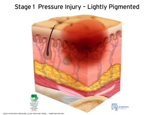
Stage 1 Pressure Injury: Non-blanchable Erythema of Intact Skin[edit | edit source]
- Intact skin with a localized area of non-blanchable erythema, which may appear differently in darkly pigmented skin.
- Presence of blanchable erythema or changes in sensation, temperature, or firmness may precede visual changes.
- Colour changes do not include purple or maroon discoloration; these may indicate deep tissue pressure injury.
- The area may be painful, firm, soft, warmer or cooler as compared to adjacent tissue.
- Stage I may be difficult to detect in individuals with dark skin tones. May indicate "at risk" persons (a heralding sign of risk)
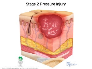
Stage 2 Pressure Injury: Partial-thickness Skin Loss with Exposed Dermis[edit | edit source]
- Partial-thickness loss of skin with exposed dermis.
- The wound bed is viable, pink or red, moist, and may also present as an intact or ruptured serum-filled blister.
- Adipose (fat) is not visible and deeper tissues are not visible.
- Granulation tissue, slough and eschar are not present.
- Presents as a shiny or dry shallow ulcer without slough or bruising (bruising indicates suspected deep tissue injury)
- These injuries commonly result from adverse microclimate and shear in the skin over the pelvis and shear in the heel.
- This stage should not be used to describe moisture associated skin damage (MASD) including incontinence associated dermatitis (IAD), intertriginous dermatitis (ITD), medical adhesive related skin injury (MARSI), or traumatic wounds (skin tears, burns, abrasions).
- This stage should not be used to describe skin tears, tape burns, perineal dermatitis, maceration or excoriation.
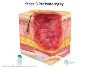
Stage 3 Pressure Injury: Full-thickness Skin Loss[edit | edit source]
- Full-thickness loss of skin, in which adipose (fat) is visible in the ulcer and granulation tissue and epibole (rolled wound edges) are often present.
- Slough and/or eschar may be visible.
- The depth of tissue damage varies by anatomical location; areas of significant adiposity can develop deep wounds.
- Undermining and tunneling may occur.
- Fascia, muscle, tendon, ligament, cartilage and/or bone are not exposed.
- If slough or eschar obscures the extent of tissue loss this is an Unstageable Pressure Injury.
- The depth of a stage III pressure ulcer varies by anatomical location.
- The bridge of the nose, ear, occiput and malleolus do not have subcutaneous tissue and stage III ulcers can be shallow.
- In contrast, areas of significant adiposity can develop extremely deep stage III pressure ulcers.
- Bone/tendon is not visible or directly palpable.
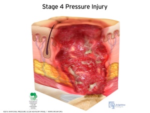
Stage 4 Pressure Injury: Full-thickness Skin and Tissue Loss[edit | edit source]
- Full-thickness skin and tissue loss with exposed or directly palpable fascia, muscle, tendon, ligament, cartilage or bone in the ulcer.
- Slough and/or eschar may be visible. Epibole (rolled edges), undermining and/or tunneling often occur.
- Depth varies by anatomical location.
- The bridge of the nose, ear, occiput and malleolus do not have subcutaneous tissue and these ulcers can be shallow.
- If slough or eschar obscures the extent of tissue loss this is an Unstageable Pressure Injury.
- Stage IV ulcers can extend into muscle and/or supporting structures (e.g., fascia, tendon or joint capsule) making osteomyelitis possible.
- Exposed bone/tendon is visible or directly palpable.
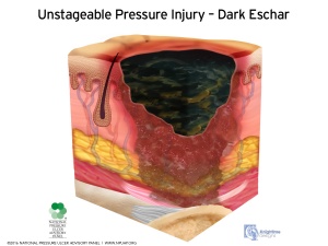
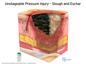
Unstageable Pressure Injury: Obscured Full-thickness Skin and Tissue Loss[edit | edit source]
- Full-thickness skin and tissue loss in which the extent of tissue damage within the ulcer cannot be confirmed because it is obscured by slough or eschar.
- If slough or eschar is removed, a Stage 3 or Stage 4 pressure injury will be revealed.
- Stable eschar (i.e. dry, adherent, intact without erythema or fluctuance) on the heel or ischemic limb should not be softened or removed.
- Until enough slough and/or eschar is removed to expose the base of the wound, the true depth, and therefore stage, cannot be determined.
Symptoms[edit | edit source]
As mentioned previously, pressure ulcers can affect any part of the body that is put under pressure. They often develop gradually, but can sometimes form in just a few hours.
Early Symptoms[edit | edit source]
- Discolouration of parts of the skin- those with pale skin tend to develop red patches, while people with darker skin tend to get purple or blue patches
- Discoloured patches not turning white when pressure is applied
- A patch of skin that is warm, spongy or hard
- Pain or itchiness in the area affected
Later Symptoms[edit | edit source]
The skin may not be broken at first, but if the pressure ulcer gets worse it may form:
- An open wound or blister (Stage 2)
- A deep wound which reaches the deeper layers of the skin (Stage 3)
- A very deep wound that may reach the muscle and bone (Stage 4)
Clinical Presentation[edit | edit source]
The severity of pressure ulceration can be estimated by observing clinical signs. A progression from least tissue damage to most severe damage is presented here.[8]
- The first clinical sign of pressure ulceration is blanchable erythemaalong with increased skin temperature. If pressure is relieved, tissues may recover in 24 hours. If pressure is unrelieved, nonblanchable erythema occurs.
- Progression to a superficial abrasion, blister, or shallow crater indicates involvement of the dermis.
- When full-thickness skin loss is apparent, the ulcer appears as a deep crater. Bleeding is minimal, and tissues are indurated and warm. Eschar formation marks full-thickness skin loss. Tunneling or undermining is often present.
- The majority of all pressure ulcers develop over six primary bony areas sacrum, coccyx, greater trochanter, ischial tuberosity, calcaneus (heel), and lateral malleolus.
Diagnosis[edit | edit source]
If an individual has a history of a period of immobility followed by the discovery of a warm, red, spot over a bony prominence, a pressure ulcer can usually be confirmed. If the spot is unnaturally soft to the touch, sometimes referred to as “boggy,” this is enough evidence to suspect that damage is deeper than the epidermis.[8]
Tests[edit | edit source]
The following tests may be performed :
- Blood tests
- Tissue cultures to diagnose a bacterial or fungal infection in a wound that doesn't heal with treatment or is already at stage IV.
- Tissue cultures to check for cancerous tissue in a chronic, non-healing wound.[9]
Treatment / Management[edit | edit source]
Managing decubitus ulcers is complicated as there is no fixed treatment regime/algorithm.
Once it has developed, there should be no delay in treatment, and management should start immediately.
- Treatment varies between site, stage, and associated complications of the ulcer. The goal of all the various treatment options is to; minimize the pressure exerted on the ulcer, minimize contact of the ulcer with a hard surface, decrease moisture, and to keep it as aseptic or least septic as possible. The choice of treatment options should be according to the stage/grade of the ulcer, and what the purpose of the treatment should be (decreasing moisture, removal of necrotic tissue, controlling bacteremia).
- Prevention is clearly the best treatment with excellent skincare, pressure dispersion cushions, and support surfaces. Support surfaces decrease the amount of pressure on the wound. Support surfaces can be either static (e.g., air, foam, and water mattress overlays) or dynamic (e.g., alternating air overlay). Repositioning and turning the patient every two hours can also lessen pressure on the area, but some patients may require more frequent repositioning, while others may require less frequent repositioning.
- In some cases, urinary and fecal diversion may be necessary depending on the site of ulcer, being prone to urine or fecal contamination.
- Hydrocolloid dressings should be used. Good antibiotic cover decreases septicemia.
- The depth and severity of the ulcer determine whether surgical management may be required.
- Some evidence exists suggesting that hyperbaric oxygen therapy can help with wound healing, as it improves oxygenation in and around the area of the wound.
In Summary - Treatment of decubitus ulcers has its basis on the following:
- Prevention of additional ulcers
- Decreasing pressure on the wound
- Wound management
- Surgical intervention
- Nutrition[1]
Prevention[edit | edit source]
Patients and their family members should have a clear idea that preventing recurrence requires commitment and responsibility. They should
- Receive education on how to manage the condition in the hospital and as well as in their homes.
- Be familiar with warning signs like skin discoloration, ulceration, discharge, or a foul smell from the ulcer site and body areas with decreased or no sensation.
The patient should
- Move or turn every 2 hours; it could not be done by themselves, or they should ask someone to help them.
- Use air or water mattress in their homes.
- Have adequate food intake adequate and it should consist of a balanced and healthy diet[1].
Reference[edit | edit source]
- ↑ 1.0 1.1 1.2 1.3 1.4 1.5 Zaidi SR, Sharma S. Decubitus Ulcer. InStatPearls [Internet] 2020 Jan 18. StatPearls Publishing. Available from:https://www.statpearls.com/kb/viewarticle/20286 (last accessed 21.9.2020)
- ↑ Bansal C, Scott R, Stewart D, Cockerell CJ. Decubitus ulcers: a review of the literature. Int J Dermatol. 2005;44(10):805-810. doi:10.1111/j.1365-4632.2005.02636.x Available from: (last accessed 21.9.2020)https://pubmed.ncbi.nlm.nih.gov/16207179/
- ↑ Bridel J. The aetiology of pressure sores. Journal of Wound Care. 1993 Jul 2;2(4):230-8.
- ↑ Defloor T. The risk of pressure sores: a conceptual scheme. Journal of clinical nursing. 1999 Mar;8(2):206-16.
- ↑ Ahn H, Cowan L, Garvan C, Lyon D, Stechmiller J. Risk factors for pressure ulcers including suspected deep tissue injury in nursing home facility residents: analysis of national minimum data set 3.0. Advances in skin & wound care. 2016 Apr 1;29(4):178-90.
- ↑ Edsberg LE, Black JM, Goldberg M, McNichol L, Moore L, Sieggreen M. Revised National Pressure Ulcer Advisory Panel pressure injury staging system: revised pressure injury staging system. Journal of Wound, Ostomy, and Continence Nursing. 2016 Nov;43(6):585.
- ↑ Black J, Baharestani MM, Cuddigan J, Dorner B, Edsberg L, Langemo D, Posthauer ME, Ratliff C, Taler G. National Pressure Ulcer Advisory Panel's updated pressure ulcer staging system. Advances in skin & wound care. 2007 May 1;20(5):269-74.
- ↑ 8.0 8.1 Susan B. O’Sullivan,Thomas J. Schmitz,George D. Fulk, Physical Rehabilitstion,6th edition,United States of America,F.A. Davis Company,2014
- ↑ http://www.mayoclinic.org/diseases-conditions/bedsores/basics/tests-diagnosis/con-20030848
