Pleurodesis: Difference between revisions
Rosie Swift (talk | contribs) (added image) |
Rosie Swift (talk | contribs) No edit summary |
||
| Line 10: | Line 10: | ||
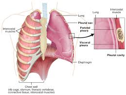
A pleurodesis is a form of pleural surgery which aims to obliterate the pleural space between the visceral and parietal pleura. This involves a chemical or surgical procedure to create an adhesion between the two pleura. | A pleurodesis is a form of pleural surgery which aims to obliterate the pleural space between the visceral and parietal pleura. This involves a chemical or surgical procedure to create an adhesion between the two pleura. | ||
* Chemical pleurodesis: This is done by inserting a sclerosing agent into the pleural cavity via a chest tube. The drug used causes the pleural surfaces to become sticky and bond together, therefore closing the pleural space. The type of sclerosing agent to be used depends on local expertise, availability and the underlying reason why the pleurodesis is required. Drugs used include: sterile medical talc, Tetracyclines (minocycline, doxycycline), Silver nitrate, Iodopovidone, Bleomycin, Corynebacterium parvum with parenteral methylprednisolone acetate, Erythromycin, Fluorouracil, Interferon beta, Autologous blood, Mitomycin C, Cisplatin, Cytarabine, Doxorubicin, Etoposide, Bevacizumab (intravenous or intrapleural) and Streptococcus pyogenes A3 (OK-432)<ref name=":0" /> | * Chemical pleurodesis: This is done by inserting a sclerosing agent into the pleural cavity via a chest tube. The drug used causes the pleural surfaces to become sticky and bond together, therefore closing the pleural space. The type of sclerosing agent to be used depends on local expertise, availability and the underlying reason why the pleurodesis is required. Drugs used include: sterile medical talc, Tetracyclines (minocycline, doxycycline), Silver nitrate, Iodopovidone, Bleomycin, Corynebacterium parvum with parenteral methylprednisolone acetate, Erythromycin, Fluorouracil, Interferon beta, Autologous blood, Mitomycin C, Cisplatin, Cytarabine, Doxorubicin, Etoposide, Bevacizumab (intravenous or intrapleural) and Streptococcus pyogenes A3 (OK-432)<ref name=":0">Ali M, Surani S. [https://www.ncbi.nlm.nih.gov/books/NBK560685/ Pleurodesis]. 2021. In: StatPearls [Internet]. Accessed 25 Mar 2022</ref> | ||
* Surgical pleurodesis: This is done through medical thoracoscopy, video-assisted thoracoscopy (VATS), or open thoracotomy, where either a sclerosis agent is placed in the pleural cavity or mechanical abrasion (also termed dry abrasion) is achieved by draining the pleural fluid using a tunnelled catheter (induces pleurodesis without instillation of a sclerosing agent). | * Surgical pleurodesis: This is done through medical thoracoscopy, video-assisted thoracoscopy (VATS), or open thoracotomy, where either a sclerosis agent is placed in the pleural cavity or mechanical abrasion (also termed dry abrasion) is achieved by draining the pleural fluid using a tunnelled catheter (induces pleurodesis without instillation of a sclerosing agent)<ref name=":0" />. | ||
Analgesia such as an NSAID or opiate, is usually given for the duration of the procedure. Patients can expect to be an inpatient for at least 24 hours following the procedure. Occasionally a single stitch may be used to close the insertion site of the chest drain. | Analgesia such as an NSAID or opiate, is usually given for the duration of the procedure. Patients can expect to be an inpatient for at least 24 hours following the procedure, longer if surgical pleurodesis is performed. Occasionally a single stitch may be used to close the insertion site of the [[Chest Drains|chest drain]]. | ||
== Indication == | == Indication == | ||
[[File:Lung pleura.jpg|thumb|Lung pleura]]Any condition which causes extra fluid to collect in the pleural cavity may require a pleurodesis. These include: heart failure, pneumonia, tuberculosis, cancer, liver and kidney disease | [[File:Lung pleura.jpg|thumb|Lung pleura]]The pleural space between the parietal and visceral pleura usually contains around 50ml of pleural fluid<ref name=":0" />. Under pathological conditions, such as [[pneumothorax]] or [[Pleural Effusion|pleural effusion]], air or excess fluid can build up in the pleural space. If this becomes recurrent and significantly symptomatic, pleurodesis may be indicated. | ||
Any condition which causes extra fluid to collect in the pleural cavity may require a pleurodesis. These include: [[Congestive Heart Failure|heart failure]], [[pneumonia]], [[tuberculosis]], cancer, liver and kidney disease and inflammation of the pancreas<ref name=":1">Healthline. [https://www.healthline.com/health/pleurodesis Pleurodesis] [online]. Accessed 31 Mar 2022.</ref>. | |||
Typically, a recurrent pleural effusion (particularly if malignant) or recurrent or persistent pneumothorax may be treated by pleurodesis. | Typically, a recurrent pleural effusion (particularly if malignant) or recurrent or persistent pneumothorax may be treated by pleurodesis. | ||
== Clinical Presentation == | == Clinical Presentation == | ||
Depending on the reason that the pleurodesis is required, patients may present with signs of [[Pleural Effusion|pleural effusion]] or [[pneumothorax]], such as: | Depending on the reason that the pleurodesis is required, patients may present with signs of [[Pleural Effusion|pleural effusion]] or [[pneumothorax]], such as: | ||
* | * Breathlessness | ||
* | * Chest pain | ||
* | * Cough | ||
* | * Fever | ||
* | * Tachycardia | ||
* | * Tachypnoea | ||
* | * Fatigue | ||
== Complications == | == Complications == | ||
| Line 39: | Line 39: | ||
* Chest pain | * Chest pain | ||
* Fever | * Fever | ||
* | * Breathlessness (localised inflammatory reaction to the procedure) | ||
* Infection | * Infection at the insertion site | ||
* Empyema | * Empyema | ||
| Line 46: | Line 46: | ||
Complications from surgical pleurodesis include: | Complications from surgical pleurodesis include: | ||
* | * Pneumothorax<ref name=":1" /> | ||
* | * Injury to the chest wall, arteries, or lungs | ||
* | * Blood clots, [[Pulmonary Embolism|pulmonary embolism]]<ref name=":1" /> | ||
In the case of failed pleurodesis, pleurectomy may be required to control malignant pleural effusions<ref name=":0" />. Patients must be good surgical candidates and have a reasonably long expected survival because total radical pleurectomy/decortication requires a thoracotomy and is a major surgical procedure associated with considerable morbidity and some mortality. | In the case of failed pleurodesis, pleurectomy may be required to control malignant pleural effusions<ref name=":0" />. Patients must be good surgical candidates and have a reasonably long expected survival because total radical pleurectomy/decortication requires a thoracotomy and is a major surgical procedure associated with considerable morbidity and some mortality. | ||
| Line 57: | Line 56: | ||
== References == | == References == | ||
<references /> | |||
Revision as of 09:41, 31 March 2022
This article or area is currently under construction and may only be partially complete. Please come back soon to see the finished work! (31/03/2022)
Description[edit | edit source]
A pleurodesis is a form of pleural surgery which aims to obliterate the pleural space between the visceral and parietal pleura. This involves a chemical or surgical procedure to create an adhesion between the two pleura.
- Chemical pleurodesis: This is done by inserting a sclerosing agent into the pleural cavity via a chest tube. The drug used causes the pleural surfaces to become sticky and bond together, therefore closing the pleural space. The type of sclerosing agent to be used depends on local expertise, availability and the underlying reason why the pleurodesis is required. Drugs used include: sterile medical talc, Tetracyclines (minocycline, doxycycline), Silver nitrate, Iodopovidone, Bleomycin, Corynebacterium parvum with parenteral methylprednisolone acetate, Erythromycin, Fluorouracil, Interferon beta, Autologous blood, Mitomycin C, Cisplatin, Cytarabine, Doxorubicin, Etoposide, Bevacizumab (intravenous or intrapleural) and Streptococcus pyogenes A3 (OK-432)[1]
- Surgical pleurodesis: This is done through medical thoracoscopy, video-assisted thoracoscopy (VATS), or open thoracotomy, where either a sclerosis agent is placed in the pleural cavity or mechanical abrasion (also termed dry abrasion) is achieved by draining the pleural fluid using a tunnelled catheter (induces pleurodesis without instillation of a sclerosing agent)[1].
Analgesia such as an NSAID or opiate, is usually given for the duration of the procedure. Patients can expect to be an inpatient for at least 24 hours following the procedure, longer if surgical pleurodesis is performed. Occasionally a single stitch may be used to close the insertion site of the chest drain.
Indication[edit | edit source]
The pleural space between the parietal and visceral pleura usually contains around 50ml of pleural fluid[1]. Under pathological conditions, such as pneumothorax or pleural effusion, air or excess fluid can build up in the pleural space. If this becomes recurrent and significantly symptomatic, pleurodesis may be indicated.Any condition which causes extra fluid to collect in the pleural cavity may require a pleurodesis. These include: heart failure, pneumonia, tuberculosis, cancer, liver and kidney disease and inflammation of the pancreas[2].
Typically, a recurrent pleural effusion (particularly if malignant) or recurrent or persistent pneumothorax may be treated by pleurodesis.
Clinical Presentation[edit | edit source]
Depending on the reason that the pleurodesis is required, patients may present with signs of pleural effusion or pneumothorax, such as:
- Breathlessness
- Chest pain
- Cough
- Fever
- Tachycardia
- Tachypnoea
- Fatigue
Complications[edit | edit source]
Complications from chemical pleurodesis include:
- Chest pain
- Fever
- Breathlessness (localised inflammatory reaction to the procedure)
- Infection at the insertion site
- Empyema
Complications from surgical pleurodesis include:
- Pneumothorax[2]
- Injury to the chest wall, arteries, or lungs
- Blood clots, pulmonary embolism[2]
In the case of failed pleurodesis, pleurectomy may be required to control malignant pleural effusions[1]. Patients must be good surgical candidates and have a reasonably long expected survival because total radical pleurectomy/decortication requires a thoracotomy and is a major surgical procedure associated with considerable morbidity and some mortality.
Resources[edit | edit source]
Oxford Centre for Respiratory Medicine. Pleurodesis Information for patients [online]. Accessed 31 March 2022
References[edit | edit source]
- ↑ 1.0 1.1 1.2 1.3 Ali M, Surani S. Pleurodesis. 2021. In: StatPearls [Internet]. Accessed 25 Mar 2022
- ↑ 2.0 2.1 2.2 Healthline. Pleurodesis [online]. Accessed 31 Mar 2022.


