Pivot Shift
Original Editor - Beth Barrett, Heleen Van Cleynenbreugel
Lead Editors - Your name will be added here if you are a lead editor on this page. Read more.
Search Strategy[edit | edit source]
add text here related to databases searched, keywords, and search timeline
Definition/Description[edit | edit source]
The pivot shift is a dynamic but passive test of knee stability, carried out by the examiner without any activity of the patient. It shows a dysregulation between rolling and gliding in the kneejoint. The patient lies in supine. The movement is a combination of axial load and valgus force, applied by the examiner, during a knee flexion from an extended position. When the test is positive, it indicates an injury of the anterior cruciate ligament. [1](A2)[2](B)
Clinically Relevant Anatomy 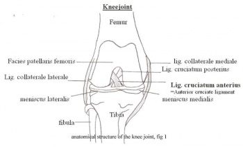 [edit | edit source]

The kneejoint or articulatio genus is the biggest and most complicated synovial joint in the human body. It’s formed by the distal end of the femur and the proximal end of the tibia. Between these two, the medial and lateral meniscus are located. The anterior part of the femur (facies patellaris femoris) articulates with the patella which forms the patellofemoral joint. The cruciate ligaments, situated in the intercondylair space, and the collateral ligaments are important ligaments in the knee.
The anterior cruciate ligament (ACL) joins the area intercondylaris anterior with the medial aspect of the lateral condyle. This ligament extends upwards, dorsally and laterally from the tibial plateau. It prevents the tibia from moving ventrally.
The posterior cruciate ligament (PCL) joins the area intercondylaris posterior with the lateral apect of the medial femurcondyle. The ligament runs medially, straight up and slightly forward. The PCL prevents posterior instability in the knee joint, this means that the tibia doesn’t displace to posteriorly. (Fig 1) [3]
The PCL is stronger than the ACL. Both cruciate ligaments run in a torsion. They stabilize the knee in the sagittal plane. When the knee is in endorotation, the cruciate ligaments are strongly crossed. When in exorotation, they run more parallel. The cruciate ligaments are primarily located in the posterior part of the joint capsule, so the flexion and extension aren’t hampered.[4][5]
Purpose
[edit | edit source]
The purpose of this test is to detect anterolateral rotary instability of the knee. The structures that could be compromised if this test is positive are the ACL, LCL, posterolateral capsule, arcuate complex and ITB.
Technique
[edit | edit source]
The patient lies supine with legs relaxed. The examiner grasps the heel of the involved leg with examiners opposite hand placed laterally on the proximal tibia just distal to the knee. The examiner then applies a valgus stress and an axial load while internally rotating the tibia as the knee is moved into flexion from a fully extended position. [6] A positive test is indicated by subluxation of the tibia while the femur rotates externally followed by a reduction of the tibia at 30-40 degrees of flexion.
Key Research[edit | edit source]
Is the pivot shift test a reliable test?
The Pivot Shift test attempts to reproduce the rotary and transalatory instability in an ACL deficient knee.
[7]
The test has a sensitivity from 0.18 to 0.48 and a specificity from 0.97 to 0.99 for diagnosing an ACL tear. The mean sensitivity and specificity are respectively 0.32 and 0.98.
[8]
(A1)
[9]
(A1)
[10]
(A1)
[11]
(A1)
[12]
(A1)
Resources
[edit | edit source]
add appropriate resources here
Clinical Bottom Line[edit | edit source]
add text here
Recent Related Research (from Pubmed)[edit | edit source]
Failed to load RSS feed from http://eutils.ncbi.nlm.nih.gov/entrez/eutils/erss.cgi?rss_guid=1houoX_LGC3R5rodk9lCaF7S5evZ4MXBE6rFjh2K9YsNYupEmB|charset=UTF-8|short|max=10: Error parsing XML for RSS
References[edit | edit source]
- ↑ Musahl V., Citak M., O’Loughlin PF., Choi D., Bedi A., Pearle AD. The effect of medial versus lateral menisectomy on the stability of the anterior cruciate ligament-deficient knee. Am J Sports Med 2010;38(8):1591-7. http://www.ncbi.nlm.nih.gov/pubmed/20530720?dopt=AbstractPlus full text: http://ajs.sagepub.com.ezproxy.vub.ac.be:2048/content/38/8/1591.full.pdf+htmlfckLR(accessed 19 Nov 2010). Level of evidence: A2
- ↑ Jakob RP., Staubli HU., Deland JT. Grading the pivot shift, objective tests with implications for treatment. VOL 69-B, No.2 1987. http://web.jbjs.org.uk/cgi/reprint/69-B/2/294.pdf (accessed 16 Nov 2010). Level of evidence: B
- ↑ http://www.taosortho.com/patientinfo/Medical/knees/ACL.htm (figure 1)
- ↑ Schünke M., Schulte E., Schumacher U., Voll M., Wesker K., Anatomische atlas Prometheus, Algemene anatomie en bewegingsapparaat. Onderste extemiteit: Botten, banden en gewrichten. Houten, 2008.p394, 396.
- ↑ Kapandji IA. Bewegingsleer, de onderste extremiteit deel 2. De functie van de kruisbanden. Houten, 2009. p135.
- ↑ Baxter R. Pocket guide to musculoskeletal assessment, second edition. Elsevier Science 2003.
- ↑ Lane CG, Warren R, Pearl AD. The pivot shift. J Am Acad Orthop Surg. 2008 Dec;16(12):679-88.
- ↑ Scholten RJPM, Opstelten W, van der Plas CG, Bijl D, Deville WLJM, Bouter LM (2003) Accuracy of physical diagnostic tests for assessing ruptures of the anterior cruciate ligament: a meta-analysis. J Fam Pract 52:689-94 http://www.ncbi.nlm.nih.gov/pubmed/12967539 full text: http://web.ebscohost.com.ezproxy.vub.ac.be:2048/ehost/pdfviewer/pdfviewer?hid=18&sid=25c1a906-13aa-4c59-ba43-a64e200c3c12%40sessionmgr13&vid=2 (accessed 18 Nov 2010. Level of evidence: A1
- ↑ Briggs KK, Lysholm J, Tegner Y, Rodkey WG, Kocher MS, Steadman JR (2009) The reliability, validity, and responsiveness of the Lysholm score and Tegner activity scale for anterior cruciate ligament injuries of the knee: 25 years later. The American journal of sports medicine 37:890-897 http://ajs.sagepub.com.ezproxy.vub.ac.be:2048/content/37/5/890.full.pdf+html (accessed 18 Nov 2010). Level of evidence: A1
- ↑ Prins M. The lachman test is the most sensitive and the pivot shift the most specific test for the diagnosis of ACL rupture. Fysiotherapeuten nr 8, 2006. http://www.fysioterapeuten.no/xp/pub/mx/filer/0806_Cap.pdf (accessed 17 Nov 2010). Level of evidence: A1
- ↑ Van der Plas CG., Opstelten W., Devillé WLJM., et al. Fysische diagnostiek - de waarde van enkele gebruikelijke tests voor het aantonen van een voorstekruisbandruptuur: meta-analyse. Ned Tijdschr Geneeskd. 2005;149:83-8. http://www.ntvg.nl/publicatie/fysische-diagnostiek-de-waarde-van-enkele-gebruikelijke-tests-voor-het-aantonen-van-een-v/volledig (accessed 16 Nov 2010) Level of evidence: A1
- ↑ Ostrowski JA. Accuracy of 3 Diagnostic Tests for Anterior Cruciate Ligament Tears.J Athl Train 2006; 41(1): 120–121. http://www.ncbi.nlm.nih.gov/pmc/articles/PMC1421494/ (accessed 16 Nov 2010) Level of evidence: A1

