Pelvic Fractures
Original Editors Rachael Lowe
Top Contributors - Descheemaeker Kari, Julie Stainier, Lynn Wright, Jentel Van De Gucht, Admin, Rachael Lowe, Lucinda hampton, 127.0.0.1, Kim Jackson, Rosie Swift, Scott Buxton, Lauren Lopez, Aminat Abolade, Siobhán Cullen, Karen Wilson, Vidya Acharya and Claire Knott
Definition/description[edit | edit source]
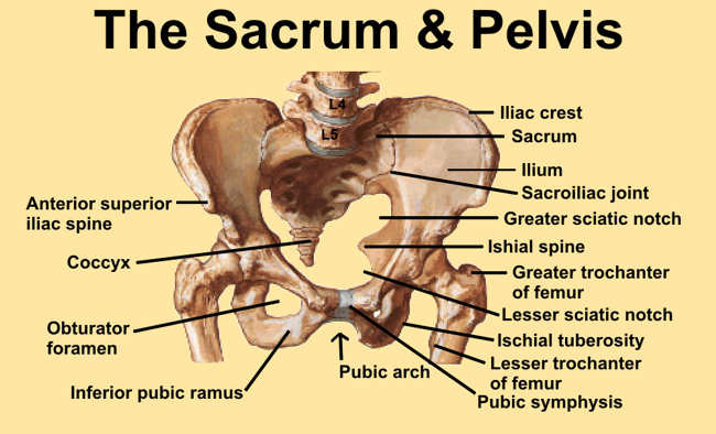
A pelvic fracture is a disruption of the bony structures of the Pelvis. The pelvis consists of the ilium, ischium and pubis. These form an anatomic ring with the sacrum. Disruption of the ring requires a lot of energy. Because in most of the cases pelvic fractures are caused by high impact, it is possible that organs in the bony pelvis are affected. Trauma to extra-pelvic organs is common. They are often associated with severe hemorrhage due to the extensive blood supply to the region. Cite error: Invalid <ref> tag; name cannot be a simple integer. Use a descriptive titleCite error: Invalid <ref> tag; name cannot be a simple integer. Use a descriptive title Pelvic fractures are associated with a high morbidity and mortality. Emergency care with primary aim reducing blood loss is always necessary. Cite error: Invalid <ref> tag; name cannot be a simple integer. Use a descriptive titleCite error: Invalid <ref> tag; name cannot be a simple integer. Use a descriptive title
Classification[edit | edit source]
There are two classification systems who are used most commonly to describe pelvic fractures:
Classification of pelvic fractures by Tile is based on the integrity of the posterior sacroiliac complex. Cite error: Invalid <ref> tag; name cannot be a simple integer. Use a descriptive titleCite error: Invalid <ref> tag; name cannot be a simple integer. Use a descriptive title
- Type A: rotationally and vertically stable, the sacroiliac complex is intact. Mostly managed nonoperatively.
- A1: avulsion fractures
- A2: stable iliac wing fractures or minimally displaced pelvic ring fractures
- A3: transverse sacral or coccyx fractures
- Type B: rotationally unstable and vertically stable, caused by external or internal rotational forces, results in partial disruption of the posterior sacroiliac complex.
- B1: open-book injuries
- B2: LC injuries
- B3: bilateral type B injuries
- Type C: rotationally unstable and vertically unstable, complete disruption of the posterior sacroiliac complex, result of great force.
- C1: unilateral injury
- C2: bilateral injuries in which one side is a type B and the controlateral side is a type C injury
- C3: bilateral injury in which both sides are type C injuries
Classification of pelvic fractures by Young and Burgess is based on mechanism of injury: lateral compression, anteroposterior compression, vertical shear or a combination of forces. Cite error: Invalid <ref> tag; name cannot be a simple integer. Use a descriptive titleCite error: Invalid <ref> tag; name cannot be a simple integer. Use a descriptive titleCite error: Invalid <ref> tag; name cannot be a simple integer. Use a descriptive title
- Grade I: associated sacral compression on side of impact. Associated widening of pubic symphysis or of the anterior sacroiliac joint, while ligaments remain intact.
- Grade II: associated posterior iliac fracture on side of impact. Associated widening of the anterior SI joint caused by disruption of the anterior SI, sacrotuberous and sacrospinous ligaments, posterior ligaments remain intact.
- Grade III: associated controlateral sacroiliac joint injury. Complete SI joint disruption with lateral displacement and disrupted anterior SI, sacrotuberous, sacrospinous and posterior SI ligaments.
Epidemiology/ etiology[edit | edit source]
Pelvic fractures have an incidence of 37 cases per 100000 person-years in the United States. The appearance of pelvic fractures is the greatest in people aged between 15-28 years. In persons younger than 35, pelvic fractures occur more in males than females. In persons older than 35, pelvic fractures occur more in females than males. In younger people pelvic fractures occur mostly as result of high-energy mechanisms, in older people they occur from minimal trauma, such as a low fall. Elderly people with Osteoporosis have a higher risk factor. Low- energy fractures are usually stable fractures of the pelvic ring. High-energy pelvic fractures occur most commonly after motor vehicle crashes, motorcycle crashes, motor vehicles striking pedestrians and falls. This are mostly avulsion fractures of the superior or inferior iliac spines or with apophyseal avulsion fractures of the iliac wing or ischial tuberosity. Cite error: Invalid <ref> tag; name cannot be a simple integer. Use a descriptive titleCite error: Invalid <ref> tag; name cannot be a simple integer. Use a descriptive title
Clinical Presentation[edit | edit source]
Patients with low-energy injuries usually present with a history of trauma like a fall from a standing or seated position onto a bony prominence or excessive strain on a muscle that inserts onto the Pelvis. Swelling, pain, ecchymosis, erythema and focal tenderness may also be present. With avulsion injuries there is often pain associated with contraction of the involved muscles. Cite error: Invalid <ref> tag; name cannot be a simple integer. Use a descriptive title
Patients with high-energy injuries present usually after motor vehicle accidents, falls and crush injuries. In severe cases, this patients complain of pain in the pelvis, lower back pain, buttocks and/or hips. Usually they are unable to stand. Concomitant distracting injuries or intoxication may limit the reliability of the history. In patients with altered mental status or spinal neurologic deficits the presence of pelvic fractures should be assumed until it can be excluded. Physical findings include abnormal position of the lower limbs, pelvic deformity or Pelvic instability, swelling and ecchymosis. The abdomen, perineum, genitals, rectum and lower back must be examined very carefully. Cite error: Invalid <ref> tag; name cannot be a simple integer. Use a descriptive title High-energy fractures are often associated with severe injuries of other organs. Cite error: Invalid <ref> tag; name cannot be a simple integer. Use a descriptive title
Physical Therapy Management[edit | edit source]
Low-energy injuries are usually managed with conservative care. This included bed rest, pain control and physical therapy. Cite error: Invalid <ref> tag; name cannot be a simple integer. Use a descriptive title. Physical therapy include gait training, stabilization exercises and mobility training. Cite error: Invalid <ref> tag; name cannot be a simple integer. Use a descriptive title. Early mobilization is very important. The patient must get out of the bed as soon as possible. Prolonged immobilization can lead to a number of complications including respiratory and circulatory compromise.
The intensity of the rehabilitation depends on whether the fracture was stable or unstable. The goals of the physical therapy program should be provide the patient with an optimal return of function by improving functional skills, self-care skills and safety awareness. Cite error: Invalid <ref> tag; name cannot be a simple integer. Use a descriptive title In people with surgical treatment (ex: ORIF), after 1 or 2 days of bed rest physical therapy is initiated to begin transfer and exercise training. The short-term goals are independence with transfers and wheelchair mobility. After leaving the hospital it is easier for the patient that the physical therapist comes at home for an exercise program. The time to achieve this goals are from 2 to 6 weeks, depending on de medical status of the patient. The home exercise program include basic ROM and strengthening exercises intended to prevent contracture and reduce atrophy. The patient performs isometric exercises of the gluteal muscle and quadriceps femoris muscle, ROM exercises and upper-extremity resistive exercises (eg. Shoulder and elbow flexion and extension) until fatigued. The number of repetitions varied with every patient. The patient is still in an non-weight-bearing status. Cite error: Invalid <ref> tag; name cannot be a simple integer. Use a descriptive title
Once weight-bearing is resumed, physical therapy consisted of gait training and resistive exercises for the trunk and extremities, along with cardiovascular exercises (eg. Treadmill or bicycle training). Aquatherapy is also good and helpful when available. Cite error: Invalid <ref> tag; name cannot be a simple integer. Use a descriptive title
Read 4 Credit[edit | edit source]
|
Would you like to earn certification to prove your knowledge on this topic? All you need to do is pass the quiz relating to this page in the Physiopedia member area. |


