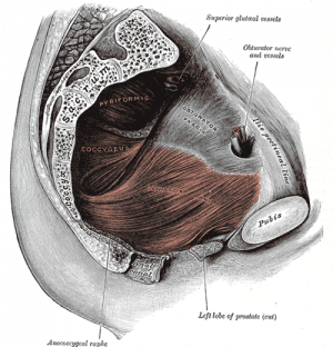
Pelvic Floor Anatomy
The Pelvic Floor - Overview and Function[edit | edit source]
The pelvic floor is an area of muscle and connective tissue that spans the area underneath the pelvis, separating the pelvic cavity above from the perineal region below. It provides support to the pelvic viscera including the bladder, intestines and uterus (in females). It also assists with continence through control of the urinary and anal sphincters. It facilitates birth by resisting the descent of the presenting part, causing the fetus to rotate forwards to navigate through the pelvic girdle. Finally , it helps to maintain optimal intraabdominal pressure.
Osteology and Ligaments[edit | edit source]
Pelvic Floor Myology[edit | edit source]
Pelvic diaphragm
- Levator ani - pubococcygeus, puborectalis, iliococcygeus
- Ischiococcygeus
Sphincter urethrae
Perineal membrane
Superficial genital muscles;
- Bulbocavernosus (bulbospongiosus in men)
- Ischiocavernosus
- Superficial transverse perineal
Associated muscles;
- Piriformis
- Obturator internus
- Adductors
- Gluteals
- Transverse abdominus
- Multifidus
- Respiratory diaphragm
| Muscle | Origin | Insertion | Action | Nerve Supply |
| Levator Ani (Pubococcygeus and Iliococcygeus) | Body of pubis, tendinous arch of obturator fascia, ischial spine | Perineal body, coccyx, anococcygeal lig, walls of prostate or vagina, rectum & anal canal | Helps to support pelvic viscera & resists increases in intra-abdominal pressure | Nerve to levator ani (branches of S4) & inferior anal (rectal) nerve & coccygeal plexus
|
| Levator Ani (Puborectalis) | Lower part of pubic symphysis and superior fascia of urogenital diaphragm | Control defecation |
| |
|
| ||||
| Ischiococcygeus | Ischial Spine | Inferior aspect of sacrum | Supports pelvic viscera, flexion of coccyx | Branches of S4 & S5 |
| Sphincter urethrae | Control flow of urine through urethra | |||


