Pelvic Floor Anatomy: Difference between revisions
No edit summary |
No edit summary |
||
| Line 75: | Line 75: | ||
<br> | <br> | ||
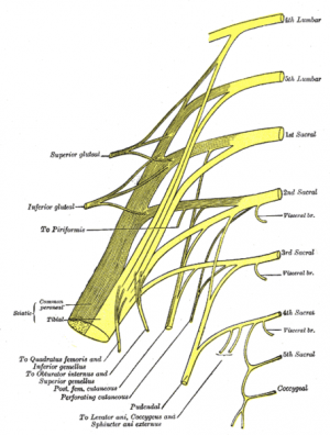
[[Image:Sacral and coccygeal plexus.png|thumb|right|300px|Sacral and Coccygeal Plexus]] | |||
Perineal membrane | Perineal membrane | ||
| Line 159: | Line 161: | ||
| Fixes perineal body | | Fixes perineal body | ||
| Deep branch of perineal nerve (branch of pudendal nerve) | | Deep branch of perineal nerve (branch of pudendal nerve) | ||
|- | |- | ||
| Male: median raphe, ventral surface of bulb of penis; Female: perineal body | | Male: median raphe, ventral surface of bulb of penis; Female: perineal body | ||
| Male: Corpora spongiosum and cavernosa, fascia of bulb of penis; Female: fascia of corpus cavernosa | | Male: Corpora spongiosum and cavernosa, fascia of bulb of penis; Female: fascia of corpus cavernosa | ||
| Male: Compression of bulb of penis and assists erection; Female: reduces lumen of vagina and assists erection of clitoris | | Male: Compression of bulb of penis and assists erection; Female: reduces lumen of vagina and assists erection of clitoris | ||
| Deep branch of perineal nerve (branch of pudendal nerve) | | Deep branch of perineal nerve (branch of pudendal nerve) | ||
|- | |- | ||
| Ischiocavernosus | | Ischiocavernosus | ||
| Ischial ramus and tuberosity | | Ischial ramus and tuberosity | ||
| Crus of penis or clitoris | | Crus of penis or clitoris | ||
| Maintains erection of penis or clitoris by compression of outflow veins | | Maintains erection of penis or clitoris by compression of outflow veins | ||
| Deep branch of perineal nerve (branch of pudendal nerve) | | Deep branch of perineal nerve (branch of pudendal nerve) | ||
|} | |} | ||
Revision as of 19:47, 6 April 2014
The Pelvic Floor - Overview and Function [1][edit | edit source]
The pelvic floor is an area of muscle and connective tissue that spans the area underneath the pelvis, separating the pelvic cavity above from the perineal region below. It provides support to the pelvic viscera including the bladder, intestines and uterus (in females). It also assists with continence through control of the urinary and anal sphincters. It facilitates birth by resisting the descent of the presenting part, causing the fetus to rotate forwards to navigate through the pelvic girdle. Finally , it helps to maintain optimal intraabdominal pressure.
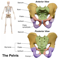
Osteology, Ligaments and Fascia
[edit | edit source]
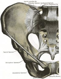
Ligaments[2]
Puboprostatic ligament
Pubovesical ligament
Sacrogenital ligaments
Superior pubic ligament - runs between pubic tubercles
Inferior arcuate ligament - runs between inferior pubic rami and blends with fibrocartilagnous disc of pubic symphysis
Anterior pubic ligament
Posterior pubic ligament - membranous structure which blends with periosteum
Ventral sacrococcygeal ligament
Dorsal sacrococcygeal ligament
Lateral sacrococcygeal ligament
Interosseous sacro-iliac ligament
Ventral sacro-iliac ligament
Sacrotuberous ligament
Sacrospinous ligament
Iliolumbar ligament
Pelvic Fascia[2]
Parietal pelvic fascia - lines the internal surface of the muscles of the pelvic floor and walls
Visceral pelvic fascia - invests each pelvic organ
The parietal and visceral fascia are continuous where organs penetrate the pelvic floor. They thicken to form the arcus tendineus, arches of fascia running adjacent to the viscera from the pubis to the sacrum.
Endopelvic fascia - "filler" material lying bewteen the parietal and visceral fascia, sometimes condensing to form fibrous fascial septa which separate the organs.
- Hypogastric sheath - separates retropubic space from presacral space; conduit for vessels and nerves
- Transverse cerical (cardinal) ligaments - part of hypogastric sheath; runs from lateral pelvic wall to uterine cervix and vagina; transmits uterine artery and provides passive support for the uterus
- Vesicovaginal septum
- Rectovesical septum
- Rectovaginal septum
Pelvic Floor Myology [2][1][edit | edit source]
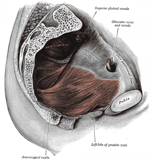
Pelvic diaphragm
- Levator ani - pubococcygeus, puborectalis, iliococcygeus
- Ischiococcygeus (Coccygeus)
Sphincter urethrae
Perineal membrane
Superficial genital muscles;
- Bulbocavernosus (bulbospongiosus in men)
- Ischiocavernosus
- Superficial transverse perineal
Associated muscles;
- Piriformis
- Obturator internus
- Adductors
- Gluteals
- Transverse abdominus
- Multifidus
- Respiratory diaphragm
| Muscle | Origin | Insertion | Action | Innervation |
| Levator Ani (Pubococcygeus) | Posterior aspect of body of pubis and anterior part of arcus tendineus | Coccyx and anococcygeal ligament | Helps to support pelvic viscera and resists increase in intra-abdominal pressure | Nerve to levator ani (branches of S4), inferior rectal nerve (from pudendal nerve - S3, S4), coccygeal plexus
|
| Levator Ani (Iliococcygeus) | Posterior aspect of arcus tendineus and the ischial spine | Anococcygeal raphe and the coccyx | Helps to support pelvic viscera and resists increase in intra-abdominal pressure | Nerve to levator ani (branches of S4), inferior rectal nerve (from pudendal nerve - S3, S4), coccygeal plexus
|
| Levator Ani (Puborectalis) | Lower part of pubic symphysis and superior fascia of urogenital diaphragm | Unites with its partner to make a U-shaped sling around the rectum | Controls defecation by pulling anorectal junction forward | Nerve to levator ani (branches of S4), branch of pudendal nerve (S2-4)
|
| Ischiococcygeus (Coccygeus) | Ischial Spine | Lower two sacral and upper two coccygeal spinal segments, blends with sacrospinous ligament on its external surface | Supports pelvic viscera, flexion of coccyx | Anterior rami of S4 and S5 |
| External anal sphincter | Skin and fascia surrounding the anus and coccyz via the anococcygeal body | Perinal body | Closes anal canal | Inferior rectal (anal) nerve |
| Sphincter urethrae | Inferior aspect of pubic ramus and ischial tuberosity | Surrounds urethra; in females, some fibres also enclose the vagina | Controls flow of urine through urethra; also compresses vagina in females | Deep branch of perineal nerve (branch of pudendal nerve) |
| Superficial transverse perineal | Ischial ramus and tuberosity | Perineal body | Supports perineal body | Deep branch of perineal nerve (branch of pudendal nerve) |
| Deep transverse perineal | Inner aspect of ischiopubic ramus | Median raphe, perineal body and external anal sphincter | Fixes perineal body | Deep branch of perineal nerve (branch of pudendal nerve) |
| Male: median raphe, ventral surface of bulb of penis; Female: perineal body | Male: Corpora spongiosum and cavernosa, fascia of bulb of penis; Female: fascia of corpus cavernosa | Male: Compression of bulb of penis and assists erection; Female: reduces lumen of vagina and assists erection of clitoris | Deep branch of perineal nerve (branch of pudendal nerve) | |
| Ischiocavernosus | Ischial ramus and tuberosity | Crus of penis or clitoris | Maintains erection of penis or clitoris by compression of outflow veins | Deep branch of perineal nerve (branch of pudendal nerve) |
References[edit | edit source]
- ↑ 1.0 1.1 Wikipedia. Pelvic floor. http://en.wikipedia.org/wiki/Pelvic_floor. (accessed 6 April 2014).
- ↑ 2.0 2.1 2.2 Netter Anatomy. Pelvis and perineum: pelvic floor and contents. http://www.netteranatomy.com/anatomylab/subregions.cfm?subregionID=R53. (accessed 6 April 2014).


