Multidimensional Nature of Pain: Difference between revisions
No edit summary |
No edit summary |
||
| Line 34: | Line 34: | ||
Nociceptors (from the latin ''nocere = ''to hurt) are sensory receptors which detect signals from damaged tissue or the threat of damage and indirectly also respond to chemicals released from the damaged tissue. There are free nerve endings present in many types of tissues, and cell bodies located in the dorsal root ganglions or in the cranial nerve ganglia.<br> | Nociceptors (from the latin ''nocere = ''to hurt) are sensory receptors which detect signals from damaged tissue or the threat of damage and indirectly also respond to chemicals released from the damaged tissue. There are free nerve endings present in many types of tissues, and cell bodies located in the dorsal root ganglions or in the cranial nerve ganglia.<br> | ||
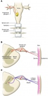
[[Image:Nociceptors.jpg| | [[Image:Nociceptors.jpg|thumb|center|100px|(A) Somatosensory neurons are located in peripheral ganglia (trigeminal and dorsal root ganglia) located alongside the spinal column and medulla. Afferent neurons project centrally to the brainstem (Vc) and dorsal horn of the spinal cord and peripherally to the skin and other organs. Vc, trigeminal brainstem sensory subnucleus caudalis. (B) Most nociceptors are unmyelinated with small diameter axons (C-fibers, red). Their peripheral afferent innervates the skin (dermis and/or epidermis) and central process projects to superficial laminae I and II of the dorsal horn. (C) A-fiber nociceptors are myelinated and usually have conduction velocities in the Aδ range (red). A-fiber nociceptors project to superficial laminae I and V. from: Dubin AE, Patapoutian A. Nociceptors: the sensors of the pain pathway. J Clin Invest. 2010 Nov 1;120(11):3760–72.]] | ||
<br> | |||
Nociceptors have unmyelinated (C-fiber) or thinly myelinated (A-fiber) axons<ref name="McCleskey 1999">McCleskey EW, Gold MS. Ion channels of nociception. Annu Rev Physiol. 1999;61:835–56.</ref>. C-fibers support conduction velocities of 0.4–1.4 m/s, while A-fibers support conduction velocities of approximately 5–30 m/s<ref name="Dubin 2010">Dubin AE, Patapoutian A. Nociceptors: the sensors of the pain pathway. J Clin Invest. 2010 Nov 1;120(11):3760–72.</ref>.<br> | |||
=== Nociception<br> === | === Nociception<br> === | ||
Revision as of 16:42, 28 March 2016
- Please do not edit unless you are involved in this project, but please come back in the near future to check out new information!!
- If you would like to get involved in this project and earn accreditation for your contributions, please get in touch!
Tips for writing this page:
Define acute and chronic pain terms of a multidimensional pain experience.
- Anatomical, physiological, and psychological basis of pain and pain relief, including pain as an 'output' from the brain
- Definition of pain and evidence of the multidimensional nature of the pain experience e.g. social and psychological infleuncing factors
Original Editor - Alberto Bertaggia.
Top Contributors - Alberto Bertaggia, Nina Myburg, Admin, Kim Jackson, Michelle Lee, Vidya Acharya, Lauren Lopez, 127.0.0.1, WikiSysop, Jess Bell and Jo Etherton
Introduction[edit | edit source]
A definition of pain is provided by the International association for the Study of Pain (IASP) as follows[1]:
"An unpleasant sensory and emotional experience associated with actual or potential tissue damage, or described in terms of such damage"
Pain is always subjective and everyone learns the use of this word through experiences related to injury in early life.
It is a sensation in a part or parts of the body, but it is also always unpleasant and therefore also emotional.
Even in the absence of tissue damage or any likely pathophysiological cause, people still report pain; usually this happens for psychological reasons. In these cases, it is challenging to distinguish whether their experience arise from a damaged tissue or not, based only upon the subjective report[2].
It is important to underline that activity induced in the nociceptor and nociceptive pathways by a noxious stimulus is not pain[2], which is always the output of a widely distributed neural network in the brain rather than one coming directly by sensory input evoked by injury, inflammation, or other pathology[3].
In the following video Karen D. Davis tries to explain why do some people react to the same painful stimulus in different ways.
Relevant anatomy and physiology[edit | edit source]
Nociceptors[edit | edit source]
Nociceptors (from the latin nocere = to hurt) are sensory receptors which detect signals from damaged tissue or the threat of damage and indirectly also respond to chemicals released from the damaged tissue. There are free nerve endings present in many types of tissues, and cell bodies located in the dorsal root ganglions or in the cranial nerve ganglia.

Nociceptors have unmyelinated (C-fiber) or thinly myelinated (A-fiber) axons[4]. C-fibers support conduction velocities of 0.4–1.4 m/s, while A-fibers support conduction velocities of approximately 5–30 m/s[5].
Nociception
[edit | edit source]
Nociception is a mechanism which comprises the processes of transduction, conduction, transmission and perception[6].
- Transduction is the conversion of a noxious thermal, mechanical, or chemical stimulus into electrical activity in the peripheral terminals of nociceptor sensory fibers. This process is mediated by specific receptor ion channels expressed only by nociceptors.
- Conduction is the passage of action potentials from the peripheral terminal along axons to the central terminal of nociceptors in the central nervous system.
- Transmission is the synaptic transfer and modulation of input from one neuron to another.
- Projection neurons in the dorsal horn transfer nociceptive input to the brainstem, hypothalamus, and thalamus and then, through relay neurons, to the cortex. Here is where perception occur as a subjective experience.
Acute and chronic pain
[edit | edit source]
Acute pain is caused by a noxious stimuli ad is mediated by nociception. It has early onset and serve to prevent tissues damages. It is also useful to learn to avoid threat of damage, because certain categories of noxious stimulii become linked to the sensation of pain. This is why this type of pain is defined as adaptive, it helps to survive and to heal[6].
References[edit | edit source]
- ↑ IASP Taxonomy - IASP [Internet]. [cited 2016 Mar 18]. Available from: http://www.iasp-pain.org/Taxonomy#Pain
- ↑ 2.0 2.1 Merskey H, Bogduk N. Classification of Chronic Pain: Descriptions of Chronic Pain Syndromes and Definitions of Pain Terms. IASP Press; 1994. 248 p.
- ↑ Melzack R. Pain and the neuromatrix in the brain. J Dent Educ. 2001 Dec;65(12):1378–82.
- ↑ McCleskey EW, Gold MS. Ion channels of nociception. Annu Rev Physiol. 1999;61:835–56.
- ↑ Dubin AE, Patapoutian A. Nociceptors: the sensors of the pain pathway. J Clin Invest. 2010 Nov 1;120(11):3760–72.
- ↑ 6.0 6.1 Woolf CJ, American College of Physicians, American Physiological Society. Pain: moving from symptom control toward mechanism-specific pharmacologic management. Ann Intern Med. 2004 Mar 16;140(6):441–51.


