McArdle's Disease
Original Editors - Ed Foring from Bellarmine University's Pathophysiology of Complex Patient Problems project.
Lead Editors - Your name will be added here if you are a lead editor on this page. Read more.
Definition/Description
[edit | edit source]
In 1951 Dr. Brain McArdle of Guy's Hospital in London England first described this disease. This disease also is refered to as myophosphorylase deficiency or Type V glycogen storage disease. This disease is a metabolic disease where skeletal muscle cells can not breakdown glycogen into glucose.
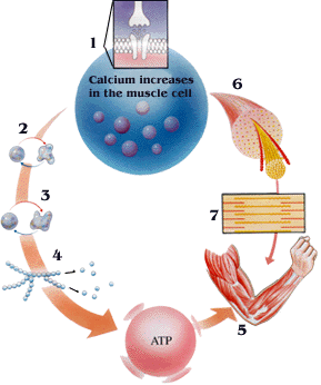
How Skeletal Muscles Normally Contract
A McArdle’s Disease Model
| 1. Acetylcholine is released from a motor nerve. This causes an entry of calcium into the muscle cell. | 1. Acetylcholine is released from a motor nerve. This causes an entry of calcium into the muscle cell. |
| 2. Calcium activates phosphorylase kinase – the first protein kinase discovered by Fischer and Krebs. | 2. Calcium activates phosphorylase kinase – the first protein kinase discovered by Fischer and Krebs. |
| 3. Phosphorylase kinase phosphorylates phosphorylase, which is activated | 3. Phosphorylase kinase phosphorylates phosphorylase, which is missing or otherwise non-functional |
| 4. Glycogen is broken to glucose. This is used to generate ATP | 4. Glycogen is unable to be broken down, creating glucose (and ATP) shortage. |
| 5. The muscle works and requires energy in the form of ATP | 5. Motor proteins attach to muscle fibers require ATP for movement. |
| 6. The muscle contains muscle cells | 6. Muscles stop responding in absence of ATP . |
| 7. Contractile proteins in the muscle are activated by calcium | 7. Because ATP is required to both contract and relax muscles, injury can occur. |
Prevalence[edit | edit source]
McArdle's disease is rare affecting approximately 1 in 100,000 people. This disease remains undiagnosed until most reach adulthood therefore the prevalence of this disease may be higher.
Characteristics/Clinical Presentation[edit | edit source]
Symptoms typically present 10 seconds after strenous exercise has begun. After 10 seconds of exercise skeletal muscle relies on the conversion of glycogen to glucose to produce ATP which is the main energy source to power muscular contraction.
Premature Exhaustion: Skeletal muscles inability to metabolize glycogen into glucose with strenous activity leads to an abrupt feeling of exhaustion or fatigue, with an increase in heart rate.
Muscle Failure: This particularly occurs under extreme stress where the muscle no longer can produce contraction regardless of the effort made. This is compaired to "wall" runners experience when doing a marathon.
Cramping: Muscle failure leads to electrically-silent contractures which are very painful and can lead to muscle damage.
Myoglobinuria: Damage to muscle tissue leads to the release of proteins,creatine kinase and myoglobin, into the blood which are than excreted with urination. These proteins are iron-rich and may cause urine to be a redish color. Myoglobinuria has lead to renal dysfunction, therefore cramping and muscle failure episodes require medical attention.
Fixed Weakness: Muscle damage caused by rhabdomyolysis can lead to muscle weakness that seems to not get physically stronger or is extremely difficult to make strength gains.
Second Wind: The phenomenon has been observed in clincal trials in patients after following a "warm up" period. This second wind does not relieve failure symptoms for intense exercise but offers some relief for light to moderate exercise.
Associated Co-morbidities[edit | edit source]
add text here
Medications[edit | edit source]
add text here
Diagnostic Tests/Lab Tests/Lab Values[edit | edit source]
Ischemic Forearm Test
This test is a valuable diagnostic test for a number of metabolic diseases. This test measures concentration of lactic acid in the blood before and after local exertion of a muscle group. The following protocol description is taken from the University of Florida School of Medicine website:
The test is performed by contracting the forearm to fatigue with a blood pressure cuff inflated to greater than systolic pressure. Antecubital blood samples for lactate and ammonia are collected before and following exercise at 0, 1, 2, 5, and 10 minutes. Ischemia blocks oxidative phosphorylation and ensures dependence on anaerobic glycogenolysis lactate normally rises at least fourfold within 1 to 2 minutes of exercise ammonia rises fivefold within 2 to 3 minutes.
Lactate concetration will rise several-fold under ischemic conditions in normal subjects (o-o-o). There will be no or minimal rise in patients with myophosphorylase deficiency (McArdle’s disease.)
The ischemic forearm test is only slightly uncomfortable to undergo, involving blood samples, a pressure cuff, and a device to squeeze with the hand for forearm muscle contraction, but takes a few hours in order to get blood samples at a resting metabolic rate.
Muscle Biopsy
The following is a description of a muscle biopsy performed in order to diagnose McArdle’s disease in an elderly patient. It involves microscopic muscle fiber examination to identify, among other characteristics, large deposits of glycogen:
Biopsy of Left quadriceps was performed. The biopsy showed subsarcolemmal vacuoles in many fibers with no abnormal contents on hematoxylin and eosin (fig. 1A), Masson’s trichrome (fig. 1B) or Gomori stains (Fig.1C). Increased glycogen content (Fig. 2A) was present in several fibers on PAS stain (diastase sensitive). Myophosphorylase activity was absent using enzyme histochemistry (fig. 2B) with a positive control (Fig. 2C). Mild to moderate myopathic features were present including increased fiber size variation and internal nucleation (Fig. 3A) with scattered myofiber hypertrophy and occasional fiber splitting (fig. 3B). Type I fiber smallness was suggested on ATPse stains (fig. 3C). Decreased oxidative enzymes staining with “moth-eaten” appearance was present, mostly in type 1 fibers (fig. 4A) with linearization of the intermyofibrillar architecture in type II fibers (Fig. 4B). Neurogenic atrophy was minimal.
The biopsy looks for the presence of myophosphorylase activity, which is absent in and can confirm diagnosis of McArdle’s disease. Click here to view biopsy images of McArdle's Disease.
Etiology/Causes[edit | edit source]
The cause of this disease is due to a missing or non-functioning enzyme that breaks down glycogen into glucose during exercise called myophosphorylase C. Several genetic mutations have been found to be the reason why this enzyme is missing or non-functioning.
Systemic Involvement[edit | edit source]
add text here
Medical Management (current best evidence)[edit | edit source]
Physical Therapy Management (current best evidence)[edit | edit source]
Be Fit
A disciplined regimen of regular physical activity of the appropriate intensity and duration can improve symptoms and reduce susceptible of injury to the muscle.
Be Flexible
Stretching following walking offers immediate and dramatic pain relief to individuals. Stretching also promotes flexibility in areas that may have become inflexible.
Be Aware, Be Patient and WARM UP
Individuals can easily over exhert themselves and become injuried without even realizing it. Indivduals need to be conscious of their energy levels, their blood sugar, their heart rate, and their general well being. Individuals should not avoid physical activity altogether, but if they are faced with a strenous task they should be instructed to take their time with it. During any type of strenous activity they should be instructed to warm up properly and periodically do several boughts of stretches.
Alternative/Holistic Management (current best evidence)[edit | edit source]
add text here
Differential Diagnosis[edit | edit source]
add text here
Case Reports/ Case Studies[edit | edit source]
add links to case studies here (case studies should be added on new pages using the case study template)
Resources
[edit | edit source]
add appropriate resources here
Recent Related Research (from Pubmed)[edit | edit source]
see tutorial on Adding PubMed Feed
Extension:RSS -- Error: Not a valid URL: Feed goes here!!|charset=UTF-8|short|max=10
References[edit | edit source]
see adding references tutorial.
McArdlesDisease.org. www.mcardledisease.org (accessed 17 March 2011).
Myoglobinuria. Wikidoc. http://www.wikidoc.org/index.php/Myoglobinuria (accessed 17 March 2011).
MedPoster 1992. nobelprize.org. http://nobelprize.org/nobel_prizes/medicine/laureates/1992/illpres/glycogen.html (accessed 3 April 2011).
Geneva Foundation for Medical Education and Research. Glycogen Storage disease V. http://www.gfmer.ch/genetic_diseases_v2/gendis_detail_list.php?cat3=912 (accessed 3 April 2011).


