Introduction to Spinal Orthotics: Difference between revisions
m (SOMI brace) |
No edit summary |
||
| Line 166: | Line 166: | ||
'''<u>1. Halo</u>''' | '''<u>1. Halo</u>''' | ||
A Halo brace is an extensive brace which is surgically applied. It is a 4-post orthotic that is attached by pins that are placed in the cranial table with a jacket fitted to torso.<ref name=":0" /> A halo fixated brace would be indicated for pre-surgical correction to post-op fusion support. Additionally, a halo brace can be used as an alternative to surgery to conserve neck mobility. <ref>Banat M, Vychopen M, Wach J, Salemdawod A, Scorzin J, Vatter H. [https://link.springer.com/article/10.1007/s00068-021-01849-z Use of halo fixation therapy for traumatic cranio-cervical instability in children: a systematic review]. European Journal of Trauma and Emergency Surgery. 2021 Dec 9:1-7.</ref>The halo brace is more efficacious than a hard collar at limiting motion in the upper cervical spine.<ref>Kumar GR. [https://www.isjonline.com/article.asp?issn=2589-5079;year=2022;volume=5;issue=1;spage=10;epage=23;aulast=Vijay Approach to upper cervical trauma. Indian Spine Journal]. 2022 Jan 1;5(1):10.</ref> | A Halo brace is an extensive brace which is surgically applied. It is a 4-post orthotic that is attached by pins that are placed in the cranial table with a jacket fitted to torso.<ref name=":0" /> A halo fixated brace would be indicated for pre-surgical correction to post-op fusion support. Additionally, a halo brace can be used as an alternative to surgery to conserve neck mobility. <ref>Banat M, Vychopen M, Wach J, Salemdawod A, Scorzin J, Vatter H. [https://link.springer.com/article/10.1007/s00068-021-01849-z Use of halo fixation therapy for traumatic cranio-cervical instability in children: a systematic review]. European Journal of Trauma and Emergency Surgery. 2021 Dec 9:1-7.</ref>The halo brace is more efficacious than a hard collar at limiting motion in the upper cervical spine.<ref>Kumar GR. [https://www.isjonline.com/article.asp?issn=2589-5079;year=2022;volume=5;issue=1;spage=10;epage=23;aulast=Vijay Approach to upper cervical trauma. Indian Spine Journal]. 2022 Jan 1;5(1):10.</ref>[[File:Halo Brace.png|thumb|Halo brace]] | ||
* Movement/Function Limitations | |||
** Flexion/Extion is limited by 96% | ** Flexion/Extion is limited by 96% | ||
** Lateral Bending is limited by 96% | ** Lateral Bending is limited by 96% | ||
| Line 194: | Line 194: | ||
** Atlanto-axial instability such as in Rheumatoid Arthritis | ** Atlanto-axial instability such as in Rheumatoid Arthritis | ||
** Neural arch fractures of C2 due to flexion instability<ref name=":0" /> | ** Neural arch fractures of C2 due to flexion instability<ref name=":0" /> | ||
'''<u>3. Minerva</u>''' | '''<u>3. Minerva</u>''' | ||
Revision as of 14:58, 11 May 2022
Top Contributors - Robin Tacchetti, Carin Hunter, Jess Bell, Kim Jackson and Tarina van der Stockt
Introduction[edit | edit source]
In the early days of orthotic making, materials such as metal and leather, which were heavy, hot and uncomfortable, were predominantly used. The materials used have progressed to light weight foams and thermoplastics facilitating new designs and more comfort for the user. The spine is a complex part of our anatomy and the key structure to our function. While it is not possible to treat all spinal issues with orthotics alone, a multidisciplinary team comprising of orthotists, surgeons and physiotherapists, along with the input of the patient often yields a good outcome.[1]
A spinal orthosis is an external aid that is used to correct and support the spine. With a good assessment, goal setting and clear expectations will lead to a well designed and appropriate orthotic device which is comfortable and functional and meets the users needs. Unlike other orthotic devices, many spinal braces are off-the-shelf, although certain situations call for a custom-made device.[1]
Principles of Spinal Bracing[edit | edit source]
Assessment[edit | edit source]
- Medical diagnosis
- Knowledge of anatomy/physiology
- Subjective assessment
- Medical history
- Social history
- Underlying conditions
- X-ray reports and findings
- Objective assessment
- Range of motion
- Muscle strength
- Sensation testing
- Determination of which motions should be restricted by the device:
- Sagittal plane
- Frontal/Coronal plane
- Transverse plane
- Combination of directional control
- Functional goals of the orthotic device and patient[1]
Aims[edit | edit source]
- Provides support and stabilization
- Maintains alignment of spine
- Prevention/correction of deformity
- Reduce pain by limiting motion
- Assist with healing post surgery
- Restriction of motion
- Reduction of axial loading of the spine
- Increasing intra-abdominal pressure may reduce axial loading of spine
- May provide heat and kinesthetic feedback, acts as a reminder[1]
Manufacturing[edit | edit source]
- Materials
- Soft fabric
- Flexible plastic
- Polyethylene
- Rigid plastic polypropylene
- Construction
- Off the shelf
- Custom made
- Suspension/strapping should be anchored above the iliac crests or over shoulders
- For cosmetic purposes the device should be worn under clothing around trunk[1]
Fitting and Evaluation[edit | edit source]
- Comfortable to wear – most important in spinal bracing as applying high forces and rejection is common.
- Good anatomical fit
- Good biomechanical function – may require in/out of brace x-rays to determine function.
- Easy to don/doff
- Cosmesis[1]
Anatomy[edit | edit source]
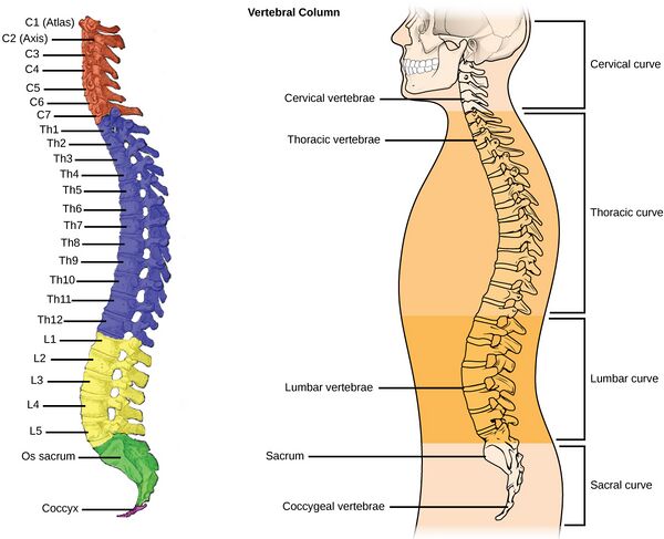
The vertebral column consists of 24 individual bones called vertebrae. The spinal column consists of this vertebral column and 2 sections of naturally fused vertebrae, the sacrum and the coccyx, located at the very bottom of the spine. Separated by Discs, intravertebral discs, fluid filled cushions between vertebrae[1]
The vertebral column can be divided into 5 regions:
- Cervical spine: 7 vertebrae of the neck (C1-C7)
- Thoracic spine: 12 vertebrae of the mid-back (T1-T12)
- Lumbar spine: 5 vertebrae of the lower back (L1-L5)
- Sacrum
- Coccyx
Cervical Region:
- There are 7 cervical vertebrae with 2 considered atypical vertebrae called the Atlas and Axis.
- Movements:
- Cervical Flexion/Extension – Atlas (Nodding)
- Cervical Rotation –Shaking head Axis
- Lateral Flexion[1]
Thoracic Region:
- There are 12 thoracic vertebrae
- Movements:
- Rotation
- Flexion/extension
- Side /Lateral flexion
- The thoracic vertebral motion is limited by the facets and ribs. The largest motion segment is at T12/L1, due to the lack of rib stabilization at this level and the facets being more medial to lateral orientation.
- Thoracic injuries are often associated with injury, trauma and degenerative changes.[1]
Lumbar Region:
- There are 5 lumbar vertebrae
- Movements:
- Primarily flexion and extension
- Partial lateral flexion
- Limited rotation[1]
Common Spinal Disorders[edit | edit source]
1. Fractures: There are three types of fractures that are commonly worked with in spinal orthotics, Compression, Dislocation and Compression/Dislocation. 5-10% of these occur in the cervical (neck) region while up to 65% occur in the thoracolumbar region, commonly at T12-L1 levels. Common causes include osteoporosis, trauma and tumors.
2. Intravertebral Disc Complications: There are many complications associated with the intervertebral disc Often complications are caused by a prolapsed or herniated disc or are degenerative in nature. A degenerative disc can be due to aging, trauma, repetitive strains or wear and tear.
3. Spondylolisthesis: This is a Latin term which means 'slipped vertebral body'. Typically, the L4 vertebral body slips forward on the L5 vertebral body. Under normal circumstances, the L4-L5 segment is the one in the lumbar spine with the most movement. Spondylolisthesis in the lumbar spine is most commonly caused by degenerative spinal disease, and is referred to as degenerative spondylolisthesis, the wear and tear of the intervertebral discs and ligament and osteoarthritis of the facet joints. Osteoarthritis can contribute instability and slippage. Degenerative spondylolisthesis usually occurs in people over 60 years of age.
4. Lordosis: A lordosis is classified as an excessive convex curvature of the lumbar spine
5. Kyphosis: Kyphosis is characterized by concave curvature of the upper spine (abnormal > 50 degrees of curvature)
6. Scoliosis: . More simply known as a lateral bend in the spine. The curve can be S-shaped or C-shaped. The cause can be varied, but if often congenital, degenerative, or due to trauma or tumors. Occasionally they are idiopathic in nature.
7. Soft Tissue Injuries: Soft tissues include the muscles, tendons, ligaments, and nerves. Injury to these tissues can often cause unnecessary stress to the spine
8. Sprains/strains: A sprain or strain is often due to poor posture or lifting a heavy object, whether it be poor lifting techniques or inadequate strength.
9. Muscle injury: These types of injuries are often caused by lifting or sport.
10. Trauma: Commonly associated with whiplash[1]
Types of Spinal Orthoses[edit | edit source]
- Cervical Orthosis
- Cervicothoracic Orthosis (HALO, SOMI, Minerva)
- Cervico-thoraco-lumbosacral Orthosis (Milwaukee)
- Thoracolumbosacral Orthosis CASH, Jewett, custom TLSO
- Lumbosacral Orthosis (Corsets, Chairback O)
- Sacral Orthosis ( Sacro-iliac bands)[1]
1. Cervical Orthosis (CO)[edit | edit source]
1. Soft Collars
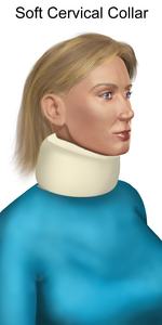
A soft cervical collar, the most basic, is made using construction foam and coated with cotton wool.[2] It provides partial support of the head reducing paraspinal contraction and spasm. If offers NO structural cervical spine support. The collar offers proprioceptive input, psychological reassurance and can provide relief through heat retention.
- Movement Limitations
- Flexion/extension is limited by ~ 8-26%,
- Lateral bending is limited by ~8%
- Rotation is limited by ~10-17%.
- Assists with
- Muscular strains/sprains
- Trauma[1]
2. Hard Collars
Hard collars are rigid/semirigid and are made of hard foam combined with plastic. The main function of this collar is support.[1] Hard collars such as the Philadelphia collar may be used initially with unstable fractures until a decision is made regarding treatment. These collars can be used for 6-8 week where nonoperative treatment is recommended.[3]
- Types of hard collars
- Miami J Collar
- VISTA Collar
- Aspen
- Headmaster
- Philadelphia
- Movement/Function Limitations
- Flexion/extension is limited by ~69-90%,
- lateral bending is limited by ~34-48%
- Rotation is limited by ~74%.
- Features
- Tracheostomy opening
- Height around chin and occiput can be adjusted
- Due to the construction most individuals report less sweating than a soft collar and increased comfort
- Indications
- Cervical trauma in unconscious patients
- Jefferson’s Fracture (C1) Hangman’s fracture
- Traumatic spondylolisthesis of C2 on C3
- Dens type I fracture
- Post op care
- Anterior discectomy
- Cervical Strain[1]
2. Cervicothoracic Orthosis (CTO)[edit | edit source]
- Types of CTO
- Halo
- Sterno-occipital mandibular orthosis (SOMI)
- Minerva
1. Halo
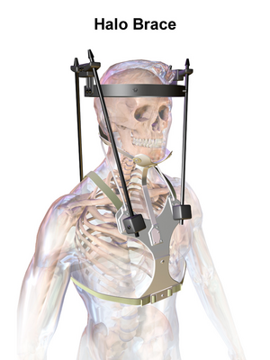
A Halo brace is an extensive brace which is surgically applied. It is a 4-post orthotic that is attached by pins that are placed in the cranial table with a jacket fitted to torso.[1] A halo fixated brace would be indicated for pre-surgical correction to post-op fusion support. Additionally, a halo brace can be used as an alternative to surgery to conserve neck mobility. [4]The halo brace is more efficacious than a hard collar at limiting motion in the upper cervical spine.[5]
- Movement/Function Limitations
- Flexion/Extion is limited by 96%
- Lateral Bending is limited by 96%
- Rotation is limited by 99%
- It offers maximum motion control to T3 level
- Indications:
- Occipital condyle fractures
- C1 ring injuries,
- Odontoid fractures
- Hangman fractures (C2)
- facet subluxations
- spinal infections
- Extradural tumor involvement that compromises the spinal alignment or bony stability
- Subaxial spine injuries[1]
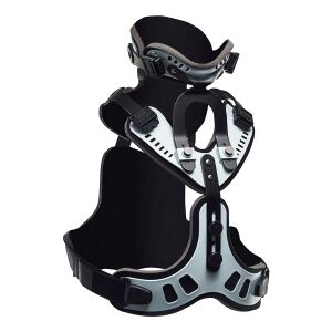
2. Sterno-occipital mandibular Orthosis (SOMI)
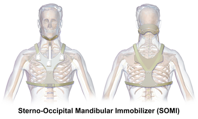
A SOMI is a 3-post, anterior chest plate that extends to the xiphoid process. It has a removeable chin strap.
- Movement/Function Limitations
- Flexion/Extension is limited by ~60-70%
- lateral bending is limited by ~20-35%
- Rotation is limited by ~30-65%
- Offers flexion control of C1-3 but controls extension less than with other cervical orthotics.
- Indications
- Atlanto-axial instability such as in Rheumatoid Arthritis
- Neural arch fractures of C2 due to flexion instability[1]
3. Minerva
The Minerva Orthosis is a removable version of the HALO. This type of orthosis is offered to a compliant patient who will remove it.
- Movement/Function Limitations
- Flexion/Extention is limited by ~96%
- Lateral Bending is limited by ~96%
- Rotation is limited by ~99%.
- This brace offers control of motion down to T3 level
- Indications
- Mid-to-lower cervical spine injuries
- Stable upper cervical spine injuries
- Can be used with skull fractures when a Halo Fixator is contraindicated
- Children due to its decreased weight and increased comfort[1]
3. Cervico-thoracolumbarsacral Orthosis (CTLSO)[edit | edit source]
The Milwaukee Brace is the classic CTLSO. It comprises of a metal vertical superstructure with a pelvic foundation, with a rigid plastic pelvic girdle connected to the neck with a ring. It has two posterior paraspinal bars. The cervical ring has mandibular and occipital bar which rest 20-30 mm inferior to the chin. The pads are positioned to apply forces to correct curvature.
- Indications
- Treatment of kyphosis
- Treatment of high thoracic curves[1]
4. Thoracolumbosacral Orthosis (TLSO)[edit | edit source]
- Types of Thoracolumbar Orthosis
- CASH
- Jewett
- Knight Taylor TLSO
- Custom-molded Spinal/Body Jacket
- Providence Night Brace
- Charleston Bending Brace
- Off-the-shelf (CASH, Jewett, Knight Taylor)
- Comfortable design
- Easy to don and doff
- Limited movement control flexion from T6 -L1
- No limitation of lateral flexion or rotation
- Indications:
- Thoracic and lumbar vertebral body fracture
- Kyphosis reduction in osteoporosis
- Cervical trauma in unconscious patients
- Off-the-shelf molded plastic Spinal Jacket
- Indications:
- Immobilization for thoracic compression fractures from osteoporosis. Immobilization after surgical stabilization for spinal fractures.
- Immobilization for unstable spinal disorders of T3-L3.
- Indications:
- Custom made Spinal Jacket
- Indications:
- Scoliosis
- Severe spinal abnormalities
- Night Bracing[1]
- Indications:
SCOLIOSIS[edit | edit source]
- Types of Scoliosis
- Neuromuscular Scoliosis
- Congenital Skeletal Scoliosis
- Idiopathic Scoliosis
- Indications for bracing in Scoliosis
- Flexible curves with Cobb angle(10°- 40°)
- 10°- 20° observe initially, if curve progresses by 5° then brace
- 30°- 40° prompt use of orthosis
- > 40◦ generally surgery
- Risser sign for remaining growth
- Boston Brace
- 1970’s, most Research and widely used
- Module from measures, type of curve
- Blueprint created from x rays
- Determine apex of curve and position of pads (3 point pressure)
- Cut outs to allow body to move
- Difficult to control rotation
- Maximum curve 45 degrees below T8
- ** Symmetrical Boston brace have a consistent success rate of over 70%[6]
- Cheneau Brace
- Relatively new type, limited research but possibly better results due to rotational control
- Fully custom made
- Type of curve, assessment, x rays
- Pads and cut outs, severe looking
- Control rotation with extensions at shoulders
- Prescription criteria for Neuromuscular Scoliosis
- Generally not Boston or Cheneau
- Different etiology, muscle weakness, underlying condition, severity
- Corrects flexible curves
- Provides stability
- Accommodates fixed deformities
- Can prevent further deformity[1]
5. Lumbo-sacral Orthosis (LSO)[edit | edit source]
Two types of lumbosacral orthosis are prescribed to patients, a soft fabric lumbar support, often constructed with metal or plastic struts to provide support for lumbar region of spine. or a moulded plastic Spinal Jacket. These can be off-the-shelf or custom-made to suit the patient needs.
- Indications
- Pain relief
- Postural support
- Reduces excessive lumbar lordosis
- Vasomotor and respiratory support in the spinal cord patient
- Increase intra abdominal pressure
- Heat
- Kinesthetic feedback[1]
6. Sacral Orthosis (SO)[edit | edit source]
A sacral orthosis is a fabric, often elasticated support
- Indications
- Sacro-iliac joint pain[1]
References[edit | edit source]
Weiss, Hans-Rudolf, Stefano Negrini, Manuel Rigo, Tomasz Kotwicki, Martha C. Hawes, Theodoros B. Grivas, Toru Maruyama, and Franz Landauer.
"Indications for conservative management of scoliosis (guidelines)."
Scoliosis 1, no. 1 (2006): 5.
Giele BM, Wiertsema SH, Beelen A, van der Schaaf M, Lucas C, Been HD, Bramer JA. No evidence for the effectiveness of bracing in patients with thoracolumbar fractures: a systematic review. Acta orthopaedica. 2009 Jan 1;80(2):226-32.
van Leeuwen PJ, Bos RP, Derksen JC, de Vries J. Assessment of spinal movement reduction by thoraco-lumbar-sacral orthoses. Journal of rehabilitation research and development. 2000 Jul 1;37(4):395.
Kawchuck, Gregory; A non-randomized clinical trial to assess the impact of nonrigid, inelastic corsets on spine function in low back pain participants and asymptomatic controls, The spine Journal 15 2015 pg 2222-2227
Harrington, Amanda: Chapter 43, Spinal Orthoses; Spinal Cord Medicine 2019 Springer Publishing Company, Editor Steven Kirshblum MD pg 744- 753
Schott, Cordelia: Effectiveness of lumbar orthoses in low back pain: Review of the literature and our results. Orthopedic Reviews 2018 Vol 10, 7791 pg 141-146
Morris: Role of the trunk in stability of the spine JBJS, 1961;43:327-351 36.
Nachemson: In Vivo Measurements of intradiscal pressure: Discometry, a method for the determination of pressure in the lower lumbar discs, JBJS American Volume 46(5) 1964 pg 1077 to 1092
Lantz SA, Schultz AB: Lumbar spine orthoses wearing: Effect on trunk muscle myoelectric activity. Spine 1986;11:838–4234. 29.
Lantz, S: Lumbar Spine Orthosis Wearing II. Effect on Trunk Muscle Myoelectric Activity Spine Vol 11, Number 8 1986 pg 838-842
References[edit | edit source]
- ↑ 1.00 1.01 1.02 1.03 1.04 1.05 1.06 1.07 1.08 1.09 1.10 1.11 1.12 1.13 1.14 1.15 1.16 1.17 1.18 1.19 1.20 1.21 1.22 1.23 Fisher, D. Introduction to Spinal Orthoses Course. Physioplus. 2022
- ↑ Xu Y, Li X, Chang Y, Wang Y, Che L, Shi G, Niu X, Wang H, Li X, He Y, Pei B. Design of Personalized Cervical Fixation Orthosis Based on 3D Printing Technology. Applied Bionics and Biomechanics. 2022 Apr 30;2022.
- ↑ Ghosh JC. Review of Management of Type-2 Odontoid Fracture in Elderly. Open Journal of Orthopedics. 2021 Jan 11;11(1):12-21.
- ↑ Banat M, Vychopen M, Wach J, Salemdawod A, Scorzin J, Vatter H. Use of halo fixation therapy for traumatic cranio-cervical instability in children: a systematic review. European Journal of Trauma and Emergency Surgery. 2021 Dec 9:1-7.
- ↑ Kumar GR. Approach to upper cervical trauma. Indian Spine Journal. 2022 Jan 1;5(1):10.
- ↑ Weiss HR, Turnbull D. Brace treatment for children and adolescents with scoliosis. InSpinal deformities in adolescents, adults and older adults 2020 Feb 27. IntechOpen.
