Intervertebral disc: Difference between revisions
No edit summary |
No edit summary |
||
| Line 5: | Line 5: | ||
</div> | </div> | ||
== Definition/Description == | == Definition/Description == | ||
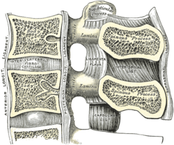
The intervertebral disc (IVD) is important in the normal functioning of the spine. It is a cushion of fibrocartilage and the principal joint between two vertebrae in the spinal column. There are 23 discs in the human spine: 6 in the cervical region (neck), 12 in the thoracic region (middle back), and 5 in the lumbar region (lower back). | [[File:Cervical_vertebrae_lig_flavum.png|right|frameless|250x250px]] | ||
The intervertebral disc (IVD) is important in the normal functioning of the spine. It is a cushion of fibrocartilage and the principal joint between two vertebrae in the spinal column. There are 23 discs in the human spine: 6 in the cervical region (neck), 12 in the thoracic region (middle back), and 5 in the lumbar region (lower back). | |||
== Clinically Relevant Anatomy == | == Clinically Relevant Anatomy == | ||
<u>Morphology</u> | === <u>Morphology</u> === | ||
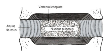
The IVD consists of three distinct components (Figure 2): <br>• a central nucleus pulposus (NP);<br>• a peripheral annulus fibrosus (AF);<br>• two vertebral endplates (VEPs). | The IVD consists of three distinct components (Figure 2): <br>• a central nucleus pulposus (NP);<br>• a peripheral annulus fibrosus (AF);<br>• two vertebral endplates (VEPs). | ||
| Line 17: | Line 16: | ||
Figure 2: Detailed structure of the IVD (adapted from Bogduk 2005) | Figure 2: Detailed structure of the IVD (adapted from Bogduk 2005) | ||
''Nucleus Pulposus'' | ''Nucleus Pulposus'': a gel-like mass composed of water and proteoglycans held by randomly arranged fibres of collagen <ref name="Bogduk">Bogduk, N., Clinical anatomy of the lumbar spine and sacrum. 4th ed. 2005, New York: Churchill Livingstone.</ref> <ref name="Gruber">Gruber, H.E. and E.N. Hanley, Jr., Recent advances in disc cell biology. Spine (Phila Pa 1976), 2003. 28(2): p. 186-93.</ref> <ref name="Humzah">Humzah, M.D. and R.W. Soames, Human intervertebral disc: structure and function. Anat Rec, 1988. 220(4): p. 337-56.</ref>. With it’s water-attracting properties, any attempt to deform the nucleus causes the applied pressure to be dispersed into various directions, similar to a person on a waterbed. | ||
''Annulus Fibrosus:'' consists of “lamellae” or concentric layers of collagen fibres <ref name="Marchand">Marchand, F. and A.M. Ahmed, Investigation of the laminate structure of lumbar disc anulus fibrosus. Spine (Phila Pa 1976), 1990. 15(5): p. 402-10.</ref>. The fibre orientation of each layer of lamellae alternate and therefore allow effective resistance of multidirectional movements. | |||
''Vertebral endplate:'' a plate of cartilage that acts as a barrier between the disc and the vertebral body. They cover the superior and inferior aspects of the annulus fibrosus and the nucleus pulposus. | |||
=== <u>Innervation</u> === | |||
The disc is innervated in the outer few millimetres of the annulus fibrosus <ref name="Roberts">Roberts, S., et al., Histology and pathology of the human intervertebral disc. J Bone Joint Surg Am, 2006. 88 Suppl 2(Supplement 2): p. 10-4.</ref>. | The disc is innervated in the outer few millimetres of the annulus fibrosus <ref name="Roberts">Roberts, S., et al., Histology and pathology of the human intervertebral disc. J Bone Joint Surg Am, 2006. 88 Suppl 2(Supplement 2): p. 10-4.</ref>. | ||
=== <u>Vascular supply and nutrition</u> === | |||
The IVD is largely avascular, with no major arterial branches to the disc <ref name="Urban4">Urban, J.P., et al., Nutrition of the intervertebral disk. An in vivo study of solute transport. Clin Orthop Relat Res, 1977(129): p. 101-14.</ref>. The outer annular layers are supplied by small branches from metaphysial arteries [1]. Due to the avascular nature of the disc, the nutrition is dependent on metabolite diffusion <ref name="Urban">Urban, J.P., et al., Nutrition of the intervertebral disk. An in vivo study of solute transport. Clin Orthop Relat Res, 1977(129): p. 101-14.</ref> <ref name="Urban 2">Urban, J., S. Holm, and A. Maroudas, Diffusion of small solutes into the intervertebral disc. Biorheology, 1978. 15(3-4): p. 203-21.</ref> <ref name="Urban 3">Urban, J.P., S. Smith, and J.C. Fairbank, Nutrition of the intervertebral disc. Spine (Phila Pa 1976), 2004. 29(23): p. 2700-9.</ref>. | The IVD is largely avascular, with no major arterial branches to the disc <ref name="Urban4">Urban, J.P., et al., Nutrition of the intervertebral disk. An in vivo study of solute transport. Clin Orthop Relat Res, 1977(129): p. 101-14.</ref>. The outer annular layers are supplied by small branches from metaphysial arteries [1]. Due to the avascular nature of the disc, the nutrition is dependent on metabolite diffusion <ref name="Urban">Urban, J.P., et al., Nutrition of the intervertebral disk. An in vivo study of solute transport. Clin Orthop Relat Res, 1977(129): p. 101-14.</ref> <ref name="Urban 2">Urban, J., S. Holm, and A. Maroudas, Diffusion of small solutes into the intervertebral disc. Biorheology, 1978. 15(3-4): p. 203-21.</ref> <ref name="Urban 3">Urban, J.P., S. Smith, and J.C. Fairbank, Nutrition of the intervertebral disc. Spine (Phila Pa 1976), 2004. 29(23): p. 2700-9.</ref>. | ||
<u>Biomechanics</u><br>''Weight bearing'' | === <u>Biomechanics</u> === | ||
<br>''Weight bearing:'' The disc is subjected to various loads, including compressive, tensile and shear stresses <ref name="Stokes">Stokes, I. and J. Iatridis, Mechanical conditions that accelerate intervertebral disc degeneration: overload versus immobilization. Spine (Phila Pa 1976), 2004. 29(23): p. 2724-32.</ref> <ref name="White">White, A.A. and M.M. Panjabi, Clinical biomechanics of the spine. Vol. 446. 1990: Lippincott Philadelphia.</ref>. During compressive loading, hydrostatic pressure develops within the NP, which thereby disperses the forces towards the endplates as well as the AF <ref name="Reuber" /> <ref name="Broberg">Broberg, K.B., On the mechanical behaviour of intervertebral discs. Spine (Phila Pa 1976), 1983. 8(2): p. 151-65.</ref> <ref name="Reuber">Reuber, M., et al., Bulging of lumbar intervertebral disks. J Biomech Eng, 1982. 104(3): p. 187-92.</ref>. This mechanism slows the rate applied loads are transmitted to the adjacent vertebra, giving the disc its shock absorbing abilities <ref name="Shah" />.<br> | |||
The disc is subjected to various loads, including compressive, tensile and shear stresses <ref name="Stokes">Stokes, I. and J. Iatridis, Mechanical conditions that accelerate intervertebral disc degeneration: overload versus immobilization. Spine (Phila Pa 1976), 2004. 29(23): p. 2724-32.</ref> <ref name="White">White, A.A. and M.M. Panjabi, Clinical biomechanics of the spine. Vol. 446. 1990: Lippincott Philadelphia.</ref>. During compressive loading, hydrostatic pressure develops within the NP, which thereby disperses the forces towards the endplates as well as the AF <ref name="Reuber" /> <ref name="Broberg">Broberg, K.B., On the mechanical behaviour of intervertebral discs. Spine (Phila Pa 1976), 1983. 8(2): p. 151-65.</ref> <ref name="Reuber">Reuber, M., et al., Bulging of lumbar intervertebral disks. J Biomech Eng, 1982. 104(3): p. 187-92.</ref>. This mechanism slows the rate applied loads are transmitted to the adjacent vertebra, giving the disc its shock absorbing abilities <ref name="Shah" /> | |||
'' | ''Movement:'' The disc is also involved in permitting movements between vertebral bodies, which include: <br>• axial compression / distraction;<br>• flexion / extension;<br>• axial rotation;<br>• lateral flexion.<br> | ||
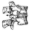
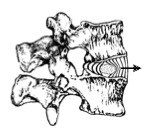
''Nuclear migration: A''symmetric compressive loading disc can cause the NP to migrate in a direction opposite to the compression <ref name="Krag">Krag, M.H., et al., Internal displacement distribution from in vitro loading of human thoracic and lumbar spinal motion segments: experimental results and theoretical predictions. Spine (Phila Pa 1976), 1987. 12(10): p. 1001-7.</ref> <ref name="Shah">Shah, J.S., W.G. Hampson, and M.I. Jayson, The distribution of surface strain in the cadaveric lumbar spine. J Bone Joint Surg Br, 1978. 60-B(2): p. 246-51.</ref> <ref name="Schnebel">Schnebel, B.E., et al., A digitizing technique for the study of movement of intradiscal dye in response to flexion and extension of the lumbar spine. Spine (Phila Pa 1976), 1988. 13(3): p. 309-12.</ref> <ref name="Fennell">Fennell, A., A. Jones, and D. Hukins, Migration of the nucleus pulposus within the intervertebral disc during flexion and extension of the spine. Spine (Phila Pa 1976), 1996. 21(23): p. 2753-7.</ref>. For example, during forward bending (or flexion) of the lumbar spine, the NP migrates posteriorly or backwards (Figure 4). Conversely, during backwards bending (or extension), the nucleus is squeezed anteriorly or forwards. This concept is known as the dynamic disc model <ref name="Kolber">Kolber, M.J. and W.J. Hanney, The dynamic disc model: a systematic review of the literature. Phys Ther Rev, 2009. 14(3): p. 181-189.</ref>. Although NP migration has been shown to behave predictably in asymptomatic discs, a variable pattern of migration occurs in people with symptomatic and/or degenerative IVDs <ref name="Kolber" />. | |||
[[Image:Discmig1.jpg]][[Image:Discmig2.jpg]] | [[Image:Discmig1.jpg]][[Image:Discmig2.jpg]] | ||
Figure 4: Direction of nuclear migration within the IVD during spinal movements (adapted from McKenzie 1981)<br> | Figure 4: Direction of nuclear migration within the IVD during spinal movements (adapted from McKenzie 1981)<br><br> | ||
{{#ev:youtube|fIIjT10eg08}}<ref>Physiotutors. The inter-body joint and the intervertebral disc. Available from: https://www.youtube.com/watch?v=fIIjT10eg08</ref> | {{#ev:youtube|fIIjT10eg08}}<ref>Physiotutors. The inter-body joint and the intervertebral disc. Available from: https://www.youtube.com/watch?v=fIIjT10eg08</ref> | ||
| Line 123: | Line 109: | ||
== Examination == | == Examination == | ||
Refer to [http://www.physio-pedia.com/Lumbar_Discogenic_Pain Lumbar discogenic pain].<br> | Refer to [http://www.physio-pedia.com/Lumbar_Discogenic_Pain Lumbar discogenic pain].<br><br> | ||
== Key Research == | == Key Research == | ||
Revision as of 21:18, 25 March 2019
Original Editors - Alexander Chan
Top Contributors - Alexander Chan
Definition/Description[edit | edit source]
The intervertebral disc (IVD) is important in the normal functioning of the spine. It is a cushion of fibrocartilage and the principal joint between two vertebrae in the spinal column. There are 23 discs in the human spine: 6 in the cervical region (neck), 12 in the thoracic region (middle back), and 5 in the lumbar region (lower back).
Clinically Relevant Anatomy[edit | edit source]
Morphology[edit | edit source]
The IVD consists of three distinct components (Figure 2):
• a central nucleus pulposus (NP);
• a peripheral annulus fibrosus (AF);
• two vertebral endplates (VEPs).
Figure 2: Detailed structure of the IVD (adapted from Bogduk 2005)
Nucleus Pulposus: a gel-like mass composed of water and proteoglycans held by randomly arranged fibres of collagen [1] [2] [3]. With it’s water-attracting properties, any attempt to deform the nucleus causes the applied pressure to be dispersed into various directions, similar to a person on a waterbed.
Annulus Fibrosus: consists of “lamellae” or concentric layers of collagen fibres [4]. The fibre orientation of each layer of lamellae alternate and therefore allow effective resistance of multidirectional movements.
Vertebral endplate: a plate of cartilage that acts as a barrier between the disc and the vertebral body. They cover the superior and inferior aspects of the annulus fibrosus and the nucleus pulposus.
Innervation[edit | edit source]
The disc is innervated in the outer few millimetres of the annulus fibrosus [5].
Vascular supply and nutrition[edit | edit source]
The IVD is largely avascular, with no major arterial branches to the disc [6]. The outer annular layers are supplied by small branches from metaphysial arteries [1]. Due to the avascular nature of the disc, the nutrition is dependent on metabolite diffusion [7] [8] [9].
Biomechanics[edit | edit source]
Weight bearing: The disc is subjected to various loads, including compressive, tensile and shear stresses [10] [11]. During compressive loading, hydrostatic pressure develops within the NP, which thereby disperses the forces towards the endplates as well as the AF [12] [13] [12]. This mechanism slows the rate applied loads are transmitted to the adjacent vertebra, giving the disc its shock absorbing abilities [14].
Movement: The disc is also involved in permitting movements between vertebral bodies, which include:
• axial compression / distraction;
• flexion / extension;
• axial rotation;
• lateral flexion.
Nuclear migration: Asymmetric compressive loading disc can cause the NP to migrate in a direction opposite to the compression [15] [14] [16] [17]. For example, during forward bending (or flexion) of the lumbar spine, the NP migrates posteriorly or backwards (Figure 4). Conversely, during backwards bending (or extension), the nucleus is squeezed anteriorly or forwards. This concept is known as the dynamic disc model [18]. Although NP migration has been shown to behave predictably in asymptomatic discs, a variable pattern of migration occurs in people with symptomatic and/or degenerative IVDs [18].
Figure 4: Direction of nuclear migration within the IVD during spinal movements (adapted from McKenzie 1981)
Pathology[edit | edit source]
Lumbar Disc[20]
| Diagnostic Category | Characteristics | |
|---|---|---|
| Normal | - Morphologically normal (not inclusive of degenerative, developmental, or adaptive changes that could in some context be considered clinically normal)
- In adults, a bilocular appearance resulting from the development of a central horizontal band of fibrous tissue is a normal sign of maturation | |
| Congenital/Developmental | - congenitally abnormal
- changes in morphology as an adaptation to the abnormal growth of the spine (i.e. scoliosis or spondylolisthesis) | |
| Degenerative/Trauma | Annular Fissures/Tear | - separation between annular fibres OR
- avulsion of fibres from their vertebral body insertions OR - breaks through the fibres that extend radially/transversely/concentrically involving one or many layers of the annular lamellae |
| Degeneration | - any or all real/apparent desiccation, fibrosis, narrowing of the disc space OR
- diffuse bulging of the annulus beyond the disc space OR - extensive fissuring (i.e. numerous annular tears) OR - mucinous degeneration of the annulus OR - defects and sclerosis of the endplates OR - osteophytes at the vertebral apophyses - A disc with one or more of these changes can be further categorised into: 1. spondylosis deformans (normal aging process) 2. intervertebral osteochondrosis (pathologic process) | |
| Herniation | - defined as a localised displacement of disc material beyond the limits of the IVD space
- Protrusion: the base is greater than the edges of the disc material beyond the disc space - Extrusion: the base is smaller than the edges of the disc material beyond the disc space - 2 types of Extrusion: 1. Sequestration: displaced disc has lost continuity with parent disc 2. Migration: displacement of material away from site of extrusion with/without sequestration - Contained: displaced portion is covered by outer annulus - Uncontained: displaced portion has no covering | |
| Inflammation/Infection | - infection, infection-like inflammatory discitis, and inflammatory response to spondyloarthropathy | |
| Morphologic Variant of Unknown Significance | - abnormal morphology without enough data to warrant diagnostic categorisation | |
Differential Diagnosis[edit | edit source]
Refer to Lumbar discogenic pain and Thoracic disc syndrome.
Diagnostic Procedures[edit | edit source]
Refer to Lumbar discogenic pain and Thoracic disc syndrome.
Outcome Measures[edit | edit source]
Examination[edit | edit source]
Refer to Lumbar discogenic pain.
Key Research[edit | edit source]
Bogduk, N., Clinical anatomy of the lumbar spine and sacrum. 4th ed. 2005, New York: Churchill Livingstone.
References[edit | edit source]
- ↑ Bogduk, N., Clinical anatomy of the lumbar spine and sacrum. 4th ed. 2005, New York: Churchill Livingstone.
- ↑ Gruber, H.E. and E.N. Hanley, Jr., Recent advances in disc cell biology. Spine (Phila Pa 1976), 2003. 28(2): p. 186-93.
- ↑ Humzah, M.D. and R.W. Soames, Human intervertebral disc: structure and function. Anat Rec, 1988. 220(4): p. 337-56.
- ↑ Marchand, F. and A.M. Ahmed, Investigation of the laminate structure of lumbar disc anulus fibrosus. Spine (Phila Pa 1976), 1990. 15(5): p. 402-10.
- ↑ Roberts, S., et al., Histology and pathology of the human intervertebral disc. J Bone Joint Surg Am, 2006. 88 Suppl 2(Supplement 2): p. 10-4.
- ↑ Urban, J.P., et al., Nutrition of the intervertebral disk. An in vivo study of solute transport. Clin Orthop Relat Res, 1977(129): p. 101-14.
- ↑ Urban, J.P., et al., Nutrition of the intervertebral disk. An in vivo study of solute transport. Clin Orthop Relat Res, 1977(129): p. 101-14.
- ↑ Urban, J., S. Holm, and A. Maroudas, Diffusion of small solutes into the intervertebral disc. Biorheology, 1978. 15(3-4): p. 203-21.
- ↑ Urban, J.P., S. Smith, and J.C. Fairbank, Nutrition of the intervertebral disc. Spine (Phila Pa 1976), 2004. 29(23): p. 2700-9.
- ↑ Stokes, I. and J. Iatridis, Mechanical conditions that accelerate intervertebral disc degeneration: overload versus immobilization. Spine (Phila Pa 1976), 2004. 29(23): p. 2724-32.
- ↑ White, A.A. and M.M. Panjabi, Clinical biomechanics of the spine. Vol. 446. 1990: Lippincott Philadelphia.
- ↑ 12.0 12.1 Reuber, M., et al., Bulging of lumbar intervertebral disks. J Biomech Eng, 1982. 104(3): p. 187-92.
- ↑ Broberg, K.B., On the mechanical behaviour of intervertebral discs. Spine (Phila Pa 1976), 1983. 8(2): p. 151-65.
- ↑ 14.0 14.1 Shah, J.S., W.G. Hampson, and M.I. Jayson, The distribution of surface strain in the cadaveric lumbar spine. J Bone Joint Surg Br, 1978. 60-B(2): p. 246-51.
- ↑ Krag, M.H., et al., Internal displacement distribution from in vitro loading of human thoracic and lumbar spinal motion segments: experimental results and theoretical predictions. Spine (Phila Pa 1976), 1987. 12(10): p. 1001-7.
- ↑ Schnebel, B.E., et al., A digitizing technique for the study of movement of intradiscal dye in response to flexion and extension of the lumbar spine. Spine (Phila Pa 1976), 1988. 13(3): p. 309-12.
- ↑ Fennell, A., A. Jones, and D. Hukins, Migration of the nucleus pulposus within the intervertebral disc during flexion and extension of the spine. Spine (Phila Pa 1976), 1996. 21(23): p. 2753-7.
- ↑ 18.0 18.1 Kolber, M.J. and W.J. Hanney, The dynamic disc model: a systematic review of the literature. Phys Ther Rev, 2009. 14(3): p. 181-189.
- ↑ Physiotutors. The inter-body joint and the intervertebral disc. Available from: https://www.youtube.com/watch?v=fIIjT10eg08
- ↑ Fardon DF. Milette PC. Nomenclature and Classification of Lumbar disc Pathology. Spine 2001;26:E93-E113.


