Hallux Rigidus: Difference between revisions
No edit summary |
No edit summary |
||
| Line 11: | Line 11: | ||
== Clinically Relevant Anatomy == | == Clinically Relevant Anatomy == | ||
The first metatarsophalangeal joint consists of several anatomic structures which, during athletic activities, must support a weight up to eight times heavier than the body. | The first metatarsophalangeal joint consists of several anatomic structures which, during athletic activities, must support a weight up to eight times heavier than the body. | ||
'''Osseous components''': | |||
* Proximal phalanx serves as muscular and ligamentous attachments | |||
* The medial (tibial) and lateral (fibular) sesamoid bones are located on the plantar surface of the first metatarsal head. | |||
'''Plantar plate complex of the great toe'''. This fibrocartilaginous pad forms a functional unit with plantar capsule, intersesamoid ligament, paired metatarsosesamoid ligaments, sesamoid phalangeal ligaments, and musculotendinous structures of the first MPJ. Its role include dispersing body weight to the sesamoids, protecting the articular surfaces, allowing gliding of the metatarsal head along the joint capsule and at the smaller sesamoid articulations, assisting propulsion during gait and sports activities, allowing effective acceleration and maintaining optimal body balance. | |||
Collateral ligaments: | |||
Dorsal extensor tendons | |||
Sagittal bands | |||
Normal resting position of the 1st metatarsophalangeal joint relative to the longitudinal axis of 1st metatarsal is 16deg DF. | Normal resting position of the 1st metatarsophalangeal joint relative to the longitudinal axis of 1st metatarsal is 16deg DF. | ||
Revision as of 01:19, 1 February 2023
Original Editor - Tracy Hall
Top Contributors - Admin, Ewa Jaraczewska, Rachael Lowe, Jess Bell, Tracy Hall, Laura Ritchie, Kim Jackson, Ilse De Bode, Simisola Ajeyalemi and Khloud Shreif
Introduction[edit | edit source]
Hallux Rigidus is an osteoarthritic degenerative condition of the 1st metatarsophalangeal joint (MPJ-1) characterised by a complete absence of the joint's sagittal plane motion, specifically dorsiflexion at the end stage of the disease. [1] Hallux Limitus (HL) is the earlier stage of this condition with restriction in the saggital plane of motion.[2] This article will discuss multiple conservative management concepts and main operative procedures in the treatment of hallux rigidus.
Clinically Relevant Anatomy[edit | edit source]
The first metatarsophalangeal joint consists of several anatomic structures which, during athletic activities, must support a weight up to eight times heavier than the body.
Osseous components:
- Proximal phalanx serves as muscular and ligamentous attachments
- The medial (tibial) and lateral (fibular) sesamoid bones are located on the plantar surface of the first metatarsal head.
Plantar plate complex of the great toe. This fibrocartilaginous pad forms a functional unit with plantar capsule, intersesamoid ligament, paired metatarsosesamoid ligaments, sesamoid phalangeal ligaments, and musculotendinous structures of the first MPJ. Its role include dispersing body weight to the sesamoids, protecting the articular surfaces, allowing gliding of the metatarsal head along the joint capsule and at the smaller sesamoid articulations, assisting propulsion during gait and sports activities, allowing effective acceleration and maintaining optimal body balance.
Collateral ligaments:
Dorsal extensor tendons
Sagittal bands
Normal resting position of the 1st metatarsophalangeal joint relative to the longitudinal axis of 1st metatarsal is 16deg DF.
Passive PF is 3-43deg & DF 40-100deg.
A normal gait cycle requires 45-60 deg of average 1st metatarsophalangeal extension (14).The base of the the first MTP specifically is where the degenerative arthritis is typically found. The joint is covered with articular cartilage, a shiny covering to protect the ends of the bones within the joint. As this covering wears, degeneration occurs until bone is against bone. Bone spurs develop as part of this degenerative process and movement decreases. Normal range of motion is between 65 to 100 degrees.
Function of the Hallux[edit | edit source]
Aetiology[edit | edit source]
Structural factors associated with HR and HL are: dorsiflexed first metatarsal relative to the second metatarsal, a plantar flexed forefoot on the rearfoot, reduced first metatarsophalangeal joint range of motion, a longer proximal phalanx, distal phalanx, medial sesamoid, and lateral sesamoid, and a wider first metatarsal and proximal phalanx[4]
Mechanism of Injury / Pathological Proces[edit | edit source]
Hallux Rigidus (HR) is a progressive disorder where the great toe’s motion is decreased over time. Some causes are faulty function or biomechanics and structural abnormalities such as displaced sesamoid bones as a result of Hallux Valgus. Over time this leads to osteoarthritis and reduced range of movement in the joint.
Clinical Presentation[edit | edit source]
Hallux Rigidus presents with various signs and symptoms including:
- Pain (Burning pain and paraesthesia might be present)
- Swelling and redness of the joint[2]
- Stiffness
- Loss of motion (A total absence of movement)[5]
- Plantar calluses[6]
- Joint enlargement[6]
The following functional limitations can be present:
- Increased pain with walking, running, or squatting
- Antalgic gait pattern: [2]
- Decreased toe off
- Shortened stride or step length
- Compensatory adaptations including:
- External rotation of the ipsilateral hip,
- Hip hiking and circumduction to allow the toe of the involved limb to clear the floor during swing phase of gait.
- Uneven shoe wear with evidence of increased wear under the MPJ-2 and lateral forefoot
The normal dorsiflexion range of motion of the first MTP joint is at least 65 degrees[7][8][9][10]. Nawoczenski, et al[11] showed a new standard of “normal” range of dorsiflexion range of motion of the great toe joint should now be set at approximately 45 degrees. However, this dorsiflexion range has only been verified for walking gait, not running.
Diagnostic Procedures[edit | edit source]
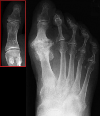
Weight bearing, anterior posterior and lateral radiographs are usually needed to examine the joint. Often non-uniform joint space narrowing, widening or flattening of the 1st MT head is seen. Subchondral sclerosis or cysts, horseshoe shaped osteophytes, lateral greater than medial osteophytes and sesamoid hypertrophy may be seen.
A clinical/radiographic grading system was described by Regnauld and appears mainly in the European literature.
Hattrup and Johnson[12] described a radiographic classification which has become standard, and in fact correlates quite well with the Regnauld grading:
- Grade 1: mild to moderate osteophytes formation but good joint space preservation
- Grade 2: moderate osteophyte formation with joint space narrowing and subchondral sclerosis
- Grade 3: marked osteophyte formation and loss of the visible joint space, with or without subchondral cyst formation
Coughlin et al[13] modified the Hattrup and Johnson classification to create the Coughlin and Shurnass[14] classification:
- Grade 0:
- Dorsiflexion 40-60°
- Normal radiography
- No pain
- Grade 1
- Dorsiflexion 30-40°
- Dorsal osteophytes
- Minimal/ no other joint changes
- Grade 2
- Dorsiflexion 10-30°
- Mild to moderate joint narrowing or sclerosis
- Osteophytes
- Grade 3
- Dorsiflexion less than 10°
- Severe radiographic changes
- Constant moderate to severe pain at extremities
- Grade 4
- Stiff joint
- Severe changes with loose bodies and osteochondritis dissecans
Examination
[edit | edit source]
- Look for other features of systemic arthropathy.
- Assess the overall foot shape, range of ankle dorsiflexion and function of the other foot joints Identify sites of tenderness – is the osteophyte symptomatic?
- Evaluate the severity of rigidity and the residual arc of movement. Is pain provoked mainly by dorsiflexion, plantarflexion or throughout the range of movement?
- Check the alignment of the great toe, looking for IPJ hyperextension or hallux rigidus with valgus. Are there any lesser ray problems?
- 1st MYP joint extension should be tested actively and passively with the patient in standing with a flexed knee
Outcome Measures[edit | edit source]
- Visual analog scales
- AOFAS (American Orthopaedic Foot and Ankle Society) scores
- Subjective self assessment score
- MTP dorsiflexion
- MTP total motion
Management / InterventionNonsurgical or conservative approaches:[edit | edit source]
Treatment for mild or moderate causes of Hallux rigidus includes anti-inflammatory NSAIDS medications that are often prescribed and usually start to relieve some symptoms with in three to four days. Glucosamine chondrointin sulfate, vitamins and minerals are recommended. Molded stiff inserts with rigid bar or rocker bottom shoes usually begin to help with in a few weeks. Shoes with a large toe box and cessation of high heels , kneeling or excessive squatting may help. Cortisone injections give relief with in 24 hours but often are only temporary.
Physical therapy to provide joint mobilizations, manipulation, range of motion, muscle reeducation, strengthening of the flexor hallucis longus muscles as well as the plantar intrinsics muscles of the feet can improve stability of the 1st MTP. Gait training for stage 1 and 2 (protection, rest, ice, compression and elevation) is often helpful to reduce the inflammation during initial stages. All of these measures can be of value to the patient even if he or she ultimately undergoes surgery.
Runners with stage II and greater hallux rigidus may need to switch to lightweight day hikers and switch from asphalt to dirt trails for long distance running.
Using rocking-soled shoes may also improve the step length of people with a hallux rigidus (level of evidence: 5). [15]
The primary goal of foot orthotic therapy or shoe modification should be blocking or shielding the hallux from dorsiflexion at the first metatarsal.
| (level of evidence: 5) [16] | (level of evidence: 5) [17] |
Surgical therapy:
[edit | edit source]
The indication for surgery is intractable pain isolated to the first metatarsophalangeal joint that is refractory to shoe modification, use of rigid shoe inserts, nonsteroidal anti-inflammatory medications, and modification of activities. Choice depends on the stage of involvement, the limitations in range of motion, the activity level of the patient and the preferences of the surgeon and patient.
Types of surgery include:
- Cheiloectomy - A procedure to remove bone spurs at the top of the joint allowing greater toe extension and improved walking. Usually beneficial for mild to moderate disease with less than 50% of joint affected usually grade 1 and grade 2[18][12][19][20].
- Dorsiflexion phalangeal osteotomy - In patients with a reasonable range of motion, a dorsal wedge osteotomy of the phalanx increases dorsiflexion at a theoretical cost of loss of plantar flexion. Mild to moderate cases occasionally require this procedure.
- Metatarsal Osteotomy – a slice is removed from the dorsal limb to slide the head down and proximally. The Place for these procedures is uncertain and more complex than cheilectomies. These procedures are intended for use in early hallux rigidus
- Excision Arthroplasty or Keller procedure[21][18]- The Keller procedure is when resection of the base of the proximal phalanx and soft-tissue reconstruction is performed with the intention to decompress the joint and improve pain and range of movement. The Keller procedure may lead to great toe weakness, cock-up deformity and metatarsalgia[22].
- MTP Arthrodesis - This is a procedure is performed to fuse the joint surfaces and is a favored procedure[23]. Suitable for most cases and severity but usually grades 3 and 4 are recommended[20] .Suitable as salvage when other procedures have failed (for example Keller procedure) Arthrodesis of the first MPJ consistently show superior results and patient satisfaction in comparison to other surgical options. While cheilectomy may be beneficial for early stages of hallux rigidus, arthrodesis of the first MPJ appears to be the best option for the relief of symptoms with stage III and stage IV hallux rigidus in active, athletic patients[24].
- Artificial joint replacement- A procedure to replace joint surfaces with a plastic or metal surface. This technique of soft-tissue interposition arthroplasty gave excellent pain relief and reliable function of the hallux, and is an alternative treatment to MTP arthrodesis in select cases of severe hallux rigidus[25]. The downside to this is the joint may not last a life time and there is currently no study documenting the long-term performance of any first MTP joint prosthesis in running athletes
Differential Diagnosis
[edit | edit source]
Turf toe, fracture, gout, rheumatoid arthritis could be some other causes of pain and stiffness in the 1st MTP joint.
Resources
[edit | edit source]
Richie D. How To Treat Hallux Rigidus In Runners. 4 April 2009. Available from: www.podiatrytoday.com/how-to-treat-hallux-rigidus-in-runners. [last accessed 5/6/9]
Foot and Ankle Center of Washington, Seattle. Available at www.footankle.com/Hallux-Rigidus.htm [last accessed 24/5/9].
References[edit | edit source]
- ↑ Massimi S, Caravelli S, Fuiano M, Pungetti C, Mosca M, Zaffagnini S. Management of high-grade hallux rigidus: a narrative review of the literature. Musculoskeletal Surgery. 2020 Dec;104:237-43.
- ↑ 2.0 2.1 2.2 Finch R. Hallux Rigidus. Plus Course 2023
- ↑ SLO Motion Shoes. Hallux Rigidus: Causes, Diagnosis, and Treatment. Available from: http://www.youtube.com/watch?v=umCSpwEvWUM [last accessed 06/01/17]
- ↑ Zammit GV, Menz HB, Munteanu SE. Structural factors associated with hallux limitus/rigidus: a systematic review of case control studies. J Orthop Sports Phys Ther. 2009 Oct;39(10):733-42.
- ↑ Colò G, Fusini F, Zoccola K, Rava A, Samaila EM, Magnan B. May footwear be a predisposing factor for the development of hallux rigidus? A review of recent findings. Acta Biomed. 2021 Jul 26;92(S3):e2021010.
- ↑ 6.0 6.1 Senga Y, Nishimura A, Ito N, Kitaura Y, Sudo A. Prevalence of and risk factors for hallux rigidus: a cross-sectional study in Japan. BMC Musculoskelet Disord. 2021 Sep 13;22(1):786.
- ↑ Root ML, Orien WP, Weed JH. Normal and abnormal function of the foot. In Clinical Biomechanics, vol II, Clinical Biomechanics Corp., Los Angeles, 1977
- ↑ Buell T, Green D, Risser J. Measurement of the first metatarsophalangeal joint range of motion. JAPMA 78:439, 1988.
- ↑ Bojsen-Moller F, Lamoreux L. Significance of free dorsiflexion of the toes in walking. Acta Orthop Scand 50: 471, 1979
- ↑ Hetherington VJ, Johnson RE, Albritton JS. Necessary dorsiflexion of the first metatarsophalangeal joint during gait. J Foot Surg 29:218, 1990
- ↑ Nawoczenski DA, Baumhauer JF, Umberger BR. Relationship between clinical measurements and motion of the first metatarsophangeal joint during gait. J Bone Joint Surg 81(3): 370-6, 1999.
- ↑ 12.0 12.1 Hattrup SJ, Johnson KA. Subjective results of hallux rigidus following treatment with cheilectomy. Clin Orthop 1988;226:182-91
- ↑ Coughlin MJ et al. Hallux rigidus. JBJS 2003; 85A:2072-88
- ↑ Coughlin MJ and Shurnas PS. Hallux rigidus: demographics, etiology, and radiographic assessment. Foot Ankle Int 2003; 24(10): 731-43.
- ↑ Cusack, G. Shtofmakher, R. L. Kilfoil Jr, S. Vu, “Improved step length symmetry and decreased low back pain with the use of a rocking-soled shoe in a patient with unilateral hallux rigidus”, BMJ Case Rep, September 2014 [Online] https://www.ncbi.nlm.nih.gov/pmc/articles/PMC4166235/pdf/bcr-2014-206408.pdf (Level of evidence: 5)
- ↑ Correct Toes. Spread Your Toes™ Series: Hallux Limitus Conservative Care vs. Conventional Care. Available from: http://www.youtube.com/watch?v=iTFn1Ldq1G8 [last accessed 06/01/17] (Level of evidence: 5)
- ↑ Joan Hope Craig. Functional hallux mobilization. Available from: http://www.youtube.com/watch?v=ZOur5I0RTVo [last accessed 06/01/17] (Level of Evidence: 5)
- ↑ 18.0 18.1 Mann RA, Clanton TO. Hallux rigidus: treatment by cheilectomy. J Bone Jt Surg 1988; 70A:400-6
- ↑ Gould N. Foot and Ankle.1981 May; 1(6):315-20.
- ↑ 20.0 20.1 Michael J. Coughlin and Paul S. Shurnas. Hallux Rigidus. Grading and Long-Term Results of Operative Treatment. J. Bone Joint Surg. Am., Nov 2003; 85: 2072 - 2088.
- ↑ Keller's arthroplasty. J Bone Jt Surg 1990; 72B:839-42
- ↑ Blewett N, Greiss ME. Long-term outcomes following Keller’s excision arthroplasty of the great toe. Foot 1993; 3:144-7
- ↑ Brodsky JW, Baum BS, Pollo FE, Mehta H. Prospective gait analysis in patients with first metatarsophalangeal joint arthrodesis for hallux rigidus. Foot Ankle International. 2007 Feb;28(2):162-5
- ↑ DP O'Doherty, IG Lowrie, PA Magnussen, and PJ Gregg. The management of the painful first metatarsophalangeal joint in the older patient. Arthrodesis or Keller's arthroplasty? Journal of Bone and Joint Surgery - British Volume, 72-B(5), 839-842
- ↑ Coughlin MJ, Shurnas PJ. Soft-tissue arthroplasty for hallux rigidus. Foot Ankle International. 2003 Sep;24(9):661-72.


