Gout
Original Editors - Students from Bellarmine University's Pathophysiology of Complex Patient Problems project.
Lead Editors - Your name will be added here if you are a lead editor on this page. Read more.
Definition/Description[edit | edit source]
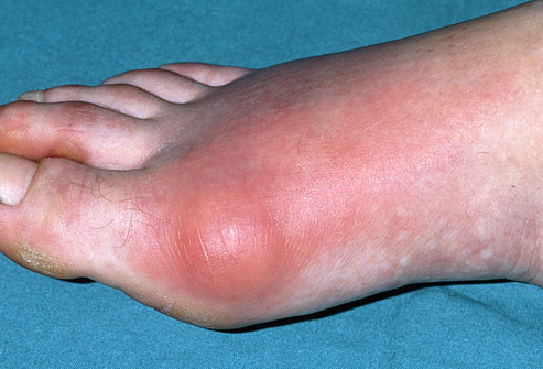
Gout is a metabolic disorder; however, because the clinical presentation closely resembles arthritis, gout is also classified as a form of crystal-induced arthritis. [1] [2]There are three main types of gout, all of which usually begin monoarticularly at the first metatarsophalangeal joint and are characterized by sudden pain, swelling, and redness.[1] [2] [3]
Prevalence[edit | edit source]
Effects over 2 million people in the US
The most common crystalopathy (in the US)
Rarely seen in children (< 10% of all cases) [1]
Predominantly seen in men (most common inflammatory disease in men over age 30)
Peak incidence in the 4th - 5th decades of life
Frequency increases in postmenopausal women (lack of estrogen) [1] [3]
Characteristics/Clinical Presentation[edit | edit source]
There are four stages of gout, although diagnosis does not require the presence or occurance of each stage. The four stages are:
- Asymptomatic hyperuricemia ( serum urate > 7mg/dl)
- Acute gouty arthritis
- Intercritical gout
- Chronic tophaceous gout[1]
- Asymtomatic hyperuricemia is the phase of gout prior to the first attack, when serum urate levels are elevated but no symptoms are present. [1]
- Acute gouty arthritis is the most common clincial presentation and is most often found (90%) at the first metatarsophalangeal joint. Symptoms generally begin with a sudden onset of localized, intense pain, often occuring at night. The pain may be great enough to awaken the patient. Redness, extreme tenderness, and swelling around the joint will occur within a few hours of the initial pain. Hypersensitivity, chills, tachycardia, malaise, and fever may also be present.[1] [2] [3] The skin may also become red or purplish, shiny, tense, and warm. [2]
Occasionally, acute gouty arthritis can also occur at the fingers, wrist, elbow, knee, ankle, and instep. Even more rarely, acute gouty arthritis may present at the cervical spine, sternoclavicular joint, shoulder, hip, and sacroilliac joint.[1] [2] [3] The initial attack may last a few days to 2 weeks, if left untreated. [2] [3] Attacks will recur; however time periods of months or years may elapse between them. As the attacks recur, they will become more intense and may spread to other joints in the body. [1] [2] [3] A patient may eventually exprience several attacks per year [1] [2]
Presentation of acute gout at the PIP joint
- The intercritical phase occurs after recovery from each episode. This phase is asymtomatic and may last for months to years at a time. It is important to note that although this is an aysmptomatic stage, serum urate levels are often still elevated and urate crystals may still be present. [1]
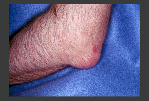
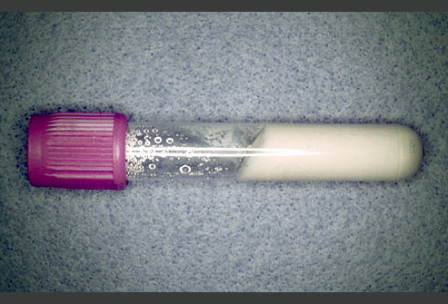
- As the gout attacks continue, tophi will develop in the joints, tendons, bursae, subchondral bone, cartilage, ligaments, subcutaneous tissue, and synovium.[1] Tophi are hard nodules of sodium urate deposits that may vary in number and location.[1] [3] [2] Although typically a painless structure, the formation of tophi can cause an acute inflammatory response within the tissue. [3] [2] As the tophi become enlarged they may cause deformities, and there is potential for them to protrude through the skin and exude a white chalky substance (urate crystals). [1] [3] [2] Some of the most common sites of enlarged tophi are the forearm, ear, knee and foot. [1]
Note: Prior to the use of urate-lowering drugs for the treatment of acute gouty arthritis, tophi developed in approximately 30 - 50% of patients. [3]
- Chronic tophaceous gout is characterized by increased pain, deformity (from tophi), decreased ROM, and subsequent functional loss.[1] [2] Due to the treatments used for gout today, chronic tophaceous gout is rare. [3]
Great pictures of the presentation of gout at various stages can also be found at:
www.skinsight.com/adult/gout.htm
[1][5]
Associated Co-morbidities[edit | edit source]
add text here
Medications[edit | edit source]
add text here
Diagnostic Tests/Lab Tests/Lab Values[edit | edit source]
add text here
Causes[edit | edit source]
add text here
Systemic Involvement[edit | edit source]
add text here
Medical Management (current best evidence)[edit | edit source]
add text here
Physical Therapy Management (current best evidence)[edit | edit source]
add text here
Alternative/Holistic Management (current best evidence)[edit | edit source]
add text here
Differential Diagnosis[edit | edit source]
add text here
Case Reports[edit | edit source]
add links to case studies here (case studies should be added on new pages using the case study template)
Resources
[edit | edit source]
add appropriate resources here
Recent Related Research (from Pubmed)[edit | edit source]
see tutorial on Adding PubMed Feed
Extension:RSS -- Error: Not a valid URL: Feed goes here!!|charset=UTF-8|short|max=10
References[edit | edit source]
see adding references tutorial.
- ↑ 1.00 1.01 1.02 1.03 1.04 1.05 1.06 1.07 1.08 1.09 1.10 1.11 1.12 1.13 1.14 1.15 Goodman CC, Fuller KS. Pathology: Implications for the Physical Therapist. 3rd ed. Saint Louis, MO: Saunders; 2009.
- ↑ 2.00 2.01 2.02 2.03 2.04 2.05 2.06 2.07 2.08 2.09 2.10 2.11 Beers MH, et. al. eds. The Merck Manual of Diagnosis and Therapy. 18th ed. Whitehouse Station, NJ: Merck Research Laboratories; 2006.
- ↑ 3.00 3.01 3.02 3.03 3.04 3.05 3.06 3.07 3.08 3.09 3.10 Goodman C, Snyder T. Differential Diagnosis for Physical Therapists: Screening for Referral. St. Louis, Missouri: Saunders Elsevier, 2007.
- ↑ 4.0 4.1 4.2 Brunilda, N. Gout Pictures Slideshow: Causes, Symptoms, and Treatments of Gout. 2008. http://arthritis.webmd.com/slideshow-gout. Accessed February 15, 2010.
- ↑ http://www.skinsight.com/adult/gout.htm


