File:Figuur 4.png: Difference between revisions
Marie Avau (talk | contribs) (...) |
(uploaded a new version of "File:Figuur 4.png": Fig. 4 Kotnis, Harish & Popowich (2011). Sonographic transducer position for examination of the tarsal tunnel structures. One end of the probe is placed on the medial malleolus (white arrow), an) |
(No difference)
| |
Latest revision as of 00:33, 10 January 2017
Summary[edit | edit source]
...
Licensing[edit | edit source]
The copyright holder has given special permission to use this work in Physiopedia.
File history
Click on a date/time to view the file as it appeared at that time.
| Date/Time | Thumbnail | Dimensions | User | Comment | |
|---|---|---|---|---|---|
| current | 00:33, 10 January 2017 | 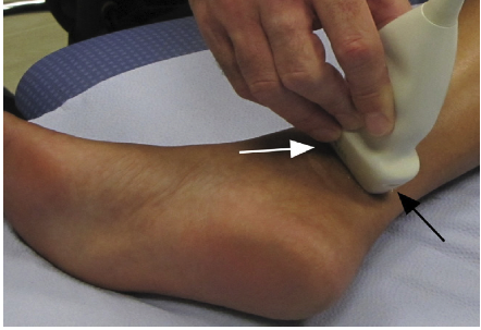 | 442 × 303 (233 KB) | Admin (talk | contribs) | Fig. 4 Kotnis, Harish & Popowich (2011). Sonographic transducer position for examination of the tarsal tunnel structures. One end of the probe is placed on the medial malleolus (white arrow), and the other end is placed as much as possible towards the ant |
| 10:11, 15 September 2015 |  | 393 × 181 (28 KB) | Marie Avau (talk | contribs) | ... |
You cannot overwrite this file.
File usage
The following file is a duplicate of this file (more details):
There are no pages that use this file.

