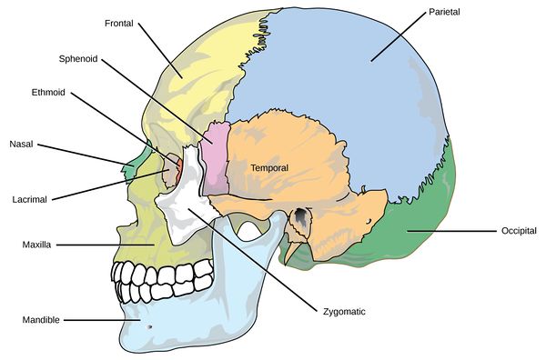
Facial Skeleton
Original Editor - User: Wendy Walker
Top Contributors - Saumya Srivastava, Wendy Walker, Angeliki Chorti, Rishika Babburu, Ines Musabyemariya, Kim Jackson, Manisha Shrestha and Ewa Jaraczewska
Description and Overview[edit | edit source]
The facial skeleton provides protection to the brain and to the special sense organs: sight, smell and taste. It is the foundation on which the facial muscles attach.
Structure[edit | edit source]
The facial skeleton consists of:
Frontal bone[edit | edit source]
This forms the forehead region of the face housing the frontal sinuses. It forms the roof of the ethmoid sinuses, nose and orbit (for the eye)[1].
Zygoma[edit | edit source]
The zygoma forms the lateral rim and wall of the orbit, and forms the anterior zygomatic arch[1].
Maxilla[edit | edit source]
This forms the roof of the oral sinus, as well as housing the upper teeth. It also forms part of the roof and lateral wall of the nasal cavity.
Nasal bones[edit | edit source]
The nasal bones are a pair of bones which form the upper part of the nasal cavity[2]. They articulate with the maxilla and the frontal bone.
Mandible[edit | edit source]
Also known as the Jaw Bone, the mandible is the only mobile bone of the facial skeleton.
It houses the lower teeth.
Small bones of the face[edit | edit source]
Inferior Conchae[edit | edit source]
This pair of small bones form part of the lateral wall of the nasal cavity.
Lacrymal Bones[edit | edit source]
These small, fragile bones lie in the antero-medial part of the orbit.
Palatine Bones[edit | edit source]
Together with the maxillae, these form the hard palate.
Vomer[edit | edit source]
This is an single bone (unpaired) which forms the inferior portion of the nasal septum.
Resources[edit | edit source]
2 part tutorial on Bones of the Facial Skeleton:


