Facial Skeleton: Difference between revisions
No edit summary |
No edit summary |
||
| Line 24: | Line 24: | ||
=== Frontal bone === | === Frontal bone === | ||
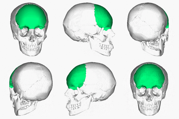
[[File:Frontal bone .gif|alt=https://commons.wikimedia.org/wiki/File:Frontal_bone_animation.gif|thumb|200x200px|Frontal bone]] | |||
This forms the forehead region of the face housing the frontal sinuses. It forms the roof of the ethmoid sinuses, nose and orbit (for the eye)<ref name=":0">Bron AJ, Tripathi RC, Tripathi BJ. | This forms the forehead region of the face housing the frontal sinuses. It forms the roof of the ethmoid sinuses, nose and orbit (for the eye)<ref name=":0">Bron AJ, Tripathi RC, Tripathi BJ. | ||
Wolff's Anatomy of the Eye and Orbit. | Wolff's Anatomy of the Eye and Orbit. | ||
| Line 37: | Line 38: | ||
===== Parts and Ossification - ===== | ===== Parts and Ossification - ===== | ||
* has 3 parts and all the 3 parts have a membranous ossification. | * has 3 parts and all the 3 parts have a membranous ossification. | ||
# Squamous portion - forms the largest portion of the frontal bone, forming majority of the forehead. | # Squamous portion - forms the largest portion of the frontal bone, including forming majority of the forehead. Also forms the supraorbital margin and the superciliary arch. | ||
# Orbital portion - forms the roof of the orbit and floor of the anterior cranial fossa. | |||
# Nasal portion - articulates with the nasal bones and the frontal process of the maxilla to form the root of the nose. The trochlea of the orbit articulates with the orbital portion. | |||
==== Zygoma ==== | ==== Zygoma ==== | ||
Revision as of 23:42, 24 October 2020
This page is under construction, come back later and check for the completed work!!!
Original Editor - User: Wendy Walker
Top Contributors - Saumya Srivastava, Wendy Walker, Angeliki Chorti, Rishika Babburu, Ines Musabyemariya, Kim Jackson, Manisha Shrestha and Ewa Jaraczewska
Description and Overview[edit | edit source]
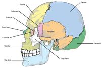
The skull is the most intricate bony structure of the body. It is made up of 28 individual bones, out of which 11 are paired and 6 are single [1]. The skull formation is divided into 2 parts:
- The Viscerocranium (the facial skeleton) - goes to develop the bones of the face. This is the portion of the skull related to the digestive and respiratory systems
- The Neurocranium (the brain case) - goes to develop the bones of the cranial base and cranial vault. This portion provides protection to the brain and to the 5 organs of special senses: Olfaction, vision, taste, vestibular function and auditory function[1].
The Viscerocranium is further divided into:
- The membranous viscerocranium, comprising the facial skeleton. It is the foundation on which the facial muscles attach.
- The cartilaginous viscerocranium, comprising the splanchnocranium.
In this article, we will cover the facial skeleton.
Structure[edit | edit source]
Since the facial skeleton is the foundation for the facial muscles, it is not inaccurate to say it helps in determining an individual's facial appearance.
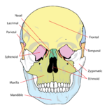
It is made up of 14 bones which integrate to house the orbits of the eyes, nasal and oral cavities along with the sinuses. The facial skeleton consists of[1]:
Frontal bone[edit | edit source]
This forms the forehead region of the face housing the frontal sinuses. It forms the roof of the ethmoid sinuses, nose and orbit (for the eye)[2]
Characteristics -[edit | edit source]
- there is only 1 frontal bone
- it contains the frontal paranasal sinuses.
- in the 2nd year, houses 2 primary centres that ossify along the frontal suture.
- helps form the foramen cecum.
Parts and Ossification -[edit | edit source]
- has 3 parts and all the 3 parts have a membranous ossification.
- Squamous portion - forms the largest portion of the frontal bone, including forming majority of the forehead. Also forms the supraorbital margin and the superciliary arch.
- Orbital portion - forms the roof of the orbit and floor of the anterior cranial fossa.
- Nasal portion - articulates with the nasal bones and the frontal process of the maxilla to form the root of the nose. The trochlea of the orbit articulates with the orbital portion.
Zygoma[edit | edit source]
The zygoma forms the lateral rim and wall of the orbit, and forms the anterior zygomatic arch[2].
Maxilla[edit | edit source]
This forms the roof of the oral sinus, as well as housing the upper teeth. It also forms part of the roof and lateral wall of the nasal cavity.
Nasal bones[edit | edit source]
The nasal bones are a pair of bones which form the upper part of the nasal cavity[3]. They articulate with the maxilla and the frontal bone.
Mandible[edit | edit source]
Also known as the Jaw Bone, the mandible is the only mobile bone of the facial skeleton.
It houses the lower teeth.
Small bones of the face[edit | edit source]
Inferior Conchae[edit | edit source]
This pair of small bones form part of the lateral wall of the nasal cavity.
Lacrymal Bones[edit | edit source]
These small, fragile bones lie in the antero-medial part of the orbit.
Palatine Bones[edit | edit source]
Together with the maxillae, these form the hard palate.
Vomer[edit | edit source]
This is an single bone (unpaired) which forms the inferior portion of the nasal septum.
Resources[edit | edit source]
2 part tutorial on Bones of the Facial Skeleton:
References[edit | edit source]
- ↑ 1.0 1.1 1.2 Neil S. Norton, Ph.D. and Frank H. Netter, MD, Netter’s Head and Neck Anatomy for Dentistry, 2nd Edition, Elsevier Saunders, 2012
- ↑ 2.0 2.1 Bron AJ, Tripathi RC, Tripathi BJ. Wolff's Anatomy of the Eye and Orbit. 8th ed. Philadelphia: Lippincott Williams & Wilkins. 1997
- ↑ Lang J. Clinical Anatomy of the Nose, Nasal Cavity, and Paranasal Sinuses. NY: Thieme Medical Publishers. 1989


