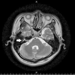
Facial Schwannoma
This article or area is currently under construction and may only be partially complete. Please come back soon to see the finished work! (28/11/2019)
Original Editor - Wendy Walker
Lead Editors
Introduction[edit | edit source]
Facial Schwannoma is a very rare tumour which grows on the 7th Cranial Nerve, the Facial Nerve.
It is also known as a Facial Neuroma.
Clinically Relevant Anatomy[edit | edit source]
For details of the path of the Cranial Nerve 7, please see the Facial Nerve page.
Location of Tumour[edit | edit source]
The majority of tumours are found on the segment of the nerve within the internal auditory canal, with one UK study[1] of a cohort of 28 Facial Schwannoma patients reporting 68% incidence in this section of the nerve.
The same study also found multi-segmental lesions in 46% of the patients.
Facial weakness was most commonly associated with involvement of the labyrinthine segment (89%).
Mechanism of Injury / Pathological Process[edit | edit source]
Schwannomas are extremely slow growing tumours, the majority of which are benign[2].
They originate from the Schwann cell sheath of the facial nerve.
Clinical Presentation[edit | edit source]
The presentation frequently comprises hearing loss and facial weakness or paralysis.
Hearing Loss[edit | edit source]
The Auditory or Acoustic Nerve, 8th Cranial Nerve, travels next to the facial nerve in the Internal Auditory Canal. A growing tumour on the portion of the Facial Nerve within the Internal Auditory Canal can therefore cause compression on the Acoustic Nerve, which results in hearing loss.
Diagnostic Procedures[edit | edit source]
In the majority of centres, a brain MRI is used as part of the evaluation of facial paralysis; however, this often misses a Facial Schwannoma because of the very small diameter of the facial nerve - approx 1 mm - and the thickness of the image slices - 5 mm.
The optimal imaging technique is an MRI of the internal auditory canals, with and without the contrast agent gadolinium. This imaging technique allows thinner slices through the temporal bone, it is much more likely to show the tumour[3].
Outcome Measures[edit | edit source]
add links to outcome measures here (see Outcome Measures Database)
Management / Interventions[edit | edit source]
add text here relating to management approaches to the condition
Differential Diagnosis[edit | edit source]
add text here relating to the differential diagnosis of this condition
Physiotherapy Management[edit | edit source]
Resources[edit | edit source]
add appropriate resources here
References[edit | edit source]
- ↑ Doshi J, Heyes R, Freeman SR, Potter G, Ward C, Rutherford S, King A, Ramsden R, Lloyd SK. Clinical and Radiological Guidance in Managing Facial Nerve Schwannomas Otology & Neurotology. 36(5):892–895, JUNE 2015
- ↑ Jayashankar N, Sankhla S. Facial schwannomas: Diagnosis and surgical perspectives. Neurol India [serial online] 2018 [cited 2019 Nov 28];66:144-6. Available from: http://www.neurologyindia.com/text.asp?2018/66/1/144/222821
- ↑ Djalilian, Hamid R. MD Symptoms: Hearing Loss and Facial Paralysis The Hearing Journal: May 2014 - Volume 67 - Issue 5 - p 11,14


