Extensor Retinaculum (Wrist): Difference between revisions
(Added an image) |
m (links) |
||
| Line 1: | Line 1: | ||
<div class="editorbox"> | <div class="editorbox"> | ||
'''Original Editor '''- [[User:Sameera Withanage| | '''Original Editor '''- [[User:Sameera Withanage|Sameera Withanage]] | ||
'''Top Contributors''' - {{Special:Contributors/{{FULLPAGENAME}}}} | '''Top Contributors''' - {{Special:Contributors/{{FULLPAGENAME}}}} | ||
| Line 6: | Line 6: | ||
== Description == | == Description == | ||
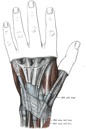
[[File:Extensor Retinaculum of the wrist.png|alt=Extensor Retinaculum of the wrist|thumb|'''Extensor Retinaculum of the wrist''']] | [[File:Extensor Retinaculum of the wrist.png|alt=Extensor Retinaculum of the wrist|thumb|'''Extensor Retinaculum of the wrist''']] | ||
Extensor Retinaculum is a fibrous, thickened band that holds the extensor tendons at the dorsum of the wrist. It is an oblique band runs downwards and medially preventing bowstringing. | Extensor Retinaculum is a fibrous, thickened band that holds the extensor tendons at the dorsum of the [[Wrist and Hand|wrist]]. It is an oblique band runs downwards and medially preventing bowstringing. | ||
=== Attachments === | === Attachments === | ||
''Laterally:'' | ''Laterally:'' | ||
# Lower part of the anterior border of the radius. | # Lower part of the anterior border of the [[radius]]. | ||
''Medially:'' | ''Medially:'' | ||
# Styloid process of the ulna. | # Styloid process of the [[ulna]]. | ||
# Triquetral. | # [[Triquetrum|Triquetral]]. | ||
# Pisiform. | # [[Pisiform]]. | ||
== Function == | == Function == | ||
Extensor Retinaculum helps to keep the extensor tendons in alignment and prevent bowstringing during movements. | Extensor Retinaculum helps to keep the extensor tendons in alignment and prevent bowstringing during [[Wrist and Hand Mobilisations|movements]]. | ||
== Clinical relevance == | == Clinical relevance == | ||
By extending fascial attachments to the underlying bones and periosteum, the retinaculum forms six osseofascial compartments over the dorsal wrist. There are several structures passing through each compartment from lateral to the medial side and each compartment is lined by a synovial sheath. | By extending fascial attachments to the underlying bones and periosteum, the retinaculum forms six osseofascial compartments over the [[Wrist and Hand Examination|dorsal wrist]]. There are several structures passing through each compartment from lateral to the medial side and each compartment is lined by a synovial sheath. | ||
{| class="wikitable" | {| class="wikitable" | ||
|+Structures passing through Extensor retinaculum | |+Structures passing through Extensor retinaculum | ||
| Line 28: | Line 28: | ||
|1 | |1 | ||
| | | | ||
* Abductor pollicis longus | * [[Abductor pollicis longus]] | ||
* Extensor pollicis brevis | * [[Extensor Pollicis Brevis|Extensor pollicis brevis]] | ||
|- | |- | ||
|2 | |2 | ||
| | | | ||
* Extensor carpi radialis longus | * [[Extensor carpi radialis longus]] | ||
* Extensor carpi radialis brevis | * [[Extensor Carpi Radialis Brevis|Extensor carpi radialis brevis]] | ||
|- | |- | ||
|3 | |3 | ||
| | | | ||
* Extensor pollicis longus | * [[Extensor Pollicis Longus|Extensor pollicis longus]] | ||
|- | |- | ||
|4 | |4 | ||
| Line 44: | Line 44: | ||
* Extensor digitorum | * Extensor digitorum | ||
* Extensor indicis | * Extensor indicis | ||
* Posterior interosseous nerve | * [[Posterior interosseous nerve syndrome|Posterior interosseous nerve]] | ||
* Anterior interosseous artery | * Anterior interosseous artery | ||
|- | |- | ||
| Line 53: | Line 53: | ||
|6 | |6 | ||
| | | | ||
* Extensor carpi ulnaris | * [[Extensor Carpi Ulnaris|Extensor carpi ulnaris]] | ||
|} | |} | ||
Revision as of 19:33, 7 October 2020
Original Editor - Sameera Withanage
Top Contributors - Sameera Withanage, Ewa Jaraczewska, Kim Jackson and Lucinda hampton
Description[edit | edit source]
Extensor Retinaculum is a fibrous, thickened band that holds the extensor tendons at the dorsum of the wrist. It is an oblique band runs downwards and medially preventing bowstringing.
Attachments[edit | edit source]
Laterally:
- Lower part of the anterior border of the radius.
Medially:
- Styloid process of the ulna.
- Triquetral.
- Pisiform.
Function[edit | edit source]
Extensor Retinaculum helps to keep the extensor tendons in alignment and prevent bowstringing during movements.
Clinical relevance[edit | edit source]
By extending fascial attachments to the underlying bones and periosteum, the retinaculum forms six osseofascial compartments over the dorsal wrist. There are several structures passing through each compartment from lateral to the medial side and each compartment is lined by a synovial sheath.
| Compartment | Structure |
|---|---|
| 1 | |
| 2 | |
| 3 | |
| 4 |
|
| 5 |
|
| 6 |


