Dial Test: Difference between revisions
Tomer Yona (talk | contribs) No edit summary |
Tomer Yona (talk | contribs) No edit summary |
||
| Line 6: | Line 6: | ||
The purpose of the Dial Test is to diagnose posterolateral instability.The test can be clinically valuable when: | The purpose of the Dial Test is to diagnose posterolateral instability.The test can be clinically valuable when: | ||
1. Three posterolateral structures (popliteus tendon, PFL, LCL) are injured. | 1. Three posterolateral structures (popliteus tendon, PFL, [http://www.physio-pedia.com/Lateral_Collateral_Ligament LCL]) are injured. | ||
2. There is combined injury to the PCL and two other posterolateral structures. | 2. There is combined injury to the PCL and two other posterolateral structures. | ||
Its important to know that when only one or two structures are injured , the dial test is not enough to diagnose the injury. For an isolated PCL tear, the posterior drawer test or sag tests are more relevant. <br> | Its important to know that when only one or two structures are injured , the dial test is not enough to diagnose the injury. For an isolated PCL tear, the posterior drawer test or sag tests are more relevant. <br> | ||
== Clinically Relevant Anatomy == | == Clinically Relevant Anatomy == | ||
Revision as of 21:00, 16 February 2016
Original Editors - Gaëlle Vertriest as part of the Vrije Universiteit Brussel's Evidence-based Practice project
Top Contributors - Tomer Yona, Gaëlle Vertriest, Rachael Lowe, Admin, Kim Jackson, Yvonne Yap, Daphne Jackson, Uchechukwu Chukwuemeka, Tony Lowe, Kai A. Sigel, WikiSysop and Wanda van NiekerkPurpose Cite error: Invalid <ref> tag; name cannot be a simple integer. Use a descriptive title[edit | edit source]
The purpose of the Dial Test is to diagnose posterolateral instability.The test can be clinically valuable when:
1. Three posterolateral structures (popliteus tendon, PFL, LCL) are injured.
2. There is combined injury to the PCL and two other posterolateral structures.
Its important to know that when only one or two structures are injured , the dial test is not enough to diagnose the injury. For an isolated PCL tear, the posterior drawer test or sag tests are more relevant.
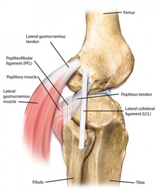
Clinically Relevant Anatomy[edit | edit source]
Relevant structure of the posterolateral corner in the knee [1]
TechniqueCite error: Invalid <ref> tag; name cannot be a simple integer. Use a descriptive titleCite error: Invalid <ref> tag; name cannot be a simple integer. Use a descriptive title
[edit | edit source]
It is possible to do the test both in a prone and supine position, and is performed in both 30° and 90° knee flexion. The dial test inspects the external rotation at the knee joint.
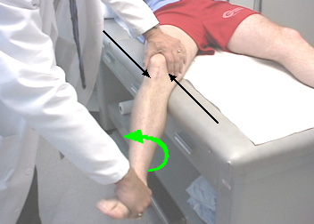
The patient is in prone: performing the test is sensitive to the notice of a PLC-injury in a PCL-injured knee. The knees are held together and bent at 30°, the therapist stands behind the table and keeps the feet in dorsiflexion. He turns the lower legs and feet outwards and compares the motion of the feet. Repeat the test with the knees at 90°.
The patient is in supine: there are 2ways to perform this test.
1) As in prone position: the knees are held together and bent at 30°, the therapist stands behind the table and keeps the feet in dorsiflexion. He turns the lower legs and feet outwards and compares the amount of rotation of the tibial tubercle. Repeat the test with the knees at 90°.
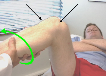
2) One leg is hanging off the edge of the table with the knee in 30° of flexion. The therapist stands beside the table and stabilizes the thigh with one hand; the other hand executes an external rotation of the foot. By observing the tibial tubercle motion, we can indicate any posterolateral knee injury. With an increase, compare to the normal contralateral side.
If the dial test at 30° is positive, perform the test when the knee is flexed on 90°. The thigh does not touch the table, hold the leg in your hands or put the foot down on the table.
Key Research[edit | edit source]
add links and reviews of high quality evidence here (case studies should be added on new pages using the case study template)
Evaluation[edit | edit source]
The evaluation of the test for both prone and supine position:
The dial test is positive when there is more than 10° of external rotation in the injured knee compared to the uninjured knee.
| Standard injury | Mild injury | Moderate injury | Severe Injury |
| <5° | 6-10° | 11-19° | >20° |
There are different injuries:
- An isolated injury to the PLC: more than 10° of external rotation in the injured knee is present at 30° of flexion, but not at 90° of flexion.
- Instability of the PCL: more than 10° of external rotation in the injured knee is present at 90° of flexion, but not at 30° of flexion.
- A combined injury: more than 10° of external rotation in the injured knee is present at 30° and 90° of flexion. This is an injury of the PCL and the PLC.
Recent Related Research (from Pubmed)[edit | edit source]
Failed to load RSS feed from https://www.ncbi.nlm.nih.gov/entrez/eutils/erss.cgi?rss_guid=1jue2dFuI_LNj0oPWak3TlgiK4yZ-f2qBZrJ3hLVgRrdLy89uW|charset=UTF-8|short|max=10: There was a problem during the HTTP request: 422 Unprocessable Entity
References[edit | edit source]
- ↑ Noyes FR. Noyes' knee disorders: surgery, rehabilitation, clinical outcomes. Elsevier Health Sciences; 2009 Aug 20.


