Classification of Peripheral Nerve Injury: Difference between revisions
Jayati Mehta (talk | contribs) No edit summary |
Kim Jackson (talk | contribs) No edit summary |
||
| (34 intermediate revisions by 8 users not shown) | |||
| Line 1: | Line 1: | ||
<div class="editorbox"> | <div class="editorbox">'''Original Editor '''- [[User:Tomer Yona|Tomer Yona]] | ||
'''Original Editor '''- [[User:Tomer Yona|Tomer Yona]] | |||
'''Top Contributors''' - {{Special:Contributors/{{FULLPAGENAME}}}} | '''Top Contributors''' - {{Special:Contributors/{{FULLPAGENAME}}}} | ||
'''Edited April 2023''' - by [[User:Emma Sewell|Emma Sewell]], [[User:Kaylee Byars|Kaylee Byars]], and [[User:Katherine Baca|Katherine Baca]] as part of the [[Arkansas Colleges of Health Education School of Physical Therapy Musculoskeletal 1 Project]]</div> | |||
== Description == | == Description == | ||
Peripheral nerves are responsible for somatic (voluntary) and autonomic (involuntary) functions. The primary functions of the peripheral nervous system are to receive general [[Sensation|sensations]] (touch, pressure, temperature, and pain), and special sensations (sight, smell, taste, and hearing), integrate sensory input from the entire body, and generate a response<ref>Radhakrishnan, R. What are the 4 main functions of the nervous system? Available from <nowiki>https://www.medicinenet.com/4_main_functions_of_the_nervous_system/article.htm</nowiki> (Accessed 18 March 2023).</ref>. Peripheral Nerve Injury can be sustained from traumatic or idiopathic mechanisms. Individuals with diagnoses of [[diabetes]], alcoholism, vascular disease, autoimmune diseases, or who have been exposed to chemotherapy drugs or infections that attack nerves have a higher likelihood of acquired peripheral neuropathy<ref name=":0">National Institute of Neurological Disorders and Stroke. Peripheral neuropathy. Available from <nowiki>https://www.ninds.nih.gov/health-information/disorders/peripheral-neuropathy</nowiki> (Accessed 18 May 2023).</ref>. The severity of peripheral nerve injury is determined with advanced imaging (CT, MRI, or MRI neurography) or nerve conduction velocity testing and classified using the Sunderland or Seddon classification systems<ref name=":0" />. Treatment of peripheral nerve damage depends on the severity of the damage and may include surgical procedures, skilled physical therapy, orthotics, or medications<ref>Johns Hopkins Medicine. Peripheral Nerve Injury. Available from <nowiki>https://www.hopkinsmedicine.org/health/conditions-and-diseases/peripheral-nerve-injury</nowiki> (Accessed 18 March 2023).</ref> . | |||
=== '''Mechanisms of Injury for Peripheral Nerves''' === | |||
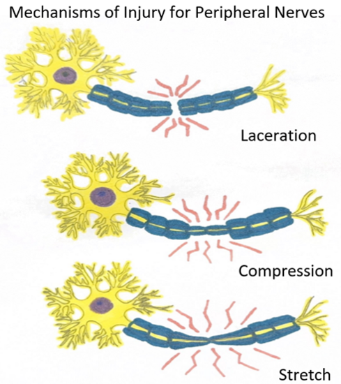
There are numerous mechanisms of injury for peripheral nerves. The three most common mechanisms of injury for peripheral nerves are stretch related, lacerations, and compressions. The most common of these three is stretch-related, followed by lacerations, and then compression<ref name="camp">Campbell WW. Evaluation and management of peripheral nerve injury. Clinical neurophysiology. 2008 Sep 30;119(9):1951-65.</ref>. Radiation, electricity, injection, crush, cold injury, and intra-neural and extra-neural pathologies could also result in peripheral nerve injuries. <ref name=":1" /> | |||
[[File:Mechanisms of Injury for Peripheral Nerves.png|center|thumb|550x550px|alt=|This visual representation of Peripheral Nerve Injury Mechanisms was created by Katherine Baca, SPT of Arkansas Colleges of Health Education.]] | |||
==== Stretch Related ==== | |||
Due to the elastic nature of peripheral nerves, stretch related injuries can occur if a traction force is too strong for the nerves elasticity. If the traction force exceeds the nerves stretch abilities, a complete tear could occur. However, it is more common that the continuity of the nerve is retained during this type of injury<ref name="burn">Burnett MG, Zager EL. Pathophysiology of peripheral nerve injury: a brief review. Neurosurgical focus. 2004 May;16(5):1-7.</ref>. | |||
===='''Lacerations'''==== | |||
Laceration injuries are the second most common types of peripheral nerve injuries. With this mechanism of injury, a nerve is severed partially or fully by some type of sharp object. Most common lacerations are from knives, broken glass, metal shards etc. <ref name=":1" />. Due to the varying nature of damage resulting from lacerations, we refer to Seddon’s and Sunderland’s classification systems which are discussed below. | |||
===='''Compressions'''==== | |||
Compression nerve injuries typically affect large-caliber nerves that cross over bony structures or between rigid surfaces. Acute compression (e.g. [[Saturday Night Palsy]]) and chronic compression injuries (e.g. [https://www.physio-pedia.com/Carpal_Tunnel_Syndrome?utm_source=physiopedia&utm_medium=search&utm_campaign=ongoing_internal carpal tunnel syndrome]) are the two main subcategories for compression injuries<ref name=":1" />. Compressive nerve injuries can result in complete functional loss of both motor and sensory function even though the nerve fibers are still intact. Two pathological mechanisms have been thought to contribute to these types of injuries: mechanical compression and ischemia<ref name="burn" />. Mechanical compression could result in secondary ischemia issues which can compromise nerve microcirculation <ref name=":1" />. | |||
== Classification == | == Classification == | ||
There are two commonly used | There are two commonly used classifications for PNI- the '''Seddon Classification''' and the '''Sunderland Classification.''' | ||
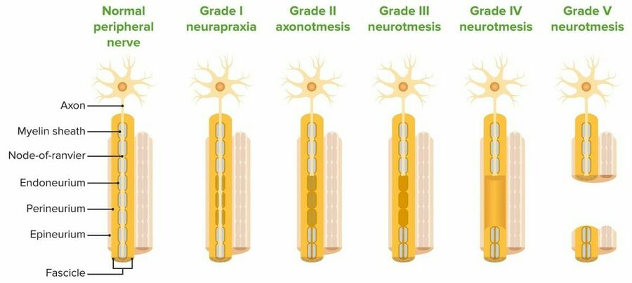
Seddon | Seddon is responsible for classifying peripheral nerve injuries into neuropraxia, axonotmesis, and neurotmesis. Sunderland expanded this idea by further classifying these into different degrees or levels of injury. It is important to note that there is a slight overlap when looking at these nerve pathologies, therefore, the degree of injury is specific to each individual patient.<br> | ||
{| width="800" border="1" cellpadding="1" cellspacing="1" | {| width="800" border="1" cellpadding="1" cellspacing="1" | ||
|+ | |||
|- | |- | ||
| '''Seddon ''' | |'''Seddon ''' | ||
| '''Process''' | |'''Process''' | ||
|'''Symptoms''' | |||
| '''Sunderland ''' | | '''Sunderland ''' | ||
|- | |- | ||
| ''Neurapraxia'' | |''Neurapraxia'' | ||
| | | This type of nerve injury is usually secondary to compression pathology. This is the mildest form of peripheral nerve injury with minimal structural damage. This allows for a complete and relatively short recovery period. In a neuropraxic injury, a focal segment of the nerve is demyelinated at the site of injury with no injury or disruption to the axon or its surroundings. This is usually due to prolonged ischemia from excess pressure or stretching of the nerve with no Wallerian degeneration <ref name=":1">Magee DJ, Manske RC. Orthopedic physical assessment. 7th Edition. St. Louis: Elsevier, 2020. </ref>. | ||
| ''First degree'' | | | ||
* pain | |||
* no muscle wasting | |||
* muscle weakness | |||
* numbness | |||
* proprioception issues | |||
|''First degree'' | |||
|- | |- | ||
| ''Axonotmesis'' | |''Axonotmesis'' | ||
| | | An axonotmesis injury involves damage to the axon and its myelin sheath. However, the endoneurium, perineurium, and epineurium remain intact. Although the internal structure is preserved, the damage of the axons does lead to [https://www.physio-pedia.com/Wallerian_Degeneration?utm_source=physiopedia&utm_medium=search&utm_campaign=ongoing_internal Wallerian degeneration]<ref name=":1" /> This type of nerve injury also results in a complete recovery although it does take longer than a neuropraxic injury. | ||
| ''Second degree'' | | | ||
* pain | |||
* muscle wasting | |||
* complete motor, sensory, and sympathetic function loss | |||
|''Second & Third degree'' | |||
|- | |- | ||
| | | Neurotmesis | ||
| | | A neurotmesis injury can occur at different levels and thus we use Sunderland’s further breakdown of PNIs. A 3rd-degree neurotmesis injury is the disruption of the axon and endoneurium. when this occurs the perineurium and epineurium remain intact<ref name=":1" />. Disruption of the axon and perineurium is considered a 4th-degree injury. And a complete disruption of the entire nerve trunk is classified as a 5th-degree injury. | ||
| | |||
* no pain (anesthesia) | |||
* muscle wasting | |||
* complete motor, sensory, and sympathetic unction loss | |||
|''Third, Fourth, & Fifth Degree'' | |||
|}<br> | |||
[[File:Peripheral Nerve Injury Classifications.jpg|center|thumb|900x900px]] | |||
<br><ref>Lecturio. Peripheral Nerve Injuries in the Upper Extremity. Sunderland classification of nerve injuries[PHOTO]. Leipzig: Lecturio, 2021.</ref> | |||
| ''Fifth | |||
|} | |||
[[ | |||
<br> | |||
[ | |||
== | <span id="1481398882317S" style="display: none;"> </span> See [[Nerve Injury Rehabilitation Physiotherapy]] for more information regarding treatment. | ||
== References == | |||
<references /><br> | <references /><br> | ||
[[Category:Primary Contact]] | |||
[[Category:Nerves]] | |||
Latest revision as of 11:21, 31 August 2023
Top Contributors - Tomer Yona, Emma Sewell, Jayati Mehta, Kaylee Byars, Katherine Baca, Kim Jackson, Naomi O'Reilly, Claire Knott, Lucinda hampton and Matt HueyEdited April 2023 - by Emma Sewell, Kaylee Byars, and Katherine Baca as part of the Arkansas Colleges of Health Education School of Physical Therapy Musculoskeletal 1 Project
Description[edit | edit source]
Peripheral nerves are responsible for somatic (voluntary) and autonomic (involuntary) functions. The primary functions of the peripheral nervous system are to receive general sensations (touch, pressure, temperature, and pain), and special sensations (sight, smell, taste, and hearing), integrate sensory input from the entire body, and generate a response[1]. Peripheral Nerve Injury can be sustained from traumatic or idiopathic mechanisms. Individuals with diagnoses of diabetes, alcoholism, vascular disease, autoimmune diseases, or who have been exposed to chemotherapy drugs or infections that attack nerves have a higher likelihood of acquired peripheral neuropathy[2]. The severity of peripheral nerve injury is determined with advanced imaging (CT, MRI, or MRI neurography) or nerve conduction velocity testing and classified using the Sunderland or Seddon classification systems[2]. Treatment of peripheral nerve damage depends on the severity of the damage and may include surgical procedures, skilled physical therapy, orthotics, or medications[3] .
Mechanisms of Injury for Peripheral Nerves[edit | edit source]
There are numerous mechanisms of injury for peripheral nerves. The three most common mechanisms of injury for peripheral nerves are stretch related, lacerations, and compressions. The most common of these three is stretch-related, followed by lacerations, and then compression[4]. Radiation, electricity, injection, crush, cold injury, and intra-neural and extra-neural pathologies could also result in peripheral nerve injuries. [5]
Stretch Related[edit | edit source]
Due to the elastic nature of peripheral nerves, stretch related injuries can occur if a traction force is too strong for the nerves elasticity. If the traction force exceeds the nerves stretch abilities, a complete tear could occur. However, it is more common that the continuity of the nerve is retained during this type of injury[6].
Lacerations[edit | edit source]
Laceration injuries are the second most common types of peripheral nerve injuries. With this mechanism of injury, a nerve is severed partially or fully by some type of sharp object. Most common lacerations are from knives, broken glass, metal shards etc. [5]. Due to the varying nature of damage resulting from lacerations, we refer to Seddon’s and Sunderland’s classification systems which are discussed below.
Compressions[edit | edit source]
Compression nerve injuries typically affect large-caliber nerves that cross over bony structures or between rigid surfaces. Acute compression (e.g. Saturday Night Palsy) and chronic compression injuries (e.g. carpal tunnel syndrome) are the two main subcategories for compression injuries[5]. Compressive nerve injuries can result in complete functional loss of both motor and sensory function even though the nerve fibers are still intact. Two pathological mechanisms have been thought to contribute to these types of injuries: mechanical compression and ischemia[6]. Mechanical compression could result in secondary ischemia issues which can compromise nerve microcirculation [5].
Classification[edit | edit source]
There are two commonly used classifications for PNI- the Seddon Classification and the Sunderland Classification.
Seddon is responsible for classifying peripheral nerve injuries into neuropraxia, axonotmesis, and neurotmesis. Sunderland expanded this idea by further classifying these into different degrees or levels of injury. It is important to note that there is a slight overlap when looking at these nerve pathologies, therefore, the degree of injury is specific to each individual patient.
| Seddon | Process | Symptoms | Sunderland |
| Neurapraxia | This type of nerve injury is usually secondary to compression pathology. This is the mildest form of peripheral nerve injury with minimal structural damage. This allows for a complete and relatively short recovery period. In a neuropraxic injury, a focal segment of the nerve is demyelinated at the site of injury with no injury or disruption to the axon or its surroundings. This is usually due to prolonged ischemia from excess pressure or stretching of the nerve with no Wallerian degeneration [5]. |
|
First degree |
| Axonotmesis | An axonotmesis injury involves damage to the axon and its myelin sheath. However, the endoneurium, perineurium, and epineurium remain intact. Although the internal structure is preserved, the damage of the axons does lead to Wallerian degeneration[5] This type of nerve injury also results in a complete recovery although it does take longer than a neuropraxic injury. |
|
Second & Third degree |
| Neurotmesis | A neurotmesis injury can occur at different levels and thus we use Sunderland’s further breakdown of PNIs. A 3rd-degree neurotmesis injury is the disruption of the axon and endoneurium. when this occurs the perineurium and epineurium remain intact[5]. Disruption of the axon and perineurium is considered a 4th-degree injury. And a complete disruption of the entire nerve trunk is classified as a 5th-degree injury. |
|
Third, Fourth, & Fifth Degree |
See Nerve Injury Rehabilitation Physiotherapy for more information regarding treatment.
References[edit | edit source]
- ↑ Radhakrishnan, R. What are the 4 main functions of the nervous system? Available from https://www.medicinenet.com/4_main_functions_of_the_nervous_system/article.htm (Accessed 18 March 2023).
- ↑ 2.0 2.1 National Institute of Neurological Disorders and Stroke. Peripheral neuropathy. Available from https://www.ninds.nih.gov/health-information/disorders/peripheral-neuropathy (Accessed 18 May 2023).
- ↑ Johns Hopkins Medicine. Peripheral Nerve Injury. Available from https://www.hopkinsmedicine.org/health/conditions-and-diseases/peripheral-nerve-injury (Accessed 18 March 2023).
- ↑ Campbell WW. Evaluation and management of peripheral nerve injury. Clinical neurophysiology. 2008 Sep 30;119(9):1951-65.
- ↑ 5.0 5.1 5.2 5.3 5.4 5.5 5.6 Magee DJ, Manske RC. Orthopedic physical assessment. 7th Edition. St. Louis: Elsevier, 2020.
- ↑ 6.0 6.1 Burnett MG, Zager EL. Pathophysiology of peripheral nerve injury: a brief review. Neurosurgical focus. 2004 May;16(5):1-7.
- ↑ Lecturio. Peripheral Nerve Injuries in the Upper Extremity. Sunderland classification of nerve injuries[PHOTO]. Leipzig: Lecturio, 2021.


