Basic Foot and Ankle Anatomy - Bones and Ligaments
Original Editor - User Name
Top Contributors - Ewa Jaraczewska, Wanda van Niekerk, Jess Bell, Tarina van der Stockt, Niha Mulla, Kim Jackson and Olajumoke Ogunleye
Description[edit | edit source]
Ankle and foot injuries are fairly common not only with the athletes or as a result of sporting activities, but they occur during routine daily activities. The typical injuries include sprains, fractures, tears, and inflammation. Good knowledge of the foot and ankle anatomy is necessary for proper diagnosis and treatment.
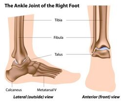
The ankle is formed by three bones: talus, tibia and fibula. The anatomic structure of the foot consists of hindfoot, midfoot and forefoot, each composed of several bones.
Structure[edit | edit source]
Ankle Bones[edit | edit source]
The ankle is composed of the lower leg and the foot and the bony elements of the ankle joint include the distal tibia, distal fibula, and talus.
Foot bones[edit | edit source]
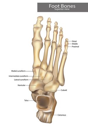
Talus and calcaneus and two tarsal bones form the posterior aspect of the foot called hindfoot. The midfoot located between the hindfoot and forefoot is made up of five tarsal bones: navicular, cuboid, and medial, intermediate, and lateral cuneiforms. The most anterior aspect of the foot including metatarsals, phalanges, and sesamoid bones is called a forefoot. Each digit, except for the great toe, consists of a metatarsal and three phalanges Great toe has two phalanges only.
Function[edit | edit source]
The foot and ankle provide various important functions which include bodyweight support, balance maintenance, shock absorption, response to ground reaction forces, or substitution for hand function in individuals with upper extremity amputation.[1]It has a crucial role in gait and posture therefore its malalignment can cause problems eg., back pain or mobility limitations.[2][3]
Ankle and Foot Articulations[edit | edit source]
Tibiotalar joint is referred to the ankle joint. The joint is framed laterally and medially by the lateral malleolus and the medial malleolus and from the top by the inferior articular surface of the tibia and the superior margin of the talus. [4]
The hindfoot has a talus and calcaneus articulation called a subtalar joint. It is made of the three facets on each of the talus and calcaneus. The talonavicular and calcaneocuboid joints known as a Chopart joint are located between the hind and midfoot. The navicular and all three cuneiform bones compose articulation distally. In addition to the navicular and cuneiforms bones, the cuboid bone has a distal articulation with the base of the forth and fifth metatarsal bones. The Lisfranc joint connects the midfoot with forefoot and is composed by lateral, intermediate and medial cuneiforms articulating with the bases of the three metatarsal bone (1st, 2nd, and 3rd). The bases of the remaining metatarsal bones(4th and 5th) connect with the cuboid bone. [4]
Tibiotalar Joint[edit | edit source]
Muscle attachments[edit | edit source]
Watch this short video on ankle and foot palpation
Vascular Supply[edit | edit source]
Nerve Supply[edit | edit source]
Clinical relevance[edit | edit source]
Resources[edit | edit source]
References[edit | edit source]
- ↑ Houglum PA, Bertotti DB. Brunnstrom's clinical kinesiology. FA Davis; 2012
- ↑ Menz HB, Dufour AB, Riskowski JL, Hillstrom HJ, Hannan MT. Foot posture, foot function and low back pain: the Framingham Foot Study. Rheumatology (Oxford). 2013 Dec;52(12):2275-82.
- ↑ Menz HB, Dufour AB, Katz P, Hannan MT. Foot Pain and Pronated Foot Type Are Associated with Self-Reported Mobility Limitations in Older Adults: The Framingham Foot Study. Gerontology. 2016;62(3):289-95.
- ↑ 4.0 4.1 Ficke J, Byerly DW. Anatomy, Bony Pelvis and Lower Limb, Foot. [Updated 2021 Aug 11]. In: StatPearls [Internet]. Treasure Island (FL): StatPearls Publishing; 2021 Jan-. Available from: https://www.ncbi.nlm.nih.gov/books/NBK546698/
- ↑ Ankle and foot palpation. 2012 Available from: https://www.youtube.com/watch?v=aJRemQbNPhk [last accessed 18/12/2021]


