Assessing Muscle Length: Difference between revisions
No edit summary |
No edit summary |
||
| Line 54: | Line 54: | ||
==== Two-Joint Muscle ==== | ==== Two-Joint Muscle ==== | ||
Unlike one-joint muscles, muscles that cross two or more joints typically do not allow full range of motion across all the joints they cover, which is known as passive insufficiency. | Unlike one-joint muscles, muscles that cross two or more joints typically do not allow full range of motion across all the joints they cover, which is known as passive insufficiency. | ||
<blockquote>"[[Active and Passive Insufficiency|Passive insufficiency]] occurs when a multi-joint muscle is lengthened to its fullest extent at both joints, thereby preventing full range of motion of each joint it crosses." <ref>Rogers M, Rogers M. Understanding Active and Passive Insufficiency [Internet]. National Federation of Professional Trainers. 2020 [cited 17 September 2020]. Available from: <nowiki>https://www.nfpt.com/blog/understanding-active-and-passive-insufficiency</nowiki></ref></blockquote>To assess and measure the length of a two-joint muscle, position one of the joints crossed by the muscle so as to lengthen the muscle across the joint. Then move the second joint through a passive range of motion until the muscle is placed on full stretch and prevents further joint motion. Assess and measure the final position of the second joint. This represents an indirect measure of the muscle length. <ref name=":4" /><ref name=":2" /> | |||
For example, [[Biceps Brachii|biceps brachii]] crosses the shoulder and the elbow. It flexes and supinates the elbow and is a weak shoulder flexor. To test the length of this muscle, we would position our patient in supine. The starting position is shoulder extension, with the elbow flexed and supinated. We then extend the elbow and measure the range of elbow extension to determine muscle length of biceps. If we want to measure elbow joint extension, we would position the shoulder joint in neutral, to prevent passive insufficiency of biceps brachii affecting our results. We can also compare the results to see the difference in range when the muscle isn’t on full stretch. | For example, [[Biceps Brachii|biceps brachii]] crosses the shoulder and the elbow. It flexes and supinates the elbow and is a weak shoulder flexor. To test the length of this muscle, we would position our patient in supine. The starting position is shoulder extension, with the elbow flexed and supinated. We then extend the elbow and measure the range of elbow extension to determine muscle length of biceps. If we want to measure elbow joint extension, we would position the shoulder joint in neutral, to prevent passive insufficiency of biceps brachii affecting our results. We can also compare the results to see the difference in range when the muscle isn’t on full stretch. | ||
Revision as of 01:05, 10 May 2023
Original Editors - Naomi O'Reilly and Jess Bell
Top Contributors - Naomi O'Reilly, Jess Bell and Ewa Jaraczewska
Introduction[edit | edit source]
Muscle length refers to the ability of a muscle crossing a joint to lengthen, allowing one or more joints to move through the full available range of motion.[1][2] The lengthening of a muscle fiber begins with the sarcomere, the basic unit of contraction in the muscle fiber. As the sarcomere contracts, the area of overlap between the thick and thin myofilaments increases. As it lengthens, this area of overlap decreases, allowing the muscle fiber to elongate. Maximal muscle length therefore is the greatest extensibility of the muscle-tendon junction. Muscle length testing is done to determine whether the muscle length is limited or excessive, i.e., whether the muscle is too short to permit normal range of motion, or stretched and allowing too much range of motion.[3]
Muscle Length Tests are are performed to determine whether the range of muscle length is normal, limited, or excessive i.e., whether the muscle is too short to permit normal range of motion, or lengthened and allowing too much range of motion and is used to identify if these changes in muscle extensibility may be contributing to movement impairment and/or symptoms. Muscle length testing consist of movements that increase the distance between the origin and insertion, thereby lengthening muscles in directions opposite to the muscles actions, while assessing its resistance to passive lengthening.[3]
Precise testing requires that one of the bony attachments of the muscle be in a fixed position while the other bony attachment is moved passively in the direction of lengthening the muscle. This means that to assess and measure the length of a muscle we need to passively stretch or lengthen the muscle across the joint or joints crossed by that muscle. For the best accuracy and precision, muscle length testing should be performed when the patient is not in acute pain in order to avoid pain inhibition and muscle guarding.[4]
The purpose of muscle length testing is to help determine whether reduced or increased range of motion at a joint is caused by the length of the muscle being tested or if it is caused by other structures. [4]
Structure of Muscles[edit | edit source]
Skeletal muscles are made up of striated muscle fibres. A muscle connects to bones or joint capsules by connective tissue structures, such as tendons or aponeuroses.[5] Muscle fibres contain smaller units called myofibrils, which in turn are made of thick and thin myofilaments. These filaments are organised longitudinally into units called sarcomeres, which is the basic contractile unit of the muscle fibre. [6]
A muscle belly generates force when the sarcomeres contract, which pulls the origin and insertion of the muscle-tendon complex closer together, so the muscle shortens during a contraction.[5] When sarcomeres contract, the amount of overlap between thick and thin myofilaments increases. As it lengthens, the amount of overlap decreases, so the muscle fibre can lengthen.
Kruse and colleagues state that “the force exerted actively by a muscle can be expressed as a function of muscle length.” The length where muscles don’t actively generate force is known as active slack length. The length at which muscles are able to generate their maximal active force is known as optimum muscle length. The difference between the two is the length range of active force exertion. [5]
Factors Effecting Muscle Length[edit | edit source]
Gender[edit | edit source]
In general evidence suggests that biological females tend to be more flexible and have increased muscle length in comparison to biological males.[7] [8]Research looking specifically at hamstrings length has found that females can have up to 8 degrees more range in their passive SLR [9] and 12 degrees in the knee active knee extension test.[10]
Age[edit | edit source]
Force production capacity is impacted by reduced muscle fibre length and altered pennation angles, which is the angle between the longitudinal axis of the muscle and its fibers.[11] Older adults experience increased fibrosis, sarcopenia, decreased force production and a general reduction in flexibility. These changes can impact independence and lead to an increase in morbidity and mortality.[12] There may also be an association between range of motion and muscle length of the lower limb and balance performance in older adults with foot deformities. [13] However, as Reese and Bandy [2] point out, there is not much research on age-related changes in muscle length using direct measurement tests, with at least one study on hamstring length using this method showing no difference in muscle length associated with age. [9]
Posture[edit | edit source]
Posture can have an impact on muscle length because our muscles and tissues adapt to how they are used. A common posture seen in clinical practice is a forward head position. [14] In a forward head position, an individual’s chin tends to come forward. They have flexion of their mid-lower cervical spine and extension of their upper cervical spine. This posture is often linked to time spent sitting at desks on computers, laptops and mobile phones. [15] Research suggests that a forward head posture can be associated with vestibular deficits, decreased proprioception, abnormal muscle activity, and altered breathing patterns [14] and it has a bearing on muscle length. The cervical flexors and occipital extensors have been found to shorten in a forward head position compared to a neutral spine, whereas the cervical extensors and occipital flexors lengthen. [15]
Measurement Methods[edit | edit source]
Review of the literature identifies primarily two methods to assess muscle length.
Composite Measurement[edit | edit source]
Muscle length is commonly assessed through the use of composite tests, which look at movement across more than one muscle or joint. Common composite tests are the Apley’s Test or the Sit and Reach Test, and while frequently used as a measure of muscle length research suggests that these composite tests do not provide accurate measurements of muscle length as they assess combinations of movements across several joints involving several muscles. Thus, they tend to provide a general idea of flexibility rather than an exact measurement of a single muscle’s length.
Direct Measurement[edit | edit source]
Direct measurement of muscle length is the alternative method where excursion between adjacent segments of one joint is measured, and typically are considered a better indicator of muscle length than composite tests. When using direct measurement techniques we need to consider that muscles are characterised by the number of joints they cross i.e. one-joint muscles, two-joint muscles and multi-joint muscles and that this will impact of on the measurement of muscle length.
- Muscles that pass over one joint only, the range of motion and range of muscle length will measure the same;
- Muscles that pass over two or more joints, the normal range of the muscle will be less than the total range of motion of the joints over which the muscle passes.[16]
One Joint Muscle[edit | edit source]
One-joint muscles typically allow full passive range of motion at the joint they cross. If a one-joint muscle is short, and it limits the range of motion, you’ll notice a firm end feel caused by muscle tension. [2][4]
As their name suggests, one-joint muscles cross just one joint. We can determine the length of a one-joint muscle by measuring the passive range of motion of the joint that it crosses, by positioning the joint where the muscle is lengthened across the joint and the position of the joint is measured. This represents an indirect measure of the muscle length. [2][16]
Effectively we measure the range in the direction opposite to its action. For example, the hip adductors (adductor longus, brevis and magnus) are one-joint muscles. So to determine their length, we measure the passive range of hip abduction. [4]
Two-Joint Muscle[edit | edit source]
Unlike one-joint muscles, muscles that cross two or more joints typically do not allow full range of motion across all the joints they cover, which is known as passive insufficiency.
To assess and measure the length of a two-joint muscle, position one of the joints crossed by the muscle so as to lengthen the muscle across the joint. Then move the second joint through a passive range of motion until the muscle is placed on full stretch and prevents further joint motion. Assess and measure the final position of the second joint. This represents an indirect measure of the muscle length. [2][16]"Passive insufficiency occurs when a multi-joint muscle is lengthened to its fullest extent at both joints, thereby preventing full range of motion of each joint it crosses." [17]
For example, biceps brachii crosses the shoulder and the elbow. It flexes and supinates the elbow and is a weak shoulder flexor. To test the length of this muscle, we would position our patient in supine. The starting position is shoulder extension, with the elbow flexed and supinated. We then extend the elbow and measure the range of elbow extension to determine muscle length of biceps. If we want to measure elbow joint extension, we would position the shoulder joint in neutral, to prevent passive insufficiency of biceps brachii affecting our results. We can also compare the results to see the difference in range when the muscle isn’t on full stretch.
Similarly, if we are considering rectus femoris length vs knee flexion range of motion. Rectus femoris crosses the hip and knee, flexing the hip and extending the knee. So to measure knee flexion, we must have the hip flexed to avoid passive insufficiency of rectus femoris. If we want to determine the length of this muscle, we can position the patient in prone, which puts the hip in some extension and then we measure the amount of knee flexion allowed by rectus femoris. If the hip flexes during the movement, we know there are length limitations in this muscle. This is also known as the Ely's Test. [4]
Multi-joint Muscle[edit | edit source]
Measurement of multi-joint muscles follows the same principles as measuring a two-joint muscle. To assess and measure the length of a multi-joint muscle, position all but one of the joints crossed by the muscle so that the muscle is lengthened across the joints. Then move the remaining joint crossed by the muscle through a passive range of motion, until the muscle is on full stretch and prevents further motion at the joint. Assess and measure the final position of the joint; the joint position represents an indirect measure of the muscle length.[2][16]
For example, the flexor digitorum superficialis crosses the elbow, wrist and hand, and inserts into the middle phalanges of digits 2-5. It primarily flexes digits 2-5 at the proximal interphalangeal (PIP) and metacarpophalangeal (MCP) joints, but it is also a wrist flexor. To assess this muscle (and the other multi-joint finger flexors), we position the patient in sitting with their forearm in pronation on a table. The hand rests over the table. We move the elbow and finger joints into extension and then passively extend the wrist. We measure the amount of wrist extension to assess the length of this muscle. [2]
Measurement Tools[edit | edit source]
Measurement of muscle length is assessed using three primary types of instruments, which include the universal goniometer and the variations of this measurement tool, the inclinometer and its variations, and linear forms of measurement such as the tape measure.
Goniometer[edit | edit source]
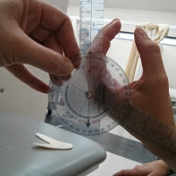
A goniometer (Fig.1) is a device that measures angles. Goniometers come in a variety of metal and plastic materials in different sizes and shapes. All universal goniometers have a central "body" with a protractor and fulcrum to center over the patient's joint, as well as two "arms" to align with the patient's body parts. [1]Generally goniometers have been shown to have good to excellent reliability, depending on the motion and joint being measured, with intra-rater reliability higher than inter-rate reliability.[18] [19] The use of standardised positions, stabilisation of the body part proximal to the joint being tested, use of bony landmarks to align the goniometer, and repeated testing conducted by the same therapist all help to improve the validity and reliability of goniometric measurements.[20][21][22]
Inclinometer;[edit | edit source]
An inclinometer or clinometer, consisting of a circular, fluid-filled disk with a bubble or weighted needle that indicates the number of degrees on the scale of a protractor, is an instrument used for measuring angles of slope, elevation, or depression of an object with respect to gravity's direction. The majority of inclinometers are calibrated or referenced to gravity, which means that gravity using gravity as a reference point means that the starting position of the inclinometer can be consistently identified and repeated.[2]
Evidence suggest that the hand held inclinometer is both a valid and reliable method for assessing muscle length.[23] [24] Intra-rater reliability for the hand-held inclinometer during SLR testing was excellent (ICCs, 0.95 to 0.98) with standard error of measurement between 0.54° and 1.22° and the minimal detectable change was between 1.50° and 3.41°.[23] While both intra- and inter-tester reliability were good during assessment of motion around the knee. [24]
Tape Measure;[edit | edit source]
A tape measure if one of the simplest measurement tools that can be used, which typically measures on a scale with centimetres (cm) or inches (in). The tape measure can be cloth, plastic or metal, is inexpensive, easy to use and readily available in most clinics. [2]The tape measure has also shown good reliability for use in muscle testing with good intra-rater reliability (ICC 0.82 0.87) shown within the same day to measure pectoralis minor muscle length.[25]
Principles of Measurement[edit | edit source]
Regardless of the measurement tool being used, the individual employing the instrument must become skilled in the use of the measurement tool to improve reliability in assessing muscle length. Practice in using an instrument should continue until the user has established a high level of intra-rater reliability. [2] Many of the steps involved in measuring muscle length are the same as though used for measuring joint range of motion. The following principles provide the basic framework for muscle length measurement.
| Description |
|---|
| Determine the type of measurement to be performed |
| Explain the purpose of the procedure to the patient |
| Position the patient in the preferred position for the measurement |
| Stabilise the proximal joint segment. |
| Instruct the patient in the specific motion that will be measured |
| Move the patient's distant joint segment passively through the available range |
| Determine the patient's end-feel at the end of the range |
| Return the patient's distal joint segment to the starting position |
| Palpate bony landmarks for measurement device alignment |
| Align the measurement device with the appropriate bony landmarks |
| Move the patient's distant joint segment passively to the end range |
| Read the scale of the measurement device and note the reading |
| Document Muscle Length |
Instructions[edit | edit source]
Patients should be provided with instructions prior to performing any assessment technique so they have an understanding of what is going to happen. Before beginning muscle length assessment, describe to the patient what will be taking place and why the measurement is being completed. Show the patient the measurement tool, and explain, in simple terms, its purpose and how it will be used. Show the patient the position they are to assume, again using simple terms and avoiding medical terminology such as supine or prone.[2][1]
Positioning[edit | edit source]
The muscle to be measured should be placed in the fully elongated position, ensuring maximal lengthening of the muscle from origin to insertion. The examiner is concerned about the final, elongated position of the muscle and not the measurement of the starting position, as would be appropriate for measurement of joint range of motion. [1]
The muscle being measured should be isolated across one joint. When measuring two or multi-joint muscles ensure to move the final joint through a passive range of motion until the muscle is placed on full stretch and prevents further joint motion. [2]
Stabilisation[edit | edit source]
To ensure accurate measurement of muscle length, firmly stabilise one end of the bony segment of the joint being measured, typically this is at the origin or proximal aspect of the bone. Without adequate stabilisation and isolation of the muscle being measured the patient may substitute motion at another joint resulting in measurement error.[2]
Speed of Movement[edit | edit source]
Once the patient is positioned and the proximal joint segment is adequately stabilised, the examiner should passively move the joint and lengthen the muscle through the available range of motion. The elongation of the muscle should be performed slowly to avoid eliciting a quick stretch of the muscle spindle and subsequently inducing a twitch response and muscle contraction. [26]
By moving the limb slowly through the range of motion to be measured, the patient is made aware of the exact movement to be performed and can cooperate more fully and accurately with the procedure. The examiner can also get an estimation of the patient's available range of motion prior to completing the final measurement, which can provide a check to minimise the possibility of gross measurement error.[2]
Determining End Feel[edit | edit source]
The most valuable clinical information during the measurement process is the end feel and the location of the end feel. Passive movement of the muscle being assessed allows the examiner to note any limitations to full elongation of the muscle, such as those caused by pain, muscle tightness, or other reasons.
Each joint has a characteristic feel to the resistance encountered at the end of normal range of motion. Typical end-feels encountered at the end of normal range of motion are the hard (bony), firm (capsular, muscular, and ligamentous) and soft (soft tissue approximation). When assessing muscle length typically we are looking for a firm end feel when the muscle is on full stretch, and the patient will report a pulling sensation, stretch or pain in the region of the muscle being lengthened. [2][3]
Align Measurement Device[edit | edit source]
Precise alignment of the measurement device relies on accurate palpation of landmarks with bony landmarks typically used for alignment of the measurement device during muscle length assessment whenever possible, since bony structures are more stable and are less subject to change in position as a result of other factors such as oedema or muscle atrophy.
Three landmarks, are typically used to align the goniometer, which is the most common measurement tool used for muscle length assessment, with two landmarks to align the arms of the goniometer;
- One landmark for the stationary arm; aligned with the midline of the stationary segment of the joint
- One landmark for the moving arm; aligned with the midline of the moving segment of the joint.
- One landmark for the fulcrum of the goniometer; aligned with a point that is near the axis of rotation of the joint.
Priority should be given to alignment of the stationary and moving arms of the goniometer for accurate alignment.[2]
Documentation[edit | edit source]
The examiner is most concerned about the final, elongated position of the muscle, with the measurement taken in this final position.
References [edit | edit source]
- ↑ 1.0 1.1 1.2 1.3 Norkin CC, White DJ. Measurement of Joint Motion: A Guide to Goniometry. FA Davis; 2016 Nov 18.
- ↑ 2.00 2.01 2.02 2.03 2.04 2.05 2.06 2.07 2.08 2.09 2.10 2.11 2.12 2.13 2.14 2.15 2.16 Reese NB, Bandy WD. Joint Range of Motion and Muscle Length Testing-E-book. Elsevier Health Sciences; 2016 Mar 31.
- ↑ 3.0 3.1 3.2 Gross JM, Fetto J, Rosen E. Musculoskeletal examination. John Wiley & Sons; 2015 Jun 29.
- ↑ 4.0 4.1 4.2 4.3 4.4 Norkin CC, White DJ. Measurement of Joint Motion: A Guide to Goniometry. FA Davis; 2016 Nov 18.
- ↑ 5.0 5.1 5.2 Kruse A, Rivares C, Weide G, Tilp M, Jaspers RT. Stimuli for adaptations in muscle length and the length range of active force exertion—a narrative review. Frontiers in Physiology. 2021:1677.
- ↑ Gash, M. C., P. F. Kandle, I. Murray, and M. Varacallo. "Physiology, Muscle Contraction. 2021 Apr 20." StatPearls [Internet]. Treasure Island (FL): StatPearls Publishing (2022).
- ↑ Allison KF, Keenan KA, Sell TC, Abt JP, Nagai T, Deluzio J, McGrail M, Lephart SM. Musculoskeletal, biomechanical, and physiological gender differences in the US military. US Army Medical Department Journal. 2015 Apr 1.
- ↑ Pawar A, Phansopkar P, Gachake A, Mandhane K, Jain R, Vaidya S. A review on impact of lower extremity muscle Length. J Pharm Res Int. 2021;33(35A):158-64.
- ↑ 9.0 9.1 Youdas JW, Krause DA, Hollman JH, Harmsen WS, Laskowski E. The influence of gender and age on hamstring muscle length in healthy adults. Journal of Orthopaedic & Sports Physical Therapy. 2005 Apr;35(4):246-52.
- ↑ Corkery M, Briscoe H, Ciccone N, Foglia G, Johnson P, Kinsman S, Legere L, Lum B, Canavan PK. Establishing normal values for lower extremity muscle length in college-age students. Physical Therapy in Sport. 2007 May 1;8(2):66-74.
- ↑ Fragala MS, Kenny AM, Kuchel GA. Muscle quality in aging: a multi-dimensional approach to muscle functioning with applications for treatment. Sports medicine. 2015 May;45:641-58.
- ↑ Zotz TG, Capriglione LG, Zotz R, Noronha L, De Azevedo ML, Martins HR, Gomes AR. Acute effects of stretching exercise on the soleus muscle of female aged rats. Acta histochemica. 2016 Jan 1;118(1):1-9.
- ↑ Justine M, Ruzali D, Hazidin E, Said A, Bukry SA, Manaf H. Range of motion, muscle length, and balance performance in older adults with normal, pronated, and supinated feet. Journal of physical therapy science. 2016;28(3):916-22.
- ↑ 14.0 14.1 Lin G, Wang W, Wilkinson T. Changes in deep neck muscle length from the neutral to forward head posture. A cadaveric study using Thiel cadavers. Clinical Anatomy. 2022 Apr;35(3):332-9.
- ↑ 15.0 15.1 Khayatzadeh S, Kalmanson OA, Schuit D, Havey RM, Voronov LI, Ghanayem AJ, Patwardhan AG. Cervical spine muscle-tendon unit length differences between neutral and forward head postures: biomechanical study using human cadaveric specimens. Physical therapy. 2017 Jul 1;97(7):756-66.
- ↑ 16.0 16.1 16.2 16.3 Conroy VM, Murray Jr BN, Alexopulos QT, McCreary J. Kendall's Muscles: Testing and Function with Posture and Pain. Lippincott Williams & Wilkins; 2022 Nov 23.
- ↑ Rogers M, Rogers M. Understanding Active and Passive Insufficiency [Internet]. National Federation of Professional Trainers. 2020 [cited 17 September 2020]. Available from: https://www.nfpt.com/blog/understanding-active-and-passive-insufficiency
- ↑ Herrero P, Carrera P, García E, Gómez-Trullén EM, Oliván-Blázquez B. Reliability of goniometric measurements in children with cerebral palsy: a comparative analysis of universal goniometer and electronic inclinometer. A pilot study. BMC musculoskeletal disorders. 2011 Dec;12:1-8.
- ↑ van Rijn SF, Zwerus EL, Koenraadt KL, Jacobs WC, van den Bekerom MP, Eygendaal D. The reliability and validity of goniometric elbow measurements in adults: A systematic review of the literature. Shoulder & elbow. 2018 Oct;10(4):274-84.
- ↑ Rothstein, JM, et al: Goniometric reliability in a clinical setting: Elbow and knee measurements. Phys Ther 63:1611, 1983. [PubMed: 6622536]
- ↑ Watkins, MA, et al: Reliability of goniometric measurements and visual estimates of knee range of motion obtained in a clinical setting. Phys Ther 71:90, 1991. [PubMed: 1989012]
- ↑ Ekstrand, J, et al: Lower extremity goniometric measurements: A study to determine their reliability. Arch Phys Med Rehabil 63:171, 1982. [PubMed: 7082141]
- ↑ 23.0 23.1 Boyd BS. Measurement properties of a hand-held inclinometer during straight leg raise neurodynamic testing. Physiotherapy. 2012 Jun 1;98(2):174-9.
- ↑ 24.0 24.1 Romero-Franco N, Montaño-Munuera JA, Jiménez-Reyes P. Validity and reliability of a digital inclinometer to assess knee joint-position sense in a closed kinetic chain. Journal of sport rehabilitation. 2017 Jan 1;26(1).
- ↑ Rosa DP, Borstad JD, Pires ED, Camargo PR. Reliability of measuring pectoralis minor muscle resting length in subjects with and without signs of shoulder impingement. Brazilian journal of physical therapy. 2016 Mar 15;20:176-83.
- ↑ Izraelski J. Assessment and treatment of muscle imbalance: The Janda approach. The Journal of the Canadian Chiropractic Association. 2012 Jun;56(2):158.


