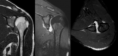
Anterior Shoulder Instability
Original Editors - Liesbeth De Feyter
Top Contributors - Laura Ritchie, Scott Cornish, Andeela Hafeez, Admin, Leana Louw, Kim Jackson, Liesbeth De Feyter, 127.0.0.1, Tony Lowe, Fasuba Ayobami, Borms Killian, Naomi O'Reilly, Kai A. Sigel and Wanda van Niekerk
Defination:[edit | edit source]
Anterior shoulder instability is an injury to the shoulder joint so that the upper arm (humerus) is displaced from its normal position in the center of the socket (glenoid) and the joint surfaces no longer touch each other.[1]
Clinically Relevant Anatomy[edit | edit source]
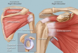
The shoulder is one of the largest and most complex joints in the body. The shoulder joint is formed where the humerus (upper arm bone) fits into the scapula (shoulder blade), like a ball and socket. Other important bones in the shoulder include:
The acromion is a bony projection off the scapula.
The clavicle (collarbone) meets the acromion in the acromioclavicular joint.
The coracoid process is a hook-like bony projection from the scapula.
The shoulder has several other important structures:
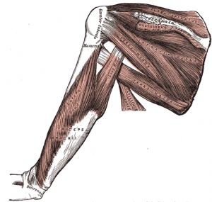
The rotator cuff is a collection of muscles(supraspinatus,infraspinatus,subscapularis and teres minor) and tendons that surround the shoulder, giving it support and allowing a wide range of motion.
The bursa is a small sac of fluid that cushions and protects the tendons of the rotator cuff.
A cuff of cartilage called the labrum forms a cup for the ball-like head of the humerus to fit into.[2]
Epidemiology /Etiology[edit | edit source]
Epidemiology
Anterior shoulder dislocations are much more common than posterior dislocations.[3]
Many investigators have shown that the incidence of recurrent shoulder dislocation is significantly higher in younger patients.[4][5] The consequences of initial anterior glenohumeral dislocation in patients over forty years of age are quite different than in the younger population, primarily because of the increased incidence of rotator cuff tears and associated neurovascular injuries. The anterior or posterior supporting structures of the shoulder can be disrupted following an anterior dislocation. In the younger patient, anterior capsuloligamentous structures most commonly fail. In the older patient with pre-existing degenerative weakening of the rotator cuff, the posterior structures are more likely to fail.[4]
Etiology
The glenohumeral joint is stabilized by dynamic and static structures.
- Dynamic stabilizers: rotator cuff muscles, biceps brachii, deltoid
- Static stabilizers: glenohumeral joint capsule, the glenohumeral ligaments, the labrum, negative pressure within the joint capsule, and the bony congruity of the joint[3][5]
- The labrum: The concavity compression mechanism plays an important role in the stability of the glenohumeral joint by maintaining the localization of the humeral head at the glenoid against translational forces. The glenoid concavity is established by the glenoid shape, the glenoid cartilage and the glenoid labrum. The glenoid labrum increases the width and depth of the glenoid. Instability increased with the size of the glenoid defect.[6]
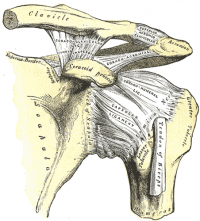
- The glenohumeral ligaments: The superior glenohumeral ligament functions primarily to resist inferior translation and external rotation of the humeral head in the adducted arm. The middle glenohumeral ligament functions primarily to resist external rotation from 0° to 90° and provides anterior stability to the moderately abducted shoulder. The inferior glenohumeral ligament is composed of two bands, anterior and posterior, and the intervening capsule. The primary function of the anterior band is to resist anteroinferior translation.[5].[7]
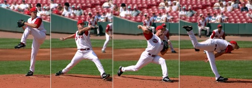
- Excessive external rotation or over-rotation of the thrower’s shoulder is purportedly associated with the development of internal impingement syndrome (which occurs when the shoulder is maximally externally rotated and the intra-articular side of the supraspinatus tendon impinges on the adjacent posterior superior glenoid and glenoid labrum). Impingement syndrome is a potential precursor to anterior instability.[7]
Characteristics/Clinical Presentation[edit | edit source]
Signs and symptoms for anterior shoulder instability:
- Anterior instability accounts for 95% of acute traumatic dislocations.[5]
- Dead-arm syndrome indicates pathologic anterior instability. It occurs when the arm is in an abducted, externally rotated position. The patient complains of a sharp anterior shoulder pain and tingling in the hand. The patient drops the arm suddenly. This syndrome can be seen in overhead sports, such as volleyball, tennis, swimming and water polo.[7]
- Rotator cuff weakness, particularly in external rotation and “empty-can” abduction, is common in athletes with anterior instability.[7]
- Bankart lesions are the most common sequelae of traumatic anterior shoulder instability.[5]
- Humeral avulsion of glenohumeral ligaments lesions are a known cause of anterior shoulder instability.[5].[5]
- During an anterior dislocation, the posterolateral aspect of the humeral head contacts the anteroinferior rim of the glenoid, often resulting in a classic Hill-Sachs defect. This defect has been observed in up to 80% of patients with initial anterior dislocation and in 100% of patients with recurrent anterior instability.[3][5]
Complaints related to recurrent anterior instability:[7]
- Glenohumeral joint pain
- Shoulder stiffness with difficulty warming up for the sport
- Rotator cuff weakness
- Sensation of popping, grinding or catching deep in the shoulder joint
- Pain when reaching backward or above shoulder height
- Apprehension when sleeping with the arm overhead in abduction and external rotation
- Neurologic complaints: tingling or burning in the lower arm and hand or localized numbness of the skin overlying the deltoid muscle
- Tenderness of the anterior glenohumeral joint line and the posterior rotator cuff
Diagnostic Procedure:
[edit | edit source]
Diagnosis by history,physical examination and radiology.
Outcome Measures[edit | edit source]
add links to outcome measures here (also see Outcome Measures Database)
Examination[edit | edit source]
Anterior instability
[edit | edit source]
- anterior Drawer Test - Pt. is supine and arm abducted over edge of couch. Examiner immobilisers scapula with one arm whilst the other grasps the arm and pulls it anteriorly.
-

- anterior Apprehension
- Jobe Relocation (Fulcrum Test)
- Rowe Test - Pt. bends forward slightly with the arm relaxed.The examiner move the arm slightly inferior and anterior by pulling on the forearm
- Throwing Test - Pt. executes a throwing motion against the examiners resistance. anterior subluxation may occur.
- Leffert Test - Examiner displaces the humeral head anteriorly holding the humeral head over the shoulder with the thumb posteriorly and index finger anteriorly. Displacement of the index finger is positive
- Surprise/Release Test - This manoeuvre is variously described but essentially is the fmal component of the apprehension and relocation tests. It is an extremely provocative test and should be used with caution. As in the Jobe relocation tests the patient's arm is maximally externally rotated with a posteriorly directed force applied to the humeral head. At the limit of range the examiner suddenly removes the posteriorly directed force from the relocation test and again a feeling of apprehension is considered a positive test.
- Dynamic anterior Jerk Test - The test combines of a compression force and a translation force, applied along the arm between the humeral head and the glenoid cavity. In so doing, a subluxation of the humeral head is provoked and it is accompanied with a jerk recognised by the patient as his instability.
- Dynamic Relocation Test
- Dynamic Rotatory Stability Test
- Bony Apprehension Test - identical to the standard apprehension test except that the arm is brought to only 45 of abduction and 45 of external rotation. A positive result should alert the examiner to the possibility of a bony lesion as the cause of symptomatic shoulder instability.
- Kinetic medial Rotation Test - used to differentiate to help determine whether symptoms are primarily impingement or instability. The subject lies supine with 90deg humeral abduction (hand to the ceiling with the humerus in the plane of the scapula). The assessor places one finger on the coracoid process and one on the humeral head. The subject is asked to actively medially rotate the humerus. The ideal is 70deg rotation without any finger movement. If the coracoid finger moves before 70deg then there is an increase in scapula relative flexibility and impingement risk. If the humeral finger moves before 70deg then there is displacing axis of rotation of the humeral head and an instability risk. If both fingers move forward then there is a combined impingement and instability risk. This test obviously needs to be used with other instability and impingement tests to confirm diagnosis but it is a good rehab indicator for where the primary focus should be[8].
Medical Management
[edit | edit source]
In patients with shoulder dislocation, stabilize and treat associated trauma as indicated.Allow the patient to assume a position of comfort while maintaining cervical spine immobilization if necessary.A pillow between the patient's arm and torso may increase comfort.Administer analgesics to decrease pain.[9]
Physical Therapy Management
[edit | edit source]
The rehab program is outlined in three phases.[10]
Phase 1[edit | edit source]
Apply modalities as needed (heat, ice, electrotherapy, etc.)
2. Perform ROM exercises (passive, active-assistive), avoid abduction, extension and external rotation
(“cocked position” for throwing).
A. Rope and Pulley
B. Wand
C. Finger Walk
3. Posterior cuff stretch in supine (cross arm adduction)
4. Manual stretching, avoiding stretching to the anterior capsule (ER in the scapular plane and no
shoulder extension)
5. Functional behind the back stretch (IR towel stretch), if needed
6. Mobilization of posterior cuff, if needed
Phase 2[edit | edit source]
Continue posterior cuff stretching
2. Continue shoulder strengthening exercises with free weights and elastic resistance (emphasize
eccentric work on the rotator cuff, progress planes of motion to the 90/90 position)
3. Add lower trap pull downs with pulley system, if available
4. Progress prone DB program by adding:
A. Horizontal abduction
B. Retraction with ER
C. Extension with palm forward
Phase 3[edit | edit source]
Focus on progressive exercises in preparation for returning to the prior activity level.
1. Continue flexibility/mobility exercises
2. Continue progressive throwing program
3. Continue with strengthening
4. May add overhead strengthening
Case Study[edit | edit source]
http://www.ncbi.nlm.nih.gov/pmc/articles/PMC2796945/
Recent Related Research (from Pubmed)[edit | edit source]
Failed to load RSS feed from http://eutils.ncbi.nlm.nih.gov/entrez/eutils/erss.cgi?rss_guid=1vKcRI80qSZZScE15PzdrfWnH_vpOM6PbzlLrtY78GPW24mkID|charset=UTF-8|short|max=10: Error parsing XML for RSS
References[edit | edit source]
- ↑ Washington Orthopaedics & Sports Medicine http://www.wosm.com/index.php/health-library/orthopaedic-conditions-and-treatments/147-anterior-shoulder-instability
- ↑ http://www.webmd.com/pain-management/picture-of-the-shoulder
- ↑ 3.0 3.1 3.2 Chen AL,Bosco JA. Glenohumeral Bone Loss and Anterior Instability. Bull Hosp Jt Dis. 2006; 64(3-4):130-138.
- ↑ 4.0 4.1 Araghi A, Prasarn M, St Clair S, Zuckerman JD. Recurrent anterior glenohumeral instability with onset after forty years of age: the role of the anterior mechanism. Bull Hosp Jt Dis. 2005;62(3-4):99-101.
- ↑ 5.0 5.1 5.2 5.3 5.4 5.5 5.6 5.7 Pope EJ, Ward JP, Rokito AS. Anterior shoulder instability - a history of arthroscopic treatment. Bull NYU Hosp Jt Dis. 2011;69(1):44-9.
- ↑ Rhee YG, Lim CT. Glenoid defect associated with anterior shoulder instability: results of open Bankart repair. Int Orthop. 2007;31(5):629-34.
- ↑ 7.0 7.1 7.2 7.3 7.4 Satterwhite YE.Evaluation and management of recurrent anterior shoulder instability. J Athl Train. 2000;35(3):273-277.
- ↑ Comerford MJ, Mottram SL. Manual Therapy 2001;6(1):15–26.
- ↑ Shoulder Dislocation in Emergency Medicine Treatment & ManagementfckLRAuthor: Sharon R Wilson, MD; Chief Editor: Trevor John Mills, MD, MPH
- ↑ http://www.shorelineortho.com/pdfs/0-StewartAnteriorShoulderDislocation.pdf
