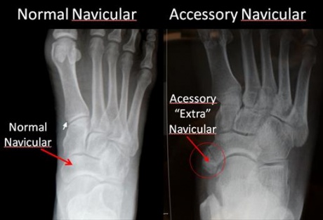
Accessory Navicular Bone
Original Editors - Carlos De Coster
Lead Editors - Your name will be added here if you are a lead editor on this page. Read more.
Search Strategy[edit | edit source]
PubMed (http://www.ncbi.nlm.nih.gov/pubmed)
MeSH terms:
• Complications
• Diagnosis
• Drug therapy
• Epidemiology
• Etiology
• Pathology
• Rehabilitation
• Surgery
• Therapy
Medline Plus
Used keyword:
• Accesory navicular bone
• Os tibiale externum
Definition/Descrition
[edit | edit source]
Also known as Prehallux, Os Tibiale Externum and Navicular Secundum. When there occurs plantar medial enlargement of tarsal navicular beyond its normal size it is termed as accessory navicular. There occurs a separate bone either in the posterior tibial tendon or with navicular as a true articulation. (1)
ITS CLASSIFICATION:
It is broadly classified in 3 types
Type I: is sesamoid bone in the posterior tibialis tendon. There is a small distance (<3m) between the sesamoid and the navicular.
File:TYPE III AN.PNG File:TYPE I AN X RAY.jpg
Type II: consists of an accessory bone, upto 1.2cm in diameter, in which a synchondrosis exist between it and the navicular.
File:Type II AN.PNG File:TypeIIAccessoryNavicular.jpg
Type III: is the fused accessory navicular to the navicular resulting in large cornuate navicular.
File:TYPE III AN.PNG File:TYPE III ACCESSORY N. X RAY.PNG
ANOTHER CLASSIFICATION PURPOSED BY DWIGHT 1920 (2)
File:Accessory navicular clsfctn by Dwight.PNG
Clinically Relevant Anatomy[edit | edit source]
The navicular bone is one of the tarsal bones found in the foot. It is located on the medial side of the foot, and articulates proximally with the talus. Distally it articulates with the three cuneiform bones. In some cases it articulates laterally with the cuboid. The tibialis posterior inserts to the os naviculare. [1] The tibialis posterior muscle also contracts to produce inversion of the foot and assists in the plantar flexion of the foot at the ankle. Tibialis posterior also has a major role in supporting the medial arch of
the foot and therefore dysfunction can lead to flat feet in adults. [2]
Epidemiology /Etiology[edit | edit source]
People who have an accessory navicular often are unaware of the condition if it causes no problems. However, some people with this extra bone develop a painful condition known as accessory navicular syndrome when the bone and/or posterior tibial tendon are aggravated. This can result from any of the following:
- Trauma, as in a foot or ankle sprain
- Chronic irritation from shoes or other footwear rubbing against the extra bone
- Excessive activity or overuse
Many people with accessory navicular syndrome also have flat feet (fallen arches). Having a flat foot puts more strain on the posterior tibial tendon, which can produce inflammation or irritation of the accessory navicular.
Characteristics/Clinical Presentation[edit | edit source]
add text here
Differential Diagnosis[edit | edit source]
add text here
Diagnostic Procedures[edit | edit source]
add text here related to medical diagnostic procedures
Outcome Measures[edit | edit source]
add links to outcome measures here (also see Outcome Measures Database)
Examination[edit | edit source]
add text here related to physical examination and assessment
Medical Management
[edit | edit source]
- Kidner operation: in which the main insertion of the tibialis posterior is re-routed. The main portion of the tendon was also rerouted under the navicular onto the medial side, with the intention of restoring the normal line of pull of the tendon. [2]
- Excision: hereby the ossicle is been cut away. However when doing this operation it is essential that the medial surface of the main navicular bone was contoured to prevent any residuel prominence.
- Occasionally, a limited fusion of the cuneiform metatarsal or talonavicular joints also was recommended. The rationale and efficacy of this operation have been questioned.
- Immobilization. Placing the foot in a cast or removable walking boot allows the affected area to rest and decreases the inflammation.
- Medications. Oral nonsteroidal anti-inflammatory drugs (NSAIDs), such as ibuprofen, may be prescribed. In some cases, oral or injected steroid medications may be used in combination with immobilization to reduce pain and inflammation. [3]
- Corticosteroid injections can be used as a treatment modality. However, this modality should be used with caution as it may weaken the posterior tibial tendon and lead to subsequent rupture. [4]
Physical Therapy Management
[edit | edit source]
If the accessory navicular bone becomes problematic physical therapy may be prescribed. This will include exercises and treatments to strengthen the intrinsic foot muscles and lateral thigh rotators muscles and decrease inflammation. Often is the accessory navicular bone linked to Posterior tibial dysfunction to a pes planus. [5] To adjust the arch of the foot, orthotic devices are required. The physician can analyze the gait of the person to see where the adjustments need to be made. Activity modification, such as limiting or stopping any strenuous activities that cause the Accessory Navicular bone to become symptomatic can be used for initial treatment. Casting can be used to prevent repetitive micro trauma either directly or from pull of the posterior tibial tendon. When the cast is being removed it the physician can start building up the ROM from the patient and counter atrophy. [6]
Key Research[edit | edit source]
add links and reviews of high quality evidence here (case studies should be added on new pages using the case study template)
Resources
[edit | edit source]
Foot health facts: http://www.footphysicians.com
Clinical Bottom Line[edit | edit source]
add text here
Recent Related Research (from Pubmed)[edit | edit source]
see tutorial on Adding PubMed Feed
Extension:RSS -- Error: Not a valid URL: Feed goes here!!|charset=UTF-8|short|max=10
References[edit | edit source]
see adding references tutorial.
- ↑ GOLANO P., ‘The anatomy of the navicular and periarticular structures.’ Foot Ankle Clinics, 2004, March, vol. 9, p. 1-23.
- ↑ 2.0 2.1 KITER E., ERDAG N., KARATOSUN V., GUNALL I., ‘Tibialis posterior tendon abnormalities in feet with accessory navicular bone and flatfoot’. Acta orthopaedica Scandinavia, 1999, December, vol. 70, p. 618-621
- ↑ MACNICOL M. F., VOUTSINAS S., ‘Surgical treatment of the symptomatic accessory navicular’, The Journal of Bone and Joint Surgery, 1984, vol. 66, p. 218-226.
- ↑ YEUNG-JEN C., WEN-WEI R., LIANG S., ‘Degeneration of the accessory navicular synchondrosis presenting as rupture of the posterior tibial tendon’. The Journal of Bone and Joint Surgery., 1997, vol. 79, p. 1791-1798.
- ↑ Cite error: Invalid
<ref>tag; no text was provided for refs namedBernaerts A. et al - ↑ LEONARD Z. C., FORTIN P. T., ‘Adolescent accessory navicular bone’ Foot Ankle Clinics, 2010, vol. 15, p. 337-347.


