Osgood-Schlatter Disease
Original Editor - Casey Kirkes, Geoffrey De Vos as part of the Vrije Universiteit Brussel Evidence-based Practice Project
Top Contributors - Geoffrey De Vos, Admin, Keta Parikh, Sam Verhelpen, Nicolas D'Hondt, Kim Jackson, Scott Buxton, Michelle Lee, Laurens Dereymaeker, Jetse De Proft, Rachael Lowe, Wanda van Niekerk, Nicolas Everaerts, Casey Kirkes, Nikhil Benhur Abburi, Rucha Gadgil, Lucinda hampton, Jess Bell, Andeela Hafeez, Hanne Velghe, Uchechukwu Chukwuemeka, Vidya Acharya, Adam Vallely Farrell, 127.0.0.1, Tony Lowe, Claire Knott, Naomi O'Reilly and Rishika Babburu
Definition/Description[edit | edit source]
Osgood-Schlatter disease is a traction apophysitis at the level of the tibial tubercle due to repetitive strain on the secondary ossification center of the tibial tuberosity.
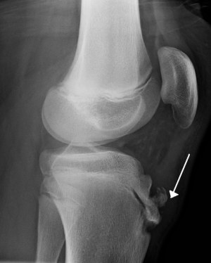
The repetitive strain is from the strong pull of the quadriceps muscle produced during sporting activities. The tibial tuberosity avulsion may occur in the preossification phase or the ossified phase of the secondary ossification center. Once the bone or cartilage is pulled away it continues to grow, ossify and enlarge. The intervening area may become fibrous, creating a separate persistent ossicle or may show complete bony union with some enlargement of the tibial tuberosity.
It occurs slightly more often in boys. These traction apophysitises are probably one the most encountered overuse injuries in children and adolescents .
[1][2]
The tibial tubercle (the tuberosity of the tibia or tibial tuberosity ) is a large oblong elevation on the proximal anterior aspect of the tibia, just distal from the anterior surfaces of the medial and lateral tibial condyles. It gives attachment to the patellar ligament or patellar tendon. The Osgood-Schlatter disease is localized at the tibial tubercle at the anterior side of the knee, but only in an adoslecent knee. At this tibial tubercle the pain can be felt by the patient, in most of the times unilateral, but more frequently also bilateral
The patellar tendon attaches to the tibial tuberosity inferior to the patella. Stress at this musculo-tendonous junction can cause pain and swelling. [3]
Epidemiology /Etiology[edit | edit source]
Children and adolescents have growth zones in both the femur and the tibia and an apophysis (cartilage bone/cartilaginous material) at the tibial tuberosity. This cartilage (flexible connective tissue that often can be found between two bones), like bones, muscles and tendons, also has a grow capacity. But during the adolescent growth spurt, bones and cartilage grow much faster than muscles and tendons. The slower elongation of the musculotendinous extensor apparatus of the knee (m.quadriceps) inflicts very strong forces on the small site of the insertion of the patellar tendon to the tibial tuberosity. These forces can cause microavulsions of the tibial tuberosity. The cartilage of the tibial tuberosity (the anterior portion of the developing ossification center of the tibial tuberosity) can resist forces but not as bone and when the child or the adolescent does some physical activities the forces at the the patellar tendon and tibial tuberosity increases, which causes pain, irritation and in some cases microavulsions or avulsions fractures of the tibial tuberosity[4].
Increased stress of the musculotendenous junction of the patellar tendon and tibial tuberosity can cause the tendon to pull away from the bone a little bit. This small amount of tearing leads to increased pain and swelling below the knee cap. The condition is worsened with activities that subject the patellar tendon to high loads such as squatting, or jumping. In some cases ossification will occur at the area of trauma leading to a bony protuberance at the tibial tuberosity.
Characteristics/Clinical Presentation[edit | edit source]
Pain is the leading symptom in this disease and it appears and aggravates during physical activities such as running, jumping, cycling, kneeling, walking up and down the stairs and kicking a ball (knee extension). In sports like basketball, volleyball, soccer, tennis, … the pain increases. The clinical picture consists of pain localized to the area of the tibial tubercle. In some cases the tubercle may be swollen and hypertrophied and there is also tightness of the m. quadriceps. Characteristics such as temperature or intra-articular swelling is not relevant (rarely the tuberosity can feel warm), but swelling, tenderness and pain of the tibial tuberosity often appears.
• Painful palpation of the tibial tuberosity.
• Pain at the tibial tuberosity that worsens with physical activity or sport.
• Increased pain at the tibial tuberosity with squating, stairs or jumping.
• In some cases increased bony protuberance at the tibial tuberosity.
Some extra information and excursions available in this video
Differential Diagnosis[edit | edit source]
Some differential diagnosis can be: jumper’s knee (patellar tendinitis) or Sinding- Larsen-Johanssen syndrome, hoffa’s syndrome, synovial plica injury, tibial tubercule fracture. These diseases are also localized at the patellar tendon and can cause similar knee problems.
Diagnostic Procedures[edit | edit source]
X-Rays may be utilized to better visualize the musculotendenous junction in severe cases or if avulsion is suspected.
- The diagnosis is based on typical clinical findings (see clinical presentation). [4]
- Radiographic examinations of both knees should always be performed, in both the anterior-posterior and lateral projections, to rule out the possibility of tumors, fractures, ruptures or infections. The lateral radiograph generally shows the characteristic picture of prominent tibial tubercle with irregularly ossific nucleus, or free bony fragment proximal to the tubercle. Imaging is also useful to exclude tuberosity epiphysiolysis or tumors. (C Reid D. et al)
- Sonographic examination can also be used. The ultrasound can be directed to demonstrate the appearance of the cartilage and bony surface, the patellar tendon, soft-tissue swelling anterior to the tibial tuberosity, and fragmentation of the tibial tuberosity.
Examination[edit | edit source]
A diagnosis can be made through a thorough history and examination. Tenderness to palpation over the tibial tuberosity that worsens with weight bearing squat or jumping is fairly indicitive ot this disease.
- Physical examination reveals pain during palpation of the tibial tubercle.
- Resisted extension of the knee from 90° flexed position will usually reproduce pain, but resisted straight leg raised test is usually painless.
- Ely's test, which proves excessive tightness of the quadriceps femoris muscle, is positive in all cases.
Outcome Measures[edit | edit source]
add links to outcome measures here (see Outcome Measures Database)
Medical Management
[edit | edit source]
Treatment should begin with rest, icing (RICE), activity modification and sometimes nonsteroidal anti-inflammatory drugs.
Physical Therapy Management
[edit | edit source]
The physiotherapist can focus on stretching and strengthening exercises for m. quadriceps, hamstrings and lower extremity muscles. [5] (+ see recent research pubmed Gholve PA et al) Non-operative treatment of this disease is based on the same principles that apply all overuse injuries. Today, there is no need for total immobilization, or for totally refraining from athletic activities. Of vital importance is that the physician inform the parents, the coach, and the child athlete of the natural course of this disease. The child should continue his normal physical activities, to the limit that the pain allows it, so lower intensity of frequency of exercising (activity modification). Also swimming, as a secondary athletic activity, is very good during this disease (no discomfort). Also knee-braces, tapes, slip-on knee support with an infrapatellar strap or pad are recommended and may help during physical activities and can reduce pain. (C Reid D. et al, but no EBP)
The symptoms tend to subside within 2 years, and the prognosis is excellent in the majority of cases.[4] So the symptoms of Osgood-Schlatter will generally decline and disappear in most patients if non operative treatment is carried out long enough, especially after bone growth is terminated. Persistent symptoms are followed by development of loose fragments above the tibial tubercle, or within the patellar ligaments. In these cases, the symptoms will disappear only after these fragments, ossicle and/or free cartilaginous material, are surgically excised (recent research Pihlajamäki HK et al). ( (M Béuima M. et al). But surgical procedures should be avoided until the child has grown up and the bone growth has been completed to avoid growth-plate arrest and the development of recurvatum and or valgus of the knee.[4]
Physiotherapy Evaluation
[edit | edit source]
Besides treatment, physiotherapists must also focus on evaluation management and follow - up principles.
During examination of the patient's knee pain, location (unilateral or bilateral) of pain and its duration must be interrogated and documented. A painful feeling during brief physical activity, or following prolonged activity, indicates severity. Answers to questions concerning presence or absence of pain while walking, running, ascending and descending stairs, and kneeling should be documented for later comparison. When the therapist examines the patient's gait pattern while walking, he/she must look for an antalgic limp or other compensatory mechanism to protect the knee from pain. Special attention should be focused on whether or not the patient flexes the involved knee during walking or attempts to maintain full extension, thereby reducing the need for quadriceps activity. [5]
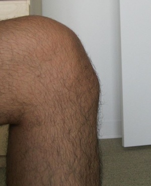
Confirmation of the diagnosis is the first task of the attending therapist. Inspection of the tubercles is performed, while the patient is holding both knees in 90° flexion and both feet in supine. By looking from the side, a silhouette image of one knee against the other reveals enlargement of the apophysis. Palpation of the tubercle is then performed and tenderness is assessed as none, slight, mild, moderate, or marked. Further, tightness of knee musculature must be checked because musculoskeletal pain may be secondary to the inability of muscles to elongate at a same rate as the human bones. With the patient still supine, hamstring length is assessed by the examiner's flexing the hip while maintaining the knee in full extension.
Comparison between involved and uninvolved limbs in unilateral problems, or comparison to normal values in the cases of bilateral involvement, helps the therapist to decide whether or not muscular tightness plays a role in the condition. Knee flexion range of motion, taking into account rectus femoris tightness, is performed with the patient prone. The knee is passively flexed by the examiner until the end of range or until pain is presented. If this stretch begins to hurt, the patient must be questioned as to the location of the pain, as this will influence treatment. If pain from this prone stretching is felt in the area of the infrapatellar tendon or tubercle area, stretching the quadriceps is contraindicated, as the pain is caused by further pulling away of the apophysis. If the strain is felt up in the muscle belly or at the proximal attachment of the muscle, quadriceps stretching will be performed as part of the treatment.
The results of muscle tightness tests along with the location of pain with stretching are recorded and documented. Manual muscle testing of the knee extensors can be performed with the patient sitting on the end of the treatment table. The knee flexors can be tested when the patient is lying face down on the table, or sideling. Muscle tone is assessed in the long sitting position as the patient performs a quadriceps set. Quadriceps atrophy should be checked in the form of girth measurements. [6][7]
Key Research[edit | edit source]
add links and reviews of high quality evidence here (case studies should be added on new pages using the case study template
Resources
[edit | edit source]
M Béuima M, Bojanic I. Overuse injuries of the musculoskeletal system. CRC press, 2004, p 230 -231, 316, 375, 378-379 (primary resources: Kyjula UM, Ivist M, Hunonen O. Osgood Schlatter disease in adolescent athletes. Am J Sports Med, 1985, 13 : 236-241)
C Reid D. Sports injuries assessment and rehabilitation. Churchill Livingstone USA, 1992, p 406 – 411
(primary resource: Mital MA, Matza RA: Osgood Schlatter disease. The painful puzzler. Physician Sports Med, 5: 60, 1997)
Suzann K. Campbell, Darl W. Vanderlinden, Robert J. Palisano; “Physical Therapy For Children”;Saunders – Elsevier 2006.
Clinical Bottom Line[edit | edit source]
add text here
Recent Related Research (from Pubmed)[edit | edit source]
Failed to load RSS feed from http://www.ncbi.nlm.nih.gov/entrez/eutils/erss.cgi?rss_guid=183xCykbTEMoHMxsx7zELmSb2WyP9DDrrMufVLkiiFv8pdrwvP|charset=UTF-8|short|max=10: Error parsing XML for RSS
References[edit | edit source]
- ↑ Kaya DO, Toprak U, Baltaci G, Yosmaoglu B, Ozer H. Long-term functional and sonographic outcomes in Osgood-Schlatter disease. Knee Surg Sports Traumatol Arthrosc. 2013 May fckLRlevel of evidence 3B
- ↑ Blankstein A, Cohen I, Heim M, Diamant L, Salai M, Chechick A, Ganel A. Ultrasonography as a diagnostic modality in Osgood-Schlatter disease. A clinical study and review of the literature. © Springer-Verlag 2001, Arch Orthop Trauma Surg (2001) 121 :536–539 www.springerlink.com/content/efbpdx4ythtqjqyu/fulltext.pdf fckLRlevel of evidence 2B
- ↑ Baltaci G, Ozer H, Tunay V. B. Rehabilitation of avulsion fracture of the tibial tuberosity following Osgood-Schlatter disease. © Springer-Verlag 2003, Knee Surg Sports Traumatol ArthroscfckLR(2004) 12 : 115–118 www.springerlink.com/content/xnbxdt65aggv30dy/fulltext.pdffckLRLevel of evidence 4
- ↑ 4.0 4.1 4.2 4.3 Cite error: Invalid
<ref>tag; no text was provided for refs namedtwo - ↑ 5.0 5.1 Bloom OJ, Mackler L, Barbee J. Clinical inquiries. What is the best treatment for Osgood-Schlatter disease? Moses Cone Health System, Greensboro, NC USA. J Fam Pract. 2004 Feb;53(2):153-6. (EBP article A1)www.jfponline.com/Pages.asp
- ↑ THE JOURNAL OF ORTHOPAEDIC AND SPORTS PHYSICAL THERAPY: The Orthopaedic and Sports Physical Therapy Sections of the American Physical Therapy Association presents: Osgood-Schlatter Disease: Review of Literature and Physical Therapy Management; T. J. ANTICH, MS, PT,* CLlVE E. BREWSTER, MS, PT (1995)
- ↑ Childhood and Adolescent Sports-Related Overuse Injuries; KYLE J. CASSAS, M.D., and AMELIA CASSETTARI-WAYHS, M.D., Methodist Health System, Dallas, Texas; This article exemplifies the AAFP 2006 Annual Clinical Focus on caring for children and adolescents


