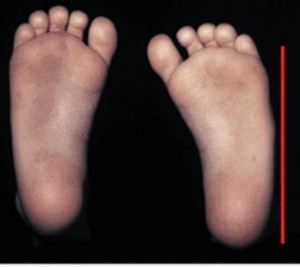
Metatarsus Adductus
Original Editor - Shaniel Walters
Description[edit | edit source]
Metatarsus Adductus ( Hooked Foot)[edit | edit source]
There occurs medial deviation of the foot from the level of the forefoot with respect to the hindfoot.
Common foot deformity is seen in children which causes the foot to turn inwards. The foot appears "c-shaped. This condition is often associated with hip dysplasia.
Types[edit | edit source]
Metatarsus Adductus may be classified as:
Flexible: Presents with adduction of the 5 metatarsal bones at the tarsometatarsal joint.
Rigid: Presents with medial subluxation of the tarsometatarsal joints. There is valgus of the hindfoot and the navicular is later to the head of the talus.
Clinically Relevant Anatomy
[edit | edit source]
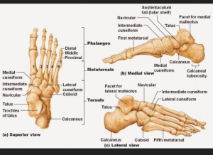
The skeleton of the foot is made of the thirty three bones, twenty six six joints and over a hundred muscles, ligaments and tendon. The foot serves primarily as a weight-bearing joint and provides a stable base of support on which to stand. Ligaments are attached to the bones which creates joints. The anatomy of the foot is divided into 3 categories: the Forefoot, the Midfoot and the Hindfoot.
Hindfoot is comprised of : Tibiofibular joint , Talocular joint and the Subtalar (Talocalcanean) joint.[1]
Midfoot (Midtarsal Joints) is comprised of: Talocalcaneonavicular Joint, Cuneonavicular Joint, Cuboideonavicular Joint, Intercuneuform Joints, Cuneocuboid joint and the Calcaneocuboid Joint.[1]
Forefoot is comprised of: Tarsometatarsal Joints, Intermaetatarsal Joint, Metatarsophalangeal Joints and Interphalangeal Joints.[1]
Epidemiology and Etiology[edit | edit source]
There is an incidence of 1 in 100 to 1 in 5,000 live births.[2] The cause of metatarsus adductus remains unknown. It is however, thought to be related to intrauterine compression. Family history may also be a causative factor. Other theories of causal relation includes abnormal tendon insertion of tibialis anterior, tibialis posterior and abductor hallucis muscles.[3]
Clinical Presentation[edit | edit source]
The forefoot is adducted and sometimes supinated , but the midfoot and hindfoot are normal. There is convexity of the lateral border of the foot, with concavity of the medial border. Older children may present with an in-toeing gait.
Diagnostic Procedures[edit | edit source]
Transmalleolar Axis Bisector[edit | edit source]
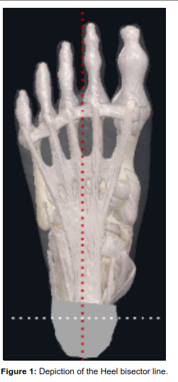
This is a geometric tool used to do the clinical assessment of Metatarsus Adductus.
Heel bisector line[edit | edit source]
Management / Interventions
[edit | edit source]
Specific treatment for metatarsus adductus is often determined by the following factors:
- Child's Age
- Medical History
- The extent of the condition
- Tolerance for the specific procedure
- Expectations for the condition
Interventions include:[edit | edit source]
- Passive Stretching
- Passive Manipulation exercises
- Stretching
- Serial Casting
- Footwear
- Surgery to release the joints
Prognosis[edit | edit source]
Generally excellent. Most cases of metatarsus adductus often resolves on its own
References[edit | edit source]
- ↑ 1.0 1.1 1.2 Magee, D. J. (2008). Orthopedic physical assessment. St. Louis, Mo: Saunders Elsevier.
- ↑ Wildhe T.Foot deformities at birth: a longitudinal prospective study over a 16 year period. J Pediatric Orthopedic. 1997;17(1):20-24
- ↑ Hassan N, Roger J (2015) Management of Metatarsus Aductus, Bean-Shaped foot, residual clubfoot adduction and Z-shaped foot in children, with conservative treatment and and double column osteotomy of the first cuneiform and cuboid. Ann Orthop Rheumatol3(3):1050.
- ↑ Alonge VO. Proposing Transmalleolar Axis Bisector (TMAB) as a Geometrically Accurate Alternative to the Heel Bisector Line for the Clinical Assessment of Metatarsus Adductus. Int J Foot Ankle. 2020;4:041.


