Basic Foot and Ankle Anatomy - Muscles and Fascia
Original Editor - User Name
Top Contributors - Ewa Jaraczewska, Wanda van Niekerk, Jess Bell, Kim Jackson, Lucinda hampton, Olajumoke Ogunleye and Tarina van der Stockt
Description[edit | edit source]
Muscles are responsible for the movement and the primary source of the ankle and foot injury is a movement performed excessively, repetitively, and for a long duration that exceeds tissue capabilities.[1]Weight bearing is a primary function of the foot and ankle and together, these two structures often have different responsibilities in order for this task to be completed. What is expected from them is a quick transformation from being flexible to adapt to the ground to becoming very rigid to propel the body forward. Other functions include maintaining balance, upright posture and recognising body position in space.[1]
Lower Leg Muscle[edit | edit source]
The lower leg muscles are divided into four compartments: the superficial posterior compartment, the deep posterior compartment, the lateral compartment, and the anterior compartment.
Posterior Compartments[edit | edit source]
The primary plantar flexors of the ankle are located in this compartment. Because of its insertion medial to the midline of the foot, they also function as supinators.
Primary responsibilities include:
- transforming the foot into a rigid lever
- assisting with push-off during the gait cycle
- controlling tibia progression over the foot during initial contact through the push-off gait cycle
- controlling foot pronation during initial contact through push-off gait cycle
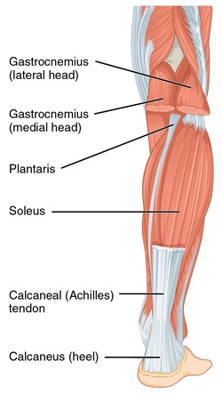
Superficial Posterior Compartment[edit | edit source]
Soleus Origin: Soleal line, medial border of tibia, head of the fibula, posterior border of the fibula. Insertion: Posterior surface of the calcaneus (via calcaneal tendon). Nerve supply: Tibial nerve (S1, S2). Vascular supply: Posterior tibial artery and vein.
Gastrocnemius Origin: Two heads, lateral: Posterolateral aspect of lateral condyle of the femur, medial: Posterior surface of the medial femoral condyle, the popliteal surface of the femoral shaft. Insertion: Posterior surface of the calcaneus via the calcaneal tendon. Nerve supply: Tibial nerve (S1, S2). Vascular supply: Medial sural artery, a branch of the popliteal artery.
Plantaris Origin: Lateral supracondylar line of the femur, oblique popliteal ligament of the knee. Insertion: Posterior surface of the calcaneus (via calcaneal tendon). Nerve supply: Tibial nerve (S1, S2). Vascular supply: lateral sural and popliteal arteries and superior lateral genicular artery
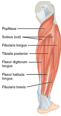
Deep Posterior Compartment[edit | edit source]
Flexor digitorum longus Origin: Posterior surface of tibia (inferior to soleal line). Insertion: Bases of distal phalanges of digits 2-5. Nerve supply: Tibial nerve (L5, S1, S2). Vascular supply: Posterior tibial artery.
Tibialis posterior Origin: Posterior surface of the tibia, posterior surface of fibula and interosseous membrane. Insertion: Tuberosity of navicular bone, all cuneiform bones, cuboid bone, bases of metatarsal bones 2-4. Nerve supply: Tibial nerve (L4, L5). Vascular supply: Branches of the posterior tibial artery.
Flexor hallucis longus Origin: Distal 2/3 of the posterior surface of fibula, interosseous membrane, the posterior intermuscular septum of the leg, fascia of tibialis posterior muscle. Insertion: Base of distal phalanx of the great toe. Nerve supply: Tibial nerve (S2, S3). Vascular supply: Posterior tibial artery, fibular artery.
Popliteus Origin: Lateral condyle of femur, posterior horn of lateral meniscus of the knee joint. Insertion: Posterior surface of the proximal tibia. Nerve supply: Tibial nerve (L4-S1). Vascular supply: Inferior medial and lateral genicular arteries (popliteal artery), posterior tibial recurrent artery, posterior tibial artery, nutrient artery of the tibia.
Lateral Compartment[edit | edit source]
Peroneus (Fibularis) Longus Origin: Head and superior 2/3 of the lateral surface of the fibula. Insertion: base of the 1st metatarsal and medial cuneiform. Nerve supply: superficial fibular nerve (L5 - S1). Vascular supply: Fibular artery.
Function:
- Ankle inversion and week plantarflexion
- Muscular control of the forefoot position
Peroneus (Fibularis) Brevis Origin: Inferior 2/3 of the lateral surface of the fibula. Insertion: base of the 5th metatarsal. Nerve supply: superficial fibular nerve (L5 - S1). Vascular supply: Anterior tibial artery.
Function:
- Ankle inversion and week plantarflexion
- Stabilises the lateral column of the foot
Anterior Compartment[edit | edit source]
Ankle dorsiflexion is performed by all the muscles within the anterior compartment. In addition
- tibialis anterior and extensor hallucis longus invert the foot during dorsiflexion
- extensor digitorum longus everts the foot during dorsiflexion
- eccentric control of foot lowering during heel strike
- concentric control of toes clearance during swing phase
Tibialis Anterior Origin:
Extensor Digitorum Longus Origin:
Extensor Hallucis Longus Origin:
Peroneus Tertius Origin:
Foot Muscle[edit | edit source]
Fascia[edit | edit source]
Arches[edit | edit source]
Clinical relevance[edit | edit source]
- Area posterior to the medial malleolus tends to get injured frequently causing tendon injury of the posterior tibialis, flexor hallucis longus or flexor digitorum and tibial nerve compression.[1]


