Basic Foot and Ankle Anatomy - Bones and Ligaments
Original Editor - User Name
Top Contributors - Ewa Jaraczewska, Wanda van Niekerk, Jess Bell, Niha Mulla, Tarina van der Stockt, Kim Jackson and Olajumoke Ogunleye
Description[edit | edit source]
Ankle and foot injuries are fairly common not only within the professional athletes or as a result of sporting activities, but they occur during routine daily activities. The typical injuries include sprains, fractures, tears, and inflammation. Good knowledge of the foot and ankle anatomy is necessary for proper diagnosis and treatment.
Structure[edit | edit source]
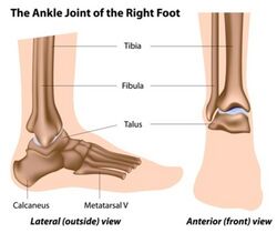
The ankle is formed by three bones: talus, tibia and fibula. The anatomic structure of the foot consists of hindfoot, midfoot and forefoot, each composed of several bones.
Ankle Bones[edit | edit source]
The ankle is composed of the lower leg and the foot and the bony elements of the ankle joint include the distal tibia, distal fibula, and talus.
Foot bones[edit | edit source]
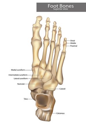
Talus and calcaneus and two tarsal bones form the posterior aspect of the foot called hindfoot. The midfoot located between the hindfoot and forefoot is made up of five tarsal bones: navicular, cuboid, and medial, intermediate, and lateral cuneiforms. The most anterior aspect of the foot including metatarsals, phalanges, and sesamoid bones is called a forefoot. Each digit, except for the great toe, consists of a metatarsal and three phalanges. Great toe has two phalanges only.
Function[edit | edit source]
The foot and ankle provide various important functions which include bodyweight support, balance maintenance, shock absorption, response to ground reaction forces, or substitution for hand function in individuals with upper extremity amputation.[1]It has a crucial role in gait and posture therefore its malalignment can cause problems eg., back pain or mobility limitations.[2][3]
Ankle and Foot Articulations[edit | edit source]
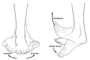
Talocrural joint (TC or sometimes called a tibiotalar joint) is referred to as the ankle joint. The joint is framed laterally and medially by the lateral and the medial malleolus and from the top by the inferior articular surface of the tibia and the superior margin of the talus. [4]Saggital plane dorsiflexion and plantarflexion are the primary movements in this joint.
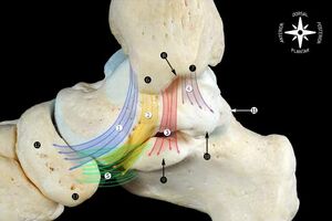
The hindfoot has a talus and calcaneus articulation called a subtalar joint (ST, also known as the talocalcaneal joint). It is made of the three facets on each of the talus and calcaneus. The main motions are inversion and eversion of the ankle and hindfoot.
The talonavicular and calcaneocuboid joints known as a Chopart joint (MT, midtarsal or transverse tarsal joint) are located between the hindfoot and midfoot. This joint allows forefoot rotation. The navicular and all three cuneiform bones compose articulation distally. In addition to the navicular and cuneiforms bones, the cuboid bone has a distal articulation with the base of the fourth and fifth metatarsal bones.
The tarsometatarsal joint (TMT or Lisfranc joint)connecting the midfoot with forefoot is composed of lateral, intermediate and medial cuneiforms articulating with the bases of the three metatarsal bone (1st, 2nd, and 3rd). The small movement that occurs in the joint is described as dorsal and plantarflexion. [5]The bases of the remaining metatarsal bones(4th and 5th) connect with the cuboid bone. [4]
The five rays, metatarsal and corresponding phalanges create forefoot medial and lateral columns where rays 1,2, and 3 belong to the middle column, ray 4 and 5 to the lateral column. Metatarsophalangeal joints (MTP joints) are the main components of the forefoot. [4] Each toe, except the great toe, has proximal and distal IP joints. The latter has only one IP joint.
Below is the summary of the ankle and foot articulation.
| Joint | Type of Joint | Plane of Movement | Motion |
|---|---|---|---|
| TC joint | Hinge | Sagittal | Dorsiflexion & Plantarflexion |
| ST joint | Condyloid |
Mainly transverse Some sagittal |
Inversion & Eversion Dorsiflexion & Plantarflexion |
| MT joint |
TN joint - Ball and socket CC joint - Modified saddle |
Largely in transverse Some sagittal |
Inversion & Eversion Flexion & Extension Forefoot rotation |
| TMT joint | Planar | ||
| MTP joint | Condyloid |
Sagittal Some Transverse |
Flexion & Extension Abduction & Adduction |
| IP joint | Hinge | Sagittal | Flexion & Extension |
Ligaments[edit | edit source]
Acute and chronic ankle pains are often associated with either ligament injury or ligament laxity.[6]The ligament of the ankle consist of the lateral ligaments, the medial side ligaments (the deltoid ligament), and the ligaments connecting the distal epiphyses of the tibia and fibula are called the tibiofibular syndesmosis. [6]
There are 4 main ligaments of the foot including plantar fascia ligament, plantar calcaneonavicular ligament (known as a spring ligament), calcaneocuboid ligament, and Lisfranc ligaments.
Medial Collateral Ligament (Deltoid Ligament)[edit | edit source]
There are various descriptions of the medial collateral ligament(MCL), but description by Milner and Soames (1998) is the most commonly accepted.[7]The authors divide the MCL into two layers: superficial and deep layers (bands that are always present are in italic):
- Superficial: tibiospring ligament, tibionavicular ligament, superficial posterior tibiotalar ligament, tibiocalcaneal ligament
- Deep: deep posterior tibiotalar ligament, deep anterior tibiotalar ligament
The characteristic of the MCL:
- multifascicular
- originates from the medial malleolus and inserts in the talus
- tendon sheath of the posterior tibial muscle covers the posterior and middle part of the deltoid ligament
- tendons covers most of the MCL as it inserts at the foot.[6]
Muscle attachments[edit | edit source]
Watch this short video on ankle and foot palpation
Vascular Supply[edit | edit source]
Nerve Supply[edit | edit source]
Clinical relevance[edit | edit source]
Resources[edit | edit source]
References[edit | edit source]
- ↑ Houglum PA, Bertotti DB. Brunnstrom's clinical kinesiology. FA Davis; 2012
- ↑ Menz HB, Dufour AB, Riskowski JL, Hillstrom HJ, Hannan MT. Foot posture, foot function and low back pain: the Framingham Foot Study. Rheumatology (Oxford). 2013 Dec;52(12):2275-82.
- ↑ Menz HB, Dufour AB, Katz P, Hannan MT. Foot Pain and Pronated Foot Type Are Associated with Self-Reported Mobility Limitations in Older Adults: The Framingham Foot Study. Gerontology. 2016;62(3):289-95.
- ↑ 4.0 4.1 4.2 Ficke J, Byerly DW. Anatomy, Bony Pelvis and Lower Limb, Foot. [Updated 2021 Aug 11]. In: StatPearls [Internet]. Treasure Island (FL): StatPearls Publishing; 2021 Jan-. Available from: https://www.ncbi.nlm.nih.gov/books/NBK546698/
- ↑ Bähler A. The Biomechanics of the Foot. CPO 1986;10(1):8-14.
- ↑ 6.0 6.1 6.2 Golanó P, Vega J, de Leeuw PA, Malagelada F, Manzanares MC, Götzens V, van Dijk CN. Anatomy of the ankle ligaments: a pictorial essay. Knee Surg Sports Traumatol Arthrosc. 2010 May;18(5):557-69.
- ↑ Milner CE, Soames RW. Anatomy of the collateral ligaments of the human ankle joint. Foot Ankle Int. 1998 Nov;19(11):757-60.
- ↑ Ankle and foot palpation. 2012 Available from: https://www.youtube.com/watch?v=aJRemQbNPhk [last accessed 18/12/2021]


