Brain Anatomy
Original Editor - Your name will be added here if you created the original content for this page.
Top Contributors - Naomi O'Reilly, Lucinda hampton, Kim Jackson, Tarina van der Stockt, Admin, Shaimaa Eldib, Olajumoke Ogunleye, Aminat Abolade, Rachael Lowe, Tony Lowe, Wendy Walker and Simisola Ajeyalemi
Introduction[edit | edit source]
Sub Heading 2[edit | edit source]
Cerebellum The cerebellum is found below the tentorial membrane in the posterior fossa. It is connected to the brainstem by 3 cerebellar peduncles. Its primary role is that of coordination and learning of movements.
There are three main systems.
- Spinocerebellum - involved in the control of axial musculature and posture.
- Pontocerebellum - coordination and planning of intended limb movement.
- Vestibulocerebellum - involved in posture and eye movements.
The cerebellum compares the intended movement originating from the motor cortex areas with the actual movement relayed back by the afferent systems and interneurons in the spinal cord. It also gains input from the vestibular systems.
Main Functions:
- To act as a comparator: comparing descending supraspinal signals with ascending afferent feedback. If there is any discrepancy, this is then fine tuned to produce the actual movement desired via descending pathways. This helps to achieve smoothness and accuracy in movement.
- To act as a timing device: it converts descending motor signals into a sequence of motor activation. This helps the movement achieve smoothness and coordination, maintaining posture and balance.(receiving input from the vestibular system).
- Initiating and Storing Movement: has ability to store and update motor information. there is a significant role played in accurate learned movement. This is due to a modifiable synapse at the purkinje cell.
Dysfunction causes:
- Hypotonia (reduced muscle tone)
- Ataxia/incoordination
- Dysarthria (the inability to articulate words properly)
- Nystagmus (rapid jerky eye movements)
- Palatal tremor/ myoclunus (tremor of the palate)
Basal Ganglia This consists of:
- Neostratum (Caudate and Putamen)
- Internal and External segments of Globus Pallidus
- Pars Reticulata
- Compacta of Substantia Nigra
- Subthalamic nucleus
The Neostratum is the main area of signal reception for the basal ganglia and receives information from the whole cortex in a somatotopic fashion. It relays signals to the thalamus which projects to the premotor cortex, supplementary motor cortex and prefrontal cortex. There is also a projection to the brainstem (pedunclopontine nucleus- involved in locomotion, and superior colliculus - eye movement).
Hypothalamus The hypothalamus lies on either side of the 3rd ventricle, below the thalamus and between the optic chiasm and the midbrain. It receives a large input from limbic structures. It has a significantly large efferent output to the ANS and has a highly significant role in the control of pituitary endocrine function.
The hypothalamus as well as inputting the ANS also has a large part to play in the homeostasis of many physiological systems such as hunger, thirst, water and sodium balance and temperature regulation. it also plays a role in memory and emotional responses, providing autonomic and endocrine responses. It also plays a role in the control of circadian rhythms via retinal input to suprachiasmatic nucleus. There may also be hypothalamus input into sexual and emotional behaviour independent of its endocrine role.
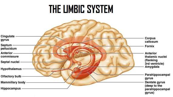
Limbic System This refers to a number of areas within the brain lying mainly on the medial side of the temporal lobe. It includes a number of structures as seen in the diagram below
The limbic system provides high level processing of sensory information. The main outflow of the limbic system is to the prefrontal cortex and the hypothalamus as well as to cortical areas. It appears to have a role in attaching behavioural significance and response to a given stimulus. Damage to this area has profound effects on emotional responses.
Long term potentiation LTP is the increase in the strength of a synaptic transmission with repetitive use, it can be seen to be effected in the hippocampus and is thought to be important for memory acquisition.
Subcortical Fibre Tracts
Overview of the Brainstem The brainstem is made up of the part of the brain that begins at the foramen magnum. It extends to the cerebral peduncles and thalamus. It consists of;
- Medulla
- Pons
- Midbrain
It contains the following:
- Nuclei for 10 of 12 pairs of cranial nerves (not olfactory or optic nerves)
- Apparatus for controlling eye movements.(3rd, 4th, 6th cranial nerves)
- Monoaminergic nuclei that project widely in the CNS
- Vital respiration and cardiovascular centres.
- Autonomic centres
- Areas important for consciousness
- Ascending and descending pathways, linking spinal cord to brain.
Visual Pathways
Sub Heading 3[edit | edit source]
Resources[edit | edit source]
References[edit | edit source]
- ↑ UBC Medicine - Educational Media. The Cerebellum - UBC Neuroanatomy - Season 1 - Ep 8. Available from: https://youtu.be/17mxfO9nklQ[last accessed 30/08/19]
- ↑ UBC Medicine - Educational Media. Basal Ganglia - UBC Flexible Neuroanatomy - Season 1 - Ep 7. Available from: https://youtu.be/InJByqg1x-0[last accessed 30/08/19]
- ↑ UBC Medicine - Educational Media. Hypothalamus and Limbic System - UBC Neuroanatomy - Season 1 - Ep 4. Available from:https://youtu.be/ErpxEwlWww4[last accessed 30/08/19]
- ↑ UBC Medicine - Educational Media. Subcortical Fiber Tracts - UBC Neuroanatomy - Season 1 - Ep 5. Available from:https://youtu.be/PazaHElk6wc[last accessed 30/08/19]
- ↑ UBC Medicine - Educational Media. Overview of the Brainstem - UBC Neuroanatomy - Season 1 - Ep 3. Available from:https://youtu.be/Mkj78h8w4a8[last accessed 30/08/19]
- ↑ UBC Medicine - Educational Media. Visual Pathways - UBC Neuroanatomy - Season 1 - Ep 6. Available from:https://youtu.be/TbDFrbXiz2s[last accessed 30/08/19]


