The Biomechanics of Pregnancy: Difference between revisions
Areeba Raja (talk | contribs) No edit summary |
Areeba Raja (talk | contribs) No edit summary |
||
| Line 27: | Line 27: | ||
* Extension of the occiput on atlas. | * Extension of the occiput on atlas. | ||
* Associated with these postural changes is a waddling gait pattern. | * Associated with these postural changes is a waddling gait pattern. | ||
{{#ev:youtube|mo-CrtgnkrA}} | |||
==== Changes in the Spine ==== | ==== Changes in the Spine ==== | ||
| Line 124: | Line 126: | ||
|Increase in knee adductors moment | |Increase in knee adductors moment | ||
|} | |} | ||
{{#ev:youtube|NpgUh5ACzW4}} | |||
== Resources == | == Resources == | ||
Revision as of 00:22, 26 November 2021
This article is currently under review and may not be up to date. Please come back soon to see the finished work! (26/11/2021)
Original Editor - User Name
Top Contributors - Areeba Raja and Kim Jackson
Introduction[edit | edit source]
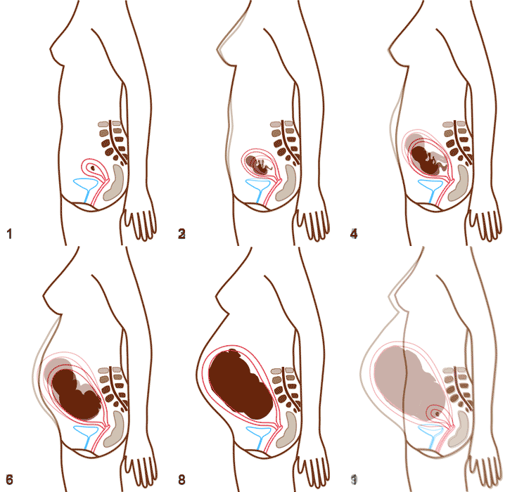
Pregnancy, as a natural and physiological process, produces in a woman a series of changes involving the motor system. Weight gain, especially changes within its distribution requires functional adaptation of the musculoskeletal system. These changes make both the posture and gait pattern of pregnant women different from non-pregnant subjects.[1]
The addition of anterior mass on the trunk in a pregnant woman changes the body's center of mass if there is not a concomitant change in posture. Consequently, kinematic adjustments are made to improve stability, allowing safer gait for the pregnant woman, but may be only mechanical in nature. The adjustments do reduce the kinetic effect of the increased mass as total mass normalized moments decrease.[2]
Biomechanical Considerations[edit | edit source]
During pregnancy, a number of biomechanical and hormonal changes occur that can alter spinal curvature, balance, and gait patterns by affecting key areas of the human body. This can greatly impact quality of life (QOL) by increasing back pain and the risk of falls.[3]
The overall postural effect of pregnancy by the final month is as follows:[2][4][5][6][7]
- Anterior tilt of pelvis with hyperextended knees
- Accentuated lumbar lordosis with a short radius curve
- Posterior gravity line
- Hyper kyphosis of the upper thoracic
- Protracted shoulders
- Anterior angulation of the cervical region
- Extension of the occiput on atlas.
- Associated with these postural changes is a waddling gait pattern.
Changes in the Spine[edit | edit source]
- Pregnant women with anterior translation of their center of mass have been shown to lack positional adjustment of lumbar lordosis, and the force of gravity, when more distant from the hip, generates a larger hip moment and an unstable upper body.[8]
- Many pregnant women demonstrate a sway-back posture, whereby the upper trunk is displaced posterior to the lower body causing the center of gravity to shift further backward, and increases the tone of head and neck muscles, causing the head to shift forward to compensate for the change in center of gravity and prevent falling.[8]
Changes in the Knee[edit | edit source]
- As pregnant women experience an anterior shift in center-of-gravity, their knees hyperextend to maintain balanced, upright posture. Knee hyperextension tenses the anterior cruciate ligament (ACL) as it impinges against the femoral notch, which may cause the ACL to adapt and lengthen throughout pregnancy.[9]
- Knee laxity during pregnancy reaches a constant level before the fifth month of pregnancy, which suggests that laxity increases early in pregnancy; it decreased significantly by about 14% 4 months postpartum.[10]
- The knee is delicately balanced to maintain stability while also allowing for a wide range-of-motion during activities. Perturbations to this balance, such as those caused by the non-uniform changes in joint laxity which persist following pregnancy, could potentially increase the risk of developing OA and other musculoskeletal disorders in women in their post-reproductive years.[9]
Changes in the Ankle and Foot[edit | edit source]
- Changes in the biomechanics of feet during pregnancy and puerperium, indicates an increase in circumference measurement and a reduction in plantar arch while the length and width of the foot increases with increased body weight and ligamentous laxity.[11]
- During pregnancy, forces exerted at the soles of the feet shift from the posterior to the anterior with consequent increases at the forefoot and, more prominently, at the midfoot.[12]
- Flat feet add stress on the foot and causes inflammation of plantar fascia and increases strain on the feet, calves and sometimes the back.[13]
Biomechanical Alterations in Gait[edit | edit source]
Pregnant women altered gait pattern to adapt the weight gain and the shift of centre of gravity. the gait of pregnant women undergoes following modifications. (Pregnancy-related changes in center of pressure during gait) ( Changes of kinematic gait parameters due to pregnancy)( Characteristics of the centre of pressure progression for pregnant women during walking)
Gait Speed[edit | edit source]
Natural locomotion of pregnant women is characterized by slower speed and lower frequency and length of steps, as compared to the pre-pregnancy and postpartum state. ( Changes of kinematic gait parameters due to pregnancy) pregnant women tend to avoid the large relative phase between pelvic and thoracic rotations that is typical for high walking velocities, possibly because the moments of inertia of their pelvis and thorax have increased which renders the control of relative phase more critical. (Gait coordination in pregnancy: transverse pelvic and thoracic rotations and their relative phase)
Gait Cycle[edit | edit source]
A significant decrease in the length of the gait cycle is observed in pregnant women. There is an increase in double support time compared to post-partum and nulliparous women. These are fine adjustments that minimize the time on one leg to reduce muscle solicitation. Thus, pregnant women exaggerate transition phases in order to increase the security of gait.
| Spatial and Temporal Parameters of Gait | |
| Step Length | Decreases |
| Step Width | Increases |
| Single Support Time | Decreases |
| Double Support Time | Increases |
| Base of Support | Increases |
| Ground Reaction Force | Decrease in late pregnancy. |
| Joint Kinematic Parameters | |
| Sagittal Plane | |
| Pelvis | Increase in anterior tilt about 5 degrees |
| Hip | Increase flexion during stance phase |
| Knee | Increase flexion during terminal stance |
| Ankle | Decrease dorsiflexion and plantarflexion |
| Frontal Plane | |
| Pelvis | Increase in pelvic separation width and reduction in the amplitude of the unilateral elevation of the pelvis. |
| Hip | Increase in abduction. |
| Joint Kinetic Parameters | |
| Sagittal Plane | |
| Hip | Significant increase in the hip extensors moment |
| Knee | Significant decrease in the knee extensor moment |
| Ankle | Significant decrease in the ankle plantar flexor moment |
| Frontal Plane | |
| Hip | Increase in hip abductors moment |
| Knee | Increase in knee adductors moment |
Resources[edit | edit source]
- bulleted list
- x
or
- numbered list
- x
References[edit | edit source]
- ↑ Forczek W;Staszkiewicz R. Changes of kinematic gait parameters due to pregnancy. Acta of bioengineering and biomechanics [Internet]. 2012 [cited 2021 Nov 25];14(4).
- ↑ 2.0 2.1 Ogamba MI, Loverro KL, Laudicina NM, Gill SV, Lewis CL. Changes in Gait with Anteriorly Added Mass: A Pregnancy Simulation Study. Journal of Applied Biomechanics [Internet]. 2016 Aug [cited 2021 Nov 21];32(4):379–87.
- ↑ Conder R, Zamani R, Akrami M. The Biomechanics of Pregnancy: A Systematic Review. Journal of Functional Morphology and Kinesiology [Internet]. 2019 Dec 2 [cited 2021 Nov 25];4(4):72.
- ↑ Fligg DB. Biomechanical and treatment considerations for the pregnant patient. The Journal of the Canadian Chiropractic Association [Internet]. 1986 [cited 2021 Nov 25];30(3):145–7.
- ↑ Yoo H, Shin D, Song C. Changes in the spinal curvature, degree of pain, balance ability, and gait ability according to pregnancy period in pregnant and nonpregnant women. Journal of Physical Therapy Science [Internet]. 2015 [cited 2021 Nov 25];27(1):279–84.
- ↑ Petrocco-Napuli K. Pregnancy and the Impact on the Lower Extremity [Internet]. [cited 2021 Nov 21].
- ↑ Franklin ME, Conner-Kerr T. An Analysis of Posture and Back Pain in the First and Third Trimesters of Pregnancy. Journal of Orthopaedic & Sports Physical Therapy [Internet]. 1998 Sep [cited 2021 Nov 25];28(3):133–8.
- ↑ 8.0 8.1 OKANISHI N, KITO N, AKIYAMA M, YAMAMOTO M. Spinal curvature and characteristics of postural change in pregnant women. Acta Obstetricia et Gynecologica Scandinavica [Internet]. 2012 Jun 18 [cited 2021 Nov 25];91(7):856–61.
- ↑ 9.0 9.1 Chu SR, Boyer EH, Beynnon B, Segal NA. Pregnancy Results in Lasting Changes in Knee Joint Laxity. PM&R [Internet]. 2019 Feb [cited 2021 Nov 25];11(2):117–24.
- ↑ Dumas GA, Reid JG. Laxity of Knee Cruciate Ligaments During Pregnancy. Journal of Orthopaedic & Sports Physical Therapy [Internet]. 1997 Jul [cited 2021 Nov 25];26(1):2–6.
- ↑ Wittkopf PG, Kretzer J, Borges DM, Santos GM, Sperandio FF. Características biomecânicas dos pés no período gravídico-puerperal: estudo de caso. Scientia Medica [Internet]. 2015 Jun 10 [cited 2021 Nov 25];25(1):19688.
- ↑ VAROL T, GÖKER A, CEZAYİRLİ E, ÖZGÜR S, TUÇ YÜCEL A. Relation between foot pain and plantar pressure in pregnancy. TURKISH JOURNAL OF MEDICAL SCIENCES [Internet]. 2017 [cited 2021 Nov 25];47:1104–8. Available from: https://pubmed.ncbi.nlm.nih.gov/29154449/
- ↑ Ghait AS, Elhosary EA, Abogazya AA. Assessment of Foot Biomechanics through Measuring of the Plantar Pressure during the Last Trimester of Pregnancy. Journal of Advances in Medicine and Medical Research [Internet]. 2018 Sep 20 [cited 2021 Nov 25];27(7):1–6.


