Extensor Retinaculum (Wrist): Difference between revisions
Kim Jackson (talk | contribs) mNo edit summary |
No edit summary |
||
| Line 6: | Line 6: | ||
== Description == | == Description == | ||
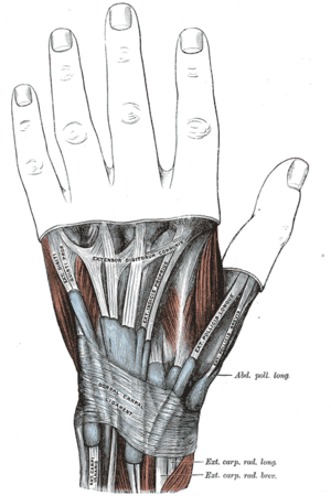
[[File:Extensor Retinaculum of the wrist.png|alt=Extensor Retinaculum of the wrist|thumb|'''Extensor Retinaculum of the wrist''']] | [[File:Extensor Retinaculum of the wrist.png|alt=Extensor Retinaculum of the wrist|thumb|'''Extensor Retinaculum of the wrist''']] | ||
Extensor Retinaculum is a fibrous, thickened band that holds the extensor tendons at the dorsum of the [[Wrist and Hand|wrist]]. It is an oblique band runs downwards and medially preventing bowstringing.<ref name=":0">Robertson BL, Jamadar DA, Jacobson JA, Kalume-Brigido M, Caoili EM, Margaliot Z, De Maeseneer MO. [https://www.ajronline.org/doi/pdf/10.2214/AJR.05.0764 Extensor retinaculum of the wrist: sonographic characterization and pseudotenosynovitis appearance]. American Journal of Roentgenology. 2007 Jan;188(1):198-202.</ref><ref name=":1">Chaurasia BD. BD Chaurasia's Human Anatomy. CBS Publishers & Distributors PVt Ltd.; 2010.</ref><ref>Taleisnik J, Gelberman RH, Miller BW, Szabo RM. The extensor retinaculum of the wrist. Journal of Hand Surgery. 1984 Jul 1;9(4):495-501.</ref> | Extensor Retinaculum is a fibrous, thickened band that holds the extensor tendons at the dorsum of the [[Wrist and Hand|wrist]]. It is an oblique band runs downwards and medially preventing bowstringing.<ref name=":0">Robertson BL, Jamadar DA, Jacobson JA, Kalume-Brigido M, Caoili EM, Margaliot Z, De Maeseneer MO. [https://www.ajronline.org/doi/pdf/10.2214/AJR.05.0764 Extensor retinaculum of the wrist: sonographic characterization and pseudotenosynovitis appearance]. American Journal of Roentgenology. 2007 Jan;188(1):198-202.</ref><ref name=":1">Chaurasia BD. BD Chaurasia's Human Anatomy. CBS Publishers & Distributors PVt Ltd.; 2010.</ref><ref>Taleisnik J, Gelberman RH, Miller BW, Szabo RM. The extensor retinaculum of the wrist. Journal of Hand Surgery. 1984 Jul 1;9(4):495-501.</ref> | ||
=== Attachments === | === Attachments === | ||
''Laterally:'' | ''Laterally:'' | ||
| Line 25: | Line 21: | ||
== Clinical relevance == | == Clinical relevance == | ||
By extending fascial attachments to the underlying bones and periosteum, the retinaculum forms six osseofascial compartments over the [[Wrist and Hand Examination|dorsal wrist]]. There are several structures passing through each compartment from lateral to the medial side and each compartment is lined by a synovial sheath.<ref name=":1" /><ref>Standring S, editor. Gray's anatomy e-book: the anatomical basis of clinical practice. Elsevier Health Sciences; 2015 Aug 7.</ref> | By extending fascial attachments to the underlying [[Bone|bones]] and [[periosteum]], the retinaculum forms six osseofascial compartments over the [[Wrist and Hand Examination|dorsal wrist]]. There are several structures passing through each compartment from lateral to the medial side and each compartment is lined by a synovial sheath.<ref name=":1" /><ref>Standring S, editor. Gray's anatomy e-book: the anatomical basis of clinical practice. Elsevier Health Sciences; 2015 Aug 7.</ref> | ||
{| class="wikitable" | {| class="wikitable" | ||
|+Structures passing through Extensor retinaculum | |+Structures passing through Extensor retinaculum | ||
Revision as of 06:43, 9 September 2022
Original Editor - Sameera Withanage
Top Contributors - Sameera Withanage, Kim Jackson, Ewa Jaraczewska and Lucinda hampton
Description[edit | edit source]
Extensor Retinaculum is a fibrous, thickened band that holds the extensor tendons at the dorsum of the wrist. It is an oblique band runs downwards and medially preventing bowstringing.[1][2][3]
Attachments[edit | edit source]
Laterally:
Medially:
Function[edit | edit source]
Extensor Retinaculum helps to keep the extensor tendons in alignment and prevent bowstringing during movements.[1]
Clinical relevance[edit | edit source]
By extending fascial attachments to the underlying bones and periosteum, the retinaculum forms six osseofascial compartments over the dorsal wrist. There are several structures passing through each compartment from lateral to the medial side and each compartment is lined by a synovial sheath.[2][5]
| Compartment | Structure |
|---|---|
| 1 | |
| 2 | |
| 3 | |
| 4 |
|
| 5 |
|
| 6 |
References[edit | edit source]
- ↑ 1.0 1.1 Robertson BL, Jamadar DA, Jacobson JA, Kalume-Brigido M, Caoili EM, Margaliot Z, De Maeseneer MO. Extensor retinaculum of the wrist: sonographic characterization and pseudotenosynovitis appearance. American Journal of Roentgenology. 2007 Jan;188(1):198-202.
- ↑ 2.0 2.1 2.2 2.3 2.4 2.5 Chaurasia BD. BD Chaurasia's Human Anatomy. CBS Publishers & Distributors PVt Ltd.; 2010.
- ↑ Taleisnik J, Gelberman RH, Miller BW, Szabo RM. The extensor retinaculum of the wrist. Journal of Hand Surgery. 1984 Jul 1;9(4):495-501.
- ↑ Extensor retinaculum of wrist. Available from:https://www.youtube.com/watch?v=tM6rmLBhrwc&ab_channel=AW2N
- ↑ Standring S, editor. Gray's anatomy e-book: the anatomical basis of clinical practice. Elsevier Health Sciences; 2015 Aug 7.


