Hip Osteoarthritis: Difference between revisions
Kim Presiaux (talk | contribs) No edit summary |
Kim Presiaux (talk | contribs) No edit summary |
||
| Line 71: | Line 71: | ||
== Examination == | == Examination == | ||
<!--StartFragment--> | <!--StartFragment--> <span lang="EN-US" style="font-family:Arial;mso-bidi-font-family: Arial;mso-ansi-language:EN-US">The beginning of OA is characterized by limited | ||
<span lang="EN-US" style="font-family:Arial;mso-bidi-font-family: | abduction and<span style="mso-spacerun:yes"> </span>rotation in the hip joint. Later on flexion, extension, adduction,.. will become more difficult.<br> Physiotherapeutic examination <sup>1</sup><br> <br> 1) Palpation of M. gluteus medius.<br> Position: patient lies on his side. Upper leg in adduction and flexion<br> OA: Zone of greater Trochanter is sensitive and painful.</span> | ||
Arial;mso-ansi-language:EN-US">The beginning of OA is characterized by limited | |||
abduction and<span style="mso-spacerun:yes"> </span>rotation in the hip joint. Later on flexion, extension, adduction,.. will become more difficult.<br> Physiotherapeutic examination <sup>1</sup><br> <br> 1) Palpation of M. gluteus medius.<br> Position: patient lies on his side. Upper leg in adduction and flexion<br> OA: Zone of greater Trochanter is sensitive and painful. | |||
<span lang="EN-US" style="font-family:Arial;mso-bidi-font-family: | <span lang="EN-US" style="font-family:Arial;mso-bidi-font-family: Arial;mso-ansi-language:EN-US">2)Flexion and forced flexion | ||
Arial;mso-ansi-language:EN-US">2)Flexion and forced flexion | </span> | ||
Position: patient lies on his back. | Position: patient lies on his back. | ||
OA: Flexion is limited. | OA: Flexion is limited. | ||
<span lang="EN-US" style="font-family:Arial;mso-bidi-font-family: | <span lang="EN-US" style="font-family:Arial;mso-bidi-font-family: Arial;mso-ansi-language:EN-US">3) Extension | ||
Arial;mso-ansi-language:EN-US">3) Extension | </span> | ||
Position: Patient in prone. Physiotherapist stabilizes the pelvis and raises | Position: Patient in prone. Physiotherapist stabilizes the pelvis and raises the leg. | ||
the leg. | |||
OA: Amplitude is limited | OA: Amplitude is limited | ||
<span lang="EN-US" style="font-family:Arial;mso-bidi-font-family: | <span lang="EN-US" style="font-family:Arial;mso-bidi-font-family: Arial;mso-ansi-language:EN-US">4) Abduction and adduction | ||
Arial;mso-ansi-language:EN-US">4) Abduction and adduction | </span> | ||
Position: Patient lies on his back. Physiotherapist stabilizes the pelvis and | Position: Patient lies on his back. Physiotherapist stabilizes the pelvis and performs abduction and adduction. | ||
performs abduction and adduction. | |||
OA: abduction is limited, adduction keeps normal amplitude. | OA: abduction is limited, adduction keeps normal amplitude. | ||
== Medical Management <br> == | == Medical Management <br> == | ||
Revision as of 16:18, 30 December 2010
Original Editors - Eric Robertson, Kim Presiaux
Lead Editors - Your name will be added here if you are a lead editor on this page. Read more.
== Search Strategy ==
Database: Pubmed
Keywords: Treatment OA, Exercise OA, OA
Database: Website Library VUB
Keywords: Treatment OA, Exercise OA, OA
Definition/Description[edit | edit source]
Hip osteoarthritis is a common type of osteoarthritis. Since the hip is a weight-bearing joint, osteoarthritis can cause significant problems.
Hip osteoarthritis is caused by deterioration of articular cartilage of the hip joint.
There are several reasons this can develop:
• Previous hip injury
• Previous fracture, which changes hip alignment
• Genetics
• Congenital and developmental hip disease
• subchondral bone that is too soft or too hard 5
Clinically Relevant Anatomy[edit | edit source]
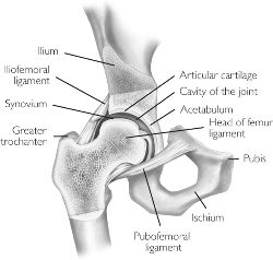
The hip joint is a synovial ball and socket joint, with the convex femoral head articulating with the concave acetabulum. Stability of the joint is achieved through a combination of muscle action and several ligaments forming a loose, but strong joint capsule, the iliofemoral ligament, the ischialfemoral ligament and the pubofemoral ligament. Another ligament, the ligamentum teres, does not provide stability to the hip but offers a portion of blood supply to the femoral head in some individuals.
The femoral head and acetablum are covered by smooth hyaline cartilage, and the acetabulum contains a labrum, which functions to facilitate movement and support the forces passed through the joint.
The hip, despite the requirement to support the weight of the body, has the second largest exursion of motion of any joint in the body.
External Link: [Hip Anatomy Video]
<== Epidemiology /Etiology ==
add text here
Characteristics/Clinical Presentation[edit | edit source]
add text here
Differential Diagnosis[edit | edit source]
add text here
Diagnostic Procedures[edit | edit source]
Altman et al have established guidelines by which clinical diagnosis of hip osteoarthritis can be made. The guidelines, established in 1991, present a 3 pronged approach to diagnosis of hip osteoarthritis including clinical, radiological, and laboratory findings. According to these guidlelines, a patient was considered to have osteoarthritis if they presented with:
- Hip Pain and...
- Hip Internal Rotation < 15 degrees and Hip Flexion less than or equal to 115 degrees
or, hip pain in combination with:
- Hip Rotation < 15 degrees or...
- Pain with Hip Internal Rotation or...
- Hip stiffness in the AM less than 60 minutes or...
- Age > 50 years
More recently, Sutlive et al have proposed a clinical prediction rule to identify individuals with hip osteoarthritis presenting with unilateral hip pain.
add text here related to medical diagnostic procedures
Outcome Measures[edit | edit source]
add links to outcome measures here (also see Outcome Measures Database)
Examination[edit | edit source]
The beginning of OA is characterized by limited
abduction and rotation in the hip joint. Later on flexion, extension, adduction,.. will become more difficult.
Physiotherapeutic examination 1
1) Palpation of M. gluteus medius.
Position: patient lies on his side. Upper leg in adduction and flexion
OA: Zone of greater Trochanter is sensitive and painful.
2)Flexion and forced flexion
Position: patient lies on his back.
OA: Flexion is limited.
3) Extension
Position: Patient in prone. Physiotherapist stabilizes the pelvis and raises the leg.
OA: Amplitude is limited
4) Abduction and adduction
Position: Patient lies on his back. Physiotherapist stabilizes the pelvis and performs abduction and adduction.
OA: abduction is limited, adduction keeps normal amplitude.
Medical Management
[edit | edit source]
add text here
Physical Therapy Management
[edit | edit source]
add text here
Key Research[edit | edit source]
add links and reviews of high quality evidence here (case studies should be added on new pages using the case study template)
Resources
[edit | edit source]
add appropriate resources here
Clinical Bottom Line[edit | edit source]
add text here
Recent Related Research (from Pubmed)[edit | edit source]
see tutorial on Adding PubMed Feed
Extension:RSS -- Error: Not a valid URL: Feed goes here!!|charset=UTF-8|short|max=10
References[edit | edit source]
see adding references tutorial.


