Spinal cord anatomy: Difference between revisions
No edit summary |
No edit summary |
||
| Line 8: | Line 8: | ||
</div><div align="justify"> | </div><div align="justify"> | ||
== Introduction == | == Introduction == | ||
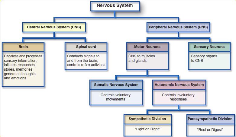
The | The nervous system is divided into the central nervous system (brain and spinal cord considered upper motor neurons) and the peripheral nervous system (nerves that enter and exit the spinal cord considered lower motor neurons).<ref name="Barker">Barker; Barasi; Neal. Neuroscience at a glance; Blackwell science Ltd; 1999</ref> Information to and from the muscles, glands, organs and sensory receptors are carried through the peripheral nervous system, which is divided into the autonomic nervous system carrying information to and from the organs, and the somatic nervous system carrying information to and from the muscles and the external environment. [5]The autonomic nervous system consists of the parasympathetic nervous system that governs resting function and the sympathetic nervous system that governs excitatory functions. The spinal cord and peripheral nerves provide all impulses to control muscle contraction, cardiac rhythm, pain and other bodily functions so therefore any lesion to the spinal cord prevents or reduces transmission of this information to and from the brain to the peripheries, affecting movement, sensation and visceral function. [6] | ||
# | |||
# | |||
[[Image:Nervous System.jpg|center|Nervous System]] | [[Image:Nervous System.jpg|center|Nervous System]] | ||
== '''Structure''' == | |||
=== Spinal Column === | |||
<div align="justify"> | |||
The spine or vertebral column bears the weight of the head, neck, trunk and upper extremities. The adult vertebral column typically consists of 33 vertebrae arranged in five regions, which provide support and protection for the spinal cord'''.''' It consists of seven cervical, twelve thoracic, five lumbar, five sacral and four coccygeal vertebrae, The five sacral vertebrae are fused in adults to form the sacrum, and the four coccygeal vertebrae are fused to form the coccyx. Its uppermost vertebrae incorporating the atlas and axis articulate with the head, and the lower most portion, the sacrum articulates with the pelvis. Each pair of adjacent vertebrae is separated by a semi-rigid intervertebral disk, which provides a level of flexibility to the vertebral column. Its flexibility is greatest in the cervical region, and lowest in the thoracic region. (Colour Atlas of Neurology - Reinhard Rohkamm) | |||
== | === Spinal Canal === | ||
<div align="justify"> | |||
= | Formed by the vertebral foramina of the vertebral bodies the spinal canal is bound anteriorly by the vertebral bodies and posteriorly by the laminae (vertebral arches) with reinforcement at the walls through the intervertebral disks and the anterior and posterior longitudinal ligaments. Containing the spinal cord, meninges, blood vessels, spinal nerve roots and surrounding fatty and connective tissues the diameter varies from 12 to 22 mm in the cervical region and from 22 to 25 mm in the lumbar region. (Colour Atlas of Neurology - Reinhard Rohkamm) | ||
=== | === Spinal Cord === | ||
<div align="justify"> | |||
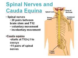
The spinal cord is the major conduit and reflex centre between the peripheral nerves and the brain, and transmits motor information from the brain to the muscles, tissues and organs, and sensory information from these areas back to the brain. [5][6 ]The spinal cord in an adult is approximately a 45 cm long, cylindrical structure that is slightly flattened anteriorly and posteriorly. (Essential Clinical Anatomy) The spinal cord lies within the vertebral canal, extending from the foramen magnum to the lowest border of the first lumbar vertebra. It is enlarged at two sites, the cervical and lumbar region. Its upper end is continuous with the medulla; the transition is defined to occur just above the level of exit of the first pair of cervical nerves. Its tapering lower end, the conus medullaris, terminates at the level of the L3 vertebra in neonates, and at the level of the L1-2 intervertebral disk in adults. The conus medullaris is continuous at its lower end with the threadlike filum terminale, composed mainly of glial and connective tissue, which, in turn, runs through the lumbar sac amidst the posterior and anterior roots of the spinal nerves, collectively called the cauda equina (“horse’s tail”), and then attaches to the dorsal surface of the coccyx. (Colour Atlas of Neurology - Reinhard Rohkamm) | |||
* Sensory Nerve Fibres enter the Spinal Cord via the Posterior (Dorsal) Root. The cell bodies for these neurons are situated in the Dorsal Root Ganglia. | |||
*Motor and Preganglionic Autonomic Fibres exit via the Anterior (Ventral) Root. | |||
=== | === Spinal Nerves === | ||
<div align="justify"> | |||
Emerging from the spinal cord are 31 pairs of anterior and posterior nerve roots. The cervical, thoracic, lumbar, and sacral portions of the spinal cord are defined according to the segmental division of the vertebral column and spinal nerves. There are eight cervical*, twelve thoracic, five lumbar, five sacral and one coccygeal. At each level an anterior pair of nerve roots carries motor nerves, while a posterior pair of nerve roots carries only sensory nerves. The anterior and posterior roots join to form two spinal nerves, one on either side of the spine, which then exit the vertebral canal through the intervertebral foramina. Once outside the intervertebral foramina they form peripheral nerves. (Colour Atlas of Neurology - Reinhard Rohkamm) | |||
[[File:Spinalnerves.jpg|frameless]]*While there are eight pairs of cervical spinal nerves there are only seven cervical vertebrae. This disparity occurs because the first pair of cervical spinal nerves exits ''above'' the first cervical vertebra just below the skull. However, the eighth pair of cervical spinal nerves exits ''below'' the last cervical vertebra.* | |||
This short video clip gives an overview of spinal cord anatomy. | |||
==== | {| width="400" cellspacing="1" cellpadding="1" border="0" align="center" | ||
|- | |||
| {{#ev:youtube|5B87zsAKmWc|350}}<ref>Handwritten tutorials. Spinal Pathways 1 - Spinal Cord Anatomy and Organisation. Available from: http://www.youtube.com/watch?v=5B87zsAKmWc [last accessed 29/08/16] </ref> | |||
|} | |||
== References == | |||
<references /> | <references /> | ||
[[Category:Spinal Cord Injuries]] | [[Category:Spinal Cord Injuries]] | ||
[[Category:SCI Content Project]] | [[Category:SCI Content Project]] | ||
Revision as of 15:35, 2 December 2018
- Please do not edit unless you are involved in this project, but please come back in the near future to check out new information!!
- If you would like to get involved in this project and earn accreditation for your contributions, [[[Special:Contact|please get in touch]]]!
Original Editor - Add a link to your Physiopedia profile here.
Top Contributors - Naomi O'Reilly, Lucinda hampton, Kim Jackson, Nikhil Benhur Abburi, Vidya Acharya, Admin, Tarina van der Stockt, Aminat Abolade, Ewa Jaraczewska, Rucha Gadgil and Jess Bell
Introduction[edit | edit source]
The nervous system is divided into the central nervous system (brain and spinal cord considered upper motor neurons) and the peripheral nervous system (nerves that enter and exit the spinal cord considered lower motor neurons).[1] Information to and from the muscles, glands, organs and sensory receptors are carried through the peripheral nervous system, which is divided into the autonomic nervous system carrying information to and from the organs, and the somatic nervous system carrying information to and from the muscles and the external environment. [5]The autonomic nervous system consists of the parasympathetic nervous system that governs resting function and the sympathetic nervous system that governs excitatory functions. The spinal cord and peripheral nerves provide all impulses to control muscle contraction, cardiac rhythm, pain and other bodily functions so therefore any lesion to the spinal cord prevents or reduces transmission of this information to and from the brain to the peripheries, affecting movement, sensation and visceral function. [6]
Structure[edit | edit source]
Spinal Column[edit | edit source]
The spine or vertebral column bears the weight of the head, neck, trunk and upper extremities. The adult vertebral column typically consists of 33 vertebrae arranged in five regions, which provide support and protection for the spinal cord. It consists of seven cervical, twelve thoracic, five lumbar, five sacral and four coccygeal vertebrae, The five sacral vertebrae are fused in adults to form the sacrum, and the four coccygeal vertebrae are fused to form the coccyx. Its uppermost vertebrae incorporating the atlas and axis articulate with the head, and the lower most portion, the sacrum articulates with the pelvis. Each pair of adjacent vertebrae is separated by a semi-rigid intervertebral disk, which provides a level of flexibility to the vertebral column. Its flexibility is greatest in the cervical region, and lowest in the thoracic region. (Colour Atlas of Neurology - Reinhard Rohkamm)
Spinal Canal[edit | edit source]
Formed by the vertebral foramina of the vertebral bodies the spinal canal is bound anteriorly by the vertebral bodies and posteriorly by the laminae (vertebral arches) with reinforcement at the walls through the intervertebral disks and the anterior and posterior longitudinal ligaments. Containing the spinal cord, meninges, blood vessels, spinal nerve roots and surrounding fatty and connective tissues the diameter varies from 12 to 22 mm in the cervical region and from 22 to 25 mm in the lumbar region. (Colour Atlas of Neurology - Reinhard Rohkamm)
Spinal Cord[edit | edit source]
The spinal cord is the major conduit and reflex centre between the peripheral nerves and the brain, and transmits motor information from the brain to the muscles, tissues and organs, and sensory information from these areas back to the brain. [5][6 ]The spinal cord in an adult is approximately a 45 cm long, cylindrical structure that is slightly flattened anteriorly and posteriorly. (Essential Clinical Anatomy) The spinal cord lies within the vertebral canal, extending from the foramen magnum to the lowest border of the first lumbar vertebra. It is enlarged at two sites, the cervical and lumbar region. Its upper end is continuous with the medulla; the transition is defined to occur just above the level of exit of the first pair of cervical nerves. Its tapering lower end, the conus medullaris, terminates at the level of the L3 vertebra in neonates, and at the level of the L1-2 intervertebral disk in adults. The conus medullaris is continuous at its lower end with the threadlike filum terminale, composed mainly of glial and connective tissue, which, in turn, runs through the lumbar sac amidst the posterior and anterior roots of the spinal nerves, collectively called the cauda equina (“horse’s tail”), and then attaches to the dorsal surface of the coccyx. (Colour Atlas of Neurology - Reinhard Rohkamm)
- Sensory Nerve Fibres enter the Spinal Cord via the Posterior (Dorsal) Root. The cell bodies for these neurons are situated in the Dorsal Root Ganglia.
- Motor and Preganglionic Autonomic Fibres exit via the Anterior (Ventral) Root.
Spinal Nerves[edit | edit source]
Emerging from the spinal cord are 31 pairs of anterior and posterior nerve roots. The cervical, thoracic, lumbar, and sacral portions of the spinal cord are defined according to the segmental division of the vertebral column and spinal nerves. There are eight cervical*, twelve thoracic, five lumbar, five sacral and one coccygeal. At each level an anterior pair of nerve roots carries motor nerves, while a posterior pair of nerve roots carries only sensory nerves. The anterior and posterior roots join to form two spinal nerves, one on either side of the spine, which then exit the vertebral canal through the intervertebral foramina. Once outside the intervertebral foramina they form peripheral nerves. (Colour Atlas of Neurology - Reinhard Rohkamm)
 *While there are eight pairs of cervical spinal nerves there are only seven cervical vertebrae. This disparity occurs because the first pair of cervical spinal nerves exits above the first cervical vertebra just below the skull. However, the eighth pair of cervical spinal nerves exits below the last cervical vertebra.*
This short video clip gives an overview of spinal cord anatomy.
| [2] |
References[edit | edit source]
- ↑ Barker; Barasi; Neal. Neuroscience at a glance; Blackwell science Ltd; 1999
- ↑ Handwritten tutorials. Spinal Pathways 1 - Spinal Cord Anatomy and Organisation. Available from: http://www.youtube.com/watch?v=5B87zsAKmWc [last accessed 29/08/16]


