Wound Assessment
Original Editor - Lucinda hampton
Top Contributors - Lucinda hampton, Abbey Wright and Kim Jackson
Introduction[edit | edit source]

A wound is damaged or disruption to the skin and, before treatment, the exact cause, location, and type of wound must be assessed to provide appropriate treatment[1]
Wound healing: complex physiological process occurring after an injury in the cells and tissues of our bodies to restore function of the tissue.
The healing process is affected by the
- Severity of the wound
- Location,
- Extent of injury
- External and internal factors that will either inhibit or promote wound healing.
A health care provider must understand how to assess a wound, assess external and internal factors, and determine treatment to optimize the healing process[2].
Why Assess?[edit | edit source]
- Wound assessment is performed as a means for determining the appropriate treatment for an extremely diverse grouping of disease processes. Each of the underlying etiologies of the given wound must be identified and treated as if it were its own disease (not a blanket classification of "wound").
- While there are many excellent biologics, skin grafts, and other options available, without the appropriate understanding of the nature of the wound the chances of healing decline significantly.[1]
Areas Of Concern[edit | edit source]
While some wounds are simple, the majority of wounds we encounter are caused by or complicated by some other issue. Eg:
- A chronic wound will have a different makeup than that of an acute wound, requiring conversion for healing.
- An underlying infection will prevent wound healing even if the infection is subacute.
- A damaged or constricted arterial supply will prevent appropriate blood flow to the wound.
- A damaged venous supply will cause venous stasis.
- Physical pressure on chronic ulceration will cause repeated damage, preventing healing[1].
Types of Wounds[edit | edit source]
Wounds are classified as:
- Acute (healing occurs in a short time frame without complications) eg a surgical incision or a traumatic wound (e.g., a gunshot wound).
- Chronic (healing occurs over weeks to years, and treatment is usually complex) eg venous and arterial ulcers, diabetic ulcers, and pressure ulcers.
Below table lists the six main types of wounds.
| Surgical | Healing occurs by primary, secondary, or tertiary intention. | ||
| Traumatic | Examples are gunshot wounds, stab wounds, or abrasions. These wounds may be acute or chronic. | ||
| Diabetic/neuropathic ulcer | These wounds generally occur as a result of damage to the autonomic, sensory, or motor nerves and have an arterial perfusion deficit. They are usually located in the lower extremity on the foot. Diabetic/neuropathic ulcers are often small with a calloused edge. Pain may be absent or severe depending on the neuropathy. | ||
| Arterial ulcer | Arterial ulcers occur when tissue ischemia occurs due to arterial insufficiency from the narrowing of an artery by an obstruction (atherosclerosis). They are located on the distal aspects of the arterial circulation, and can be anywhere on the legs, including feet or toes. Wound margins are well defined with a pale wound bed with little or no granulation. Necrotic tissue is often present. There is minimal to no exudate present. Pedal pulses are usually absent or diminished. Pain occurs in limb at rest, at night, or when limb is elevated.
Account for 5% to 20% of all leg ulcers. Perfusion must be assessed prior to initiating treatment. | ||
| Venous ulcer | Lower extremity wound. Tissue ischemia occurs due to the failure of the venous valve function to return blood from the lower extremities to the heart. It is usually located in the ankle to mid-calf region, usually medial or lateral, and can be circumferential. Drainage can be moderate to heavy. A venous ulcer can be irregularly shaped, large, and shallow with generalized edema to lower limbs. Pulse may be difficult to palpate.
Account for 70% to 90% of all leg ulcers. Perfusion must be assessed prior to initiating treatment. | ||
| Pressure ulcer | Localized area of tissue damage that results from compression of soft tissue between a hard surface and a bony prominence (coccyx, ankle, shoulder blade, or hip). As blood supply decreases to the area of compression, tissue anoxia occurs, which can lead to eventual tissue death. Wounds are usually circular and may have viable or necrotic tissue, and exudate can vary from none to heavy. Pressure ulcers are classified depending on the level of tissue damage (stages 1 to 4). Treatment is based on stage, exudate, type of available dressing, and frequency of dressing changes. |
Wound Assessment[edit | edit source]

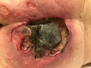
Wound assessment: Should include a comprehensive patient history, etiology of the wound, condition of the wound bed and periwound area including the amount, colour, and consistency of exudate as well as signs of infection. Images R 1. diabetic ulcer 2. Pressure Ulcer
- Identify the location of the wound
- Determine the cause of the wound
- Determine the stage of the wound
- Stage I: Superficial involving only the epidermal layer
- Stage II: Partial-thickness affects the epidermis, and may extend into the dermis
- Stage III: Full-thickness extends through the dermis and into the adipose tissues
- Stage IV: Full-thickness extends through the dermis, and adipose exposing muscle or bone
- Evaluate and measure the depth, length, and width of the wound
- Measure the amount of undermining and tunneling
- Evaluate the wound bed
- Assess for presents, type, and amount of exudate:
- Serous, serosanguineous, sanguineous, or purulent
- Minimal, light, moderate, or heavy
- Access surrounding skin tissue
- Assess wound margins for tunneling, rolled, undermining, fibrotic changes, and if unattached
- Evaluate for signs and symptoms of infection (warm, pain, odor, delayed healing)
- Assess pain[1]

Patient Assessment: A holistic assessment of the patient is essential to identify the causative or contributory factors and to highlight issues that could delay wound healing. The following factors must be considered when choosing an appropriate plan of care.
- Age
- Medication and allergies
- Other diseases such as
- Diabetes
- Vascular diseases
- Immune systems that are compromised
- Nutritional status and fluid intake
- Smoking/alcohol
- Obesity
- Mobility
- Continence
- Pressure sore risk
- Oxygen deficit/anaemia
- Skin sensitivity and integrity[3]
These factors above will either delay or promote the wound healing process. Successful wound healing is dependent on the patient’s ability to heal, not the dressing used.[3]
Most chronic wounds are complex and best managed by an interprofessional team that includes a wound care nurse, general and vascular surgeon, hyperbaric specialist, infectious disease consultant, dietitian, and physical therapist. The key is to first find out the cause of wound breakdown. Without resolving the primary cause, wounds cannot heal.[1]
Conclusion[edit | edit source]
Accurate wound assessment is vital to give a successful resolution and uncomplicated wound healing. It provides valuable information to enable informed decisions about a patient and their wound.[3]
References[edit | edit source]
- ↑ 1.0 1.1 1.2 1.3 1.4 Nagle SM, Waheed A, Wilbraham SC. Wound Assessment InStatPearls [Internet] 2020 Apr 28. StatPearls Publishing.Available from:https://www.ncbi.nlm.nih.gov/books/NBK482198/ (last accessed 9.10.2020)
- ↑ 2.0 2.1 BC campus CLINICAL PROCEDURES FOR SAFER PATIENT CARE Available from:https://opentextbc.ca/clinicalskills/chapter/introduction-3/ (last accessed 9.10.2020)
- ↑ 3.0 3.1 3.2 Activheal academy Wound Assessment Available from:http://academy.activheal.com/lessons/sf-wound-assessment-simplified/ (last accessed 9.10.2020)

