Vestibular Oculomotor Motor Screening (VOMS) Assessment
Original Editor - Megyn Robertson
Top Contributors - Simisola Ajeyalemi, Kim Jackson, Rachael Lowe, Shaimaa Eldib, Mandy Roscher, Stacy Schiurring, Megyn Robertson, Samuel Adedigba, Jess Bell, Olajumoke Ogunleye and Nupur Smit Shah
Introduction[edit | edit source]
Impairments in the vestibulo-ocular system commonly manifest as symptoms of dizziness and visual instability.Nearly 30% of concussed athletes report visual problems during the first week after the injury.[1] Ocular motor impairments and symptoms may manifest as blurred vision, diplopia, impaired eye movements, difficulty in reading, dizziness, headaches, ocular pain, and poor visual-based concentration.[2] Dizziness, which may represent an underlying impairment of the vestibular and/or ocular motor systems, is reported by 50% of concussed athletes[1] and is associated with a 6.4-times greater risk, relative to any other on-field symptom, in predicting protracted (>21 days) recovery.[3]
Concussions can directly impair oculomotor function. Ocular issues (poor eye tracking) after concussion are common. Specifically cranial nerves (CN) III, IV and VI which innervate the eye muscles are susceptible to injury anywhere along the route from brainstem to eye muscles, so this needs to be assessed.
Vestibular/Ocular-Motor Screening (VOMS) for Concussion[edit | edit source]
This test is designed for use with subjects ages 9-40. When used with patients outside this age range, interpretation may vary.
Equipment: Tape measure (cm), Target with a 14 point font print.
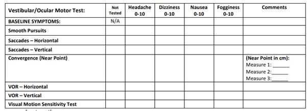
Table 1: A Brief Vestibular/Ocular Motor,Screening (VOMS) Assessment to Evaluate Concussions[4]
Baseline Symptoms – Record: Headache, Dizziness, Nausea & Fogginess on 0-10 scale prior to beginning screening
Smooth Pursuits[edit | edit source]
This tests the ability to follow a slowly moving target. The patient and the examiner are seated. The examiner holds a fingertip at a distance of 3 ft. from the patient. The patient is instructed to maintain focus on the target as the examiner moves the target smoothly in the horizontal direction 1.5 ft. to the right and 1.5 ft. to the left of midline. One repetition is complete when the target moves back and forth to the starting position, and 2 repetitions are performed. The target should be moved at a rate requiring approximately 2 seconds to go fully from left to right and 2 seconds to go fully from right to left. The test is repeated with the examiner moving the target smoothly and slowly in the vertical direction 1.5 ft. above and 1.5 ft. below midlinefor 2 complete repetitions up and down. Again, the target should be moved at a rate requiring approximately 2 seconds to move the eyes fully upward and 2 seconds to move fully downward.
Record: Headache, Dizziness, Nausea & Fogginess ratings after the test.
Saccades[edit | edit source]
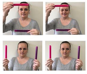
This tests the ability of the eyes to move quickly between targets. The patient and the examiner are seated.
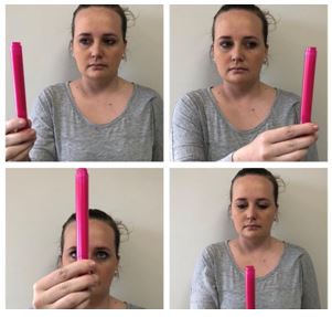
- Vertical Saccades: Repeat the test with 2 points held vertically at a distance of 3 ft. from the patient, and 1.5 feet above and 1.5 feet below midline so that the patient must gaze 30 degrees upward and 30 degrees downward. Instruct the patient to move their eyes as quickly as possible from point to point. One repetition is complete when the eyes move up and down to the starting position, and 10 repetitions are performed.
Record: Headache, Dizziness, Nausea & Fogginess ratings after the test.
- Horizontal Saccades: The examiner holds two single points (fingertips) horizontally at a distance of 3 ft. from the patient, and 1.5 ft. to the right and 1.5 ft. to the left of midline so that the patient must gaze 30 degrees to left and 30 degrees to the right. Instruct the patient to move their eyes as quickly as possible from point to point. One repetition is complete when the eyes move back and forth to the starting position, and 10 repetitions are performed.
Record: Headache, Dizziness, Nausea & Fogginess ratings after the test.
Convergence[edit | edit source]
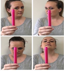
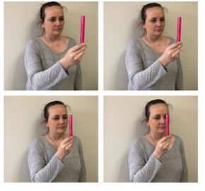
Convergence is assessed by both symptom report and objective measurement of the near point of convergence.The NPC values were averaged across 3 trials, and normal NPC values are within 5 cm.[5]
Measure the ability to view a near target without double vision. The patient is seated and wearing corrective lenses (if needed). The examiner is seated front of the patient and observes their eye movement during this test. The patient focuses on a small target (approximately 14 point font size) at arm’s length and slowly brings it toward the tip of their nose. The patient is instructed to stop moving the target when they see two distinct images or when the examiner observes an outward deviation of one eye. Blurring of the image is ignored. The distance in centimetres between target and the tip of nose is measured and recorded. This is repeated a total of 3 times with measures recorded each time.
Record: Headache, Dizziness, Nausea & Fogginess ratings after the test. Abnormal: Near Point of convergence ≥ 6 cm from the tip of the nose.
Vestibular-Ocular Reflex (VOR) Test[edit | edit source]
Assess the ability to stabilize vision as the headmoves. The patient and the examiner are seated. The examiner holds a target of approximately 14 point font size in front of the patient in midline at a distance of 3 ft.
• Horizontal VOR Test: The patient is asked to rotate their head horizontally while maintaining focus on the target. The head is moved at an amplitude of 20 degrees to each side. One repetition is complete when the head moves back and forth to the starting position, and 10 repetitions are performed.
Record: Headache, Dizziness, Nausea and Fogginess ratings 10 sec after the test is completed.
• Vertical VOR Test: The test is repeated with the patient moving their head vertically. The head is moved in an amplitude of 20 degrees up and 20 degrees down. One repetition is complete when the head moves up and down to the starting position, and 10 repetitions are performed.
Record: Headache, Dizziness, Nausea and Fogginess ratings after the test.
Visual Motion Sensitivity (VMS) Test[edit | edit source]
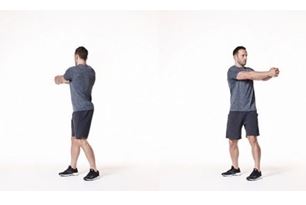
Test visual motion sensitivity and the ability to inhibit vestibular-induced eye movements using vision. The patient stands with feet shoulder width apart, facing a busy area of the clinic. The examiner stands next to and slightly behind the patient, so that the patient is guarded but the movement can be performed freely. The patient holds arm outstretched and focuses on their thumb. Maintaining focus on their thumb, the patient rotates, together as a unit, their head, eyesand trunk at an amplitude of 80 degrees to the right and 80 degrees to the left. One repetition is complete when the trunk rotates back and forth to the starting position, and 5 repetitions are performed.
Record: Headache, Dizziness, Nausea &Fogginess ratings after the test.
Resources[edit | edit source]
Vestibular/Ocular-Motor Screening (VOMS) for Concussion
References[edit | edit source]
- ↑ 1.0 1.1 Kontos AP, Elbin RJ, Schatz P, et al. A revised factor structure for the Post-Concussion Symptom Scale: baseline and postconcussion factors. Am J Sports Med. 2012;40(10):2375-2384.
- ↑ Ciuffreda KJ, Ludlam D, Thiagarajan P. Oculomotor diagnostic protocol for the mTBI population. Optometry. 2011;82(2):61-63.
- ↑ Lau BC, Kontos AP, Collins MW, Mucha A, Lovell MR. Which on-field signs/symptoms predict protracted recovery from sport-related concussion among high school football players? Am J Sports Med. 2011;39(11):2311-2318.
- ↑ Mucha A, Collins MW, Elbin RJ, Furman JS, Troutman-Enseki C, DeWolf RM, Marchetti G, Kontos AP. A Brief Vestibular/Ocular Motor,Screening (VOMS) Assessment to Evaluate Concussions. Preliminary findings. The American Journal of Sports Medicine 2014;42(10): 2479-2486
- ↑ Scheiman M, Gallaway M, Frantz KA, et al. Nearpoint of convergence: test procedure, target selection, and normative data. Optom Vis Sci80(3) 2003:214-225
