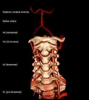
Vertebral Artery
Original Editor -Rachael Lowe
Top Contributors - Rachael Lowe, Vidya Acharya, Priyanka Chugh, Kim Jackson, Evan Thomas and Lucinda hampton
Description[edit | edit source]
The vertebral artery is a major artery in the neck[1]. It branches from the subclavian artery, where it arises from the posterosuperior portion of the subclavian artery.
Path[edit | edit source]
It ascends though the foramina of the transverse processes of the cervical vertebrae, usually starting at C6 but entering as high as C4[2]. It winds behind the superior articular process of the atlas. It enters the cranium through the foramen magnum where it unites with the opposite vertebral artery to form the basilar artery (at the lower border of the pons)[3].
Divisions[edit | edit source]
The vertebral artery can be divided into four divisions:
- The first division runs posterocranial between the longus colli and the scalenus anterior.[3] The first division is also called the ‘pre-foraminal division’.[1]
- The second division runs cranial through the foramina in the cervical transverse processes of the cervical vertebrae C2.[3] The second division is also called the foraminal division[1].
- The third division is defined as the part that rises from C2. It rises from the latter foramen on the medial side of the rectus capitis lateralis, and curves behind the superior articular process of the atlas. Then, it lies in the
- groove on the upper surface of the posterior arch of the atlas, and enters the vertebral canal by passing beneath the posterior atlantoöccipital membrane[3].
- The fourth part pierces the dura mater and inclines medial to the front of the medulla oblongata.[3]
Supply[edit | edit source]
It supplies 20% of blood to the brain (mainly hindbrain) along with the internal carotid artery (80%).
Clinical Relevance[edit | edit source]
It lies close to the vertebral bodies and facet joints where it may be compressed by osteophyte formation or injury to the facet joint.
In older individuals, artherosclerotic changes and other vascular risk facotrs (e.g. hypertension, high cholesterol, smoking, diabletes) may contribute to altered blood flow in the arteries.
The vertebral and carotid artieries are stressed primarily by rotation, extension and traction, but other movements may also stretch the artery. As little as 20% of rotation and extension have been shown to significantly decrease vertebral artery blood flow[2].
The greatest stresses are placed on the verterbal arteries in 4 places:
- on entry to the C6 transverse process
- within the bony canals of the vertebral transverse processes
- between C1 and C2
- between C1 and entry of the arteries into the skull.
The most common mechanism for non-penetrating injury to the vertebral artery is extension with or without side flexion or rotation[2].
Symptoms, which may be delayed include:
- vertigo
- nausea
- tinitus
- drop attacks
- visual disturbances
- and in rare cases strok or death


