Trochanteric Bursitis
Original Editors - Emy Van Rode
Top Contributors - Mudra Shah, Emy Van Rode, Gertjan Van Gijsegem, Lionel Geernaert, Lena Vanderaa, Wendy Snyders, Admin, Kim Jackson, Andrea Nees, Rachael Lowe, Uchechukwu Chukwuemeka, Vidya Acharya, Wanda van Niekerk, Claire Knott and WikiSysop
Definition/Description[edit | edit source]
Trochanteric bursitis is an inflammation of the trochanteric bursa. The fact that it’s a Bursitis, implicates it has an inflammatory component but we have to take into account that 3 of the 4 elements of an inflammation named rubor, calor and tumor aren’t present. The only cardinal sign of inflammation that is present is pain.
Trochanteric bursitis is an element of a greater term, hip bursitis, that envelopes 4 different types
- Trochanteric bursitis
- Iliopsoas Bursitis
- Gluteal Bursitis
- Ischial Bursitis
It’s often used as a general term to describe pain around the greater trochanteric region of the hip. Trochanteric bursitis is frequently confused with Greater Trochanter Pain Syndrome (GTPS) but is in fact a component of GTPS that also includes other conditions that cause lateral-sided hip pain.
Clinically Relevant Anatomy[edit | edit source]
A bursa is a double - membrane sac filled with fluid located near a joint. It forms a sort of cushion between to minimize friction between the soft tissue/bone interface and acts as a shock absorber during the movement of muscles and joints.
For the mechanism of injury or the pathological process of bursitis: refer to the page Bursitis
In case of Trochanteric Bursitis, two bursae are commonly involved:
- Subgluteus Medius bursa - located above the greater trochanter and underneath the insertion of the gluteus medius.
- Subgluteus Maximus bursa - located between the greater trochanter and the insertion of the gluteus medius and gluteus maximus muscles.
Epidemiology /Etiology[edit | edit source]
Inflammation of the bursa is a slow process, which progresses over time. This bursitis most often occurs because of friction, overuse, direct trauma or too much pressure.
There are two types of bursitis
- Acute bursitis occurs because of trauma or a massive overload. After a few days’ symptoms like pain, swelling and a warm feeling when touching the affected area can be noticed. It will also be very painful to move the joint.
- Chronic bursitis which is caused by overuse, too much pressure on the structures or by extreme movements. Wrong muscle strain can also be a cause of chronic bursitis. The main symptom – which is always present – is pain.
There are many predisposing factors that can cause Trochanteric Bursitis:
- Sex: Women more commonly affected than men.
- Overweight/Obesity
- Trauma: e.g. injury of the greater trochanter: this can deface the bursa.
- Overuse of the muscles around the bursa or the joint underneath the bursa.
- Incorrect position: this can cause an increase in pressure.
- Too much pressure on the bursa (caused by friction of the Iliotibial band)
- Dysfunction of the insertion of the muscle gluteus medius.
- Hip osteoarthritis
- Lumbar spondylosis
- Excessive or rapidly increased mileage
- Repetitive strain: e.g. frequent training with too much weight or training in a bad position
- Poorly cushioned shoes: results in increased pressure on the muscles, joint and bursa
- Excessive pronation/ extreme movement
- Leg length differences
- ITBS (Iliotibial Band Syndrome)
- Bacterial infection
- Other inflammatory diseases
- Hip prosthesis
Characteristics/Clinical Presentation[edit | edit source]
Following characteristics may occur
- Chronic pain and/or hip tenderness in the lateral aspect of the hip that may radiate down the thigh[1]
- A snap felt in the lateral aspect of the hip[1]
- Ascending stairs is a painful activity
- Patient is unable to lie down on the affected side
- Development of pain-related sleep disturbance
- Lower back pain (Trochanteric Bursitis can present as lumbago)[2]
Diagnostic Procedures[edit | edit source]
Diagnosing lateral hip pain is very complex since clinical presentations are variable and sometimes inconclusive. To be sure to diagnose the right affection the examination has to follow a stepwise approach, including thorough history, inspection, palpation, range of motion, stability and strength in all planes.
An important diagnostic test for lateral hip pain, particularly for trochanteric bursitis is without a doubt palpation. You have to palpate in and around the greater trochanter. This is the most provocative clinical test by physical therapists.
As additional test you can also perform the Ober's_Test. It was originally conceived for abductor muscle contracture, but it was found that the pain reproduction or the reduced range of motion was significant to diagnose trochanteric bursitis.
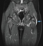
If there is still any doubt about the diagnosis it’s favorable to make an MRI, which will give more specific information.
Physical Examination[edit | edit source]
Physical examination is performed based upon the history of previous injuries and it is used to confirm the source of the pain and establish any limitations or deficits that the patient might have. It also assesses the underlying disorder or anatomical impairment that may cause a bursitis.The physical examination must have a stepwise approach which Observation, Palpation, Range of motion, Muscle Strength, Gait Assessment and the execution of special tests.[3]
The first part is the observation. The most important aspect of observation is the patient’s posture in a seated and upright position.The patient with an irritated hip will tend to stand with the joint slightly flexed. In a seated position: slouching and leaning to the uninvolved slide allows the hip to seek a slightly less flexed position. The observation is also focused on the asymmetry, the gross atrophy, the spinal alignment or the pelvic skewness.[4]
Bursae pain may be detected by palpation. We perform palpation to assess sources of the hip pain. The palpation starts with joint tenderness on the proximal and distal area of the hip. Also each part of the body that is associated with this injury must be assessed, e.g.: the bone, muscle, ligaments, etc. It is important to check the lumbar spine, sacroiliac joints, ischium, iliac crest, lateral aspect of the greater trochanteric bursa, muscle bellies and the pubic symphysis. They can determine a potential source of hip symptoms or pain.[3]
The range of motion should be checked on the actual injured hip as well as on the contralateral hip. An active hip flexion, an internal and external rotation, an abduction and adduction will reproduce pain in the injured area. The range of motion can be identified with several tests: the faber test, Trendelenburg test, Ober’s test, Thomas test and a test whereby the forced flexion combined with internal rotation could be helpful in diagnosing the cause of lateral hip pain.
Muscle strength needs to be tested for all the major muscle groups acting on the hip joint which can be assessed with resisted contraction. Hip abductor weakness is a common finding and testing the abductors can provoke lateral hip pain during the examination.
While assessing the gait, one should look for any limb length discrepancy, weakness and heel strike which contributes to the function of the gluteus maximus.[5]
Differential Diagnosis[edit | edit source]
There are many conditions which can present as lateral hip pain in a patient. This is why it is crucial to rule out other possible causes to accurately arrive at a diagnosis of Trochanteric Bursitis.
Common conditions that can cause lateral hip pain are:
- Iliotibial Band Syndrome
- Snapping Hip Syndrome
- Gluteus Medius Tendon Dysfunction and Tears
- Meralgia Paresthetica
- Referred Pain
Outcome Measures[edit | edit source]
• VAS-scale for pain
• International Hip Outcome Tool (iHot) [6]
• Oswestry Disability Index [7]
• Harris Hip score [8]
• 6 Minute Walk Test
• Hip Disability and Osteoarthritis Outcome Score
• Copenhagen Hip and Groin Outcome Score [6]
Medical Management[edit | edit source]
There are various approaches in the treatment of Trochanteric Bursitis, depending on whether or not the bursitis has an infection, and whether it is necessary to treat the lesion with or without surgery.
Aseptic trochanteric bursitis [9][10]
- In most cases trochanteric bursitis is treated without surgery. If the pain results from overuse, it is recommended to reduce the activities or modify the body mechanics in which those specific activities are performed.
- Furthermore, an exercise program of stretching and strengthening with a physiotherapist will help to bring back full range of motion in the hip, sometimes in combination with anti-inflammatory medications or heat and ice applications to calm inflammation.
- If the above treatment fails to reduce the symptoms, an injection of cortisone into the swollen bursa may be required. This anti-inflammatory injection will reduce the symptoms for months, but it will not cure the problem itself.
Septic trochanteric bursitis [9][10]
- Infectious trochanteric bursitis does occur, but only in exceptional cases.
- Further examination of the bursa fluid in the laboratory is necessary to assess which bacteria has caused the infection. Once this is known, an (intravenous) antibiotic therapy can be prescribed.
Surgical treatment [11]
Only when the nonsurgical therapy fails, and when the pain is still unbearable, it is recommended to consider surgery. The aim of surgery is to remove the thickened bursa and bone spurs that have arisen on the greater trochanter. Also the large tendon of the gluteus maximus is treated. Some doctors prefer to remove a part of the tendon that rubs against the greater trochanter while others prefer to elongate the tendon surgically.
Physical Therapy Management[edit | edit source]
There are several treatments that can be used to reduce pain and swelling on a patient with trochanteric bursitis. When pain is the main complaint, we can relieve the pain for other underlying disorders so as to treat them more effectively. Physical therapy is given to improve flexibility, muscle strengthening and joint mechanics. When these aspects are improved, pain will decrease. To heal trochanteric bursitis it is necessary to proceed to infiltration of the bursa with antiphlogistic medication (Corticosteroid-injections). In case of a persistent bursitis, surgery has to be considered as well. Other physical therapy interventions are the use of ultrasound, moist heat and educating the patient on activity modification and correcting possible training errors.
The pain of this injury can be reduced in different phases: The first phase is to manage the pain and the inflammation. Pain being the main reason for treatment of the trochanteric bursitis, we can use two common treatments to decrease the pain: the use of ice and non-steroidal anti-inflammatory drugs (NSAIDs). The bursa inflammation can be treated with ice therapy and techniques or exercises that reduce the inflammation structures. There are also other treatments that a physiotherapist can use, e.g.: electrotherapy, acupuncture, taping techniques, soft tissue massage and the temporary use of a mobility aid to off-load the affected side.
The second phase is to reinforce the patient’s strength and to restore the normal ROM. The physiotherapist will also to improve the muscle length and resting tension, the proprioception, balance and gait through a supervised and thorough exercise rehabilitation program.
The next phase of rehabilitation is the restoration of all functions. Many patients develop Trochanteric Bursitis due to their common daily activities like running, walking etc. The goal of the physiotherapist is to provide a specialized program for the patient to improve the movement and to reduce the pain, so that the patient can perform his daily activities with less difficulty.
The final phase is to prevent a relapse. It may be as simple as training your core muscles or fabricating foot orthotics to address any biomechanical faults in the lower limbs. The therapist will examine your hip stability and function by addressing any deficits in the core strength and balance. Furthermore, he will also teach the patient some self-management techniques. The ultimate goal is to see the patient safely returning to his former sporting or leisure activities.
References[edit | edit source]
- ↑ 1.0 1.1 Snider RK. Essentials of musculoskeletal care. Rosemont (IL): American Academy of Orthopaedic Surgeons. 1997.
- ↑ Margo K, Drezner J, Motzkin D. Evaluation and management of hip pain: an algorithmic approach.(Applied evidence: new research findings that are changing clinical practice). Journal of family practice. 2003 Aug 1;52(8):607-18.
- ↑ 3.0 3.1 Grumet RC, Frank RM, Slabaugh MA, Virkus WW, Bush-Joseph CA, Nho SJ. Lateral hip pain in an athletic population: differential diagnosis and treatment options. Sports Health. 2010 May;2(3):191-6.
- ↑ Byrd JT. Evaluation of the hip: history and physical examination. North American journal of sports physical therapy: NAJSPT. 2007 Nov;2(4):231.
- ↑ Woodley SJ, Nicholson HD, Livingstone V, Doyle TC, Meikle GR, Macintosh JE, Mercer SR. Lateral hip pain: findings from magnetic resonance imaging and clinical examination. journal of orthopaedic & sports physical therapy. 2008 Jun;38(6):313-28.
- ↑ 6.0 6.1 Enseki K, Harris-Hayes M, White DM, Cibulka MT, Woehrle J, Fagerson TL, Clohisy JC. Nonarthritic Hip Joint Pain: Clinical Practice Guidelines Linked to the International Classifiation of Functioning, Disability and Health From the Orthopaedic Section of the American Physical Therapy Association. Journal of Orthopaedic & Sports Physical Therapy. 2014 Jun;44(6):A1-32.
- ↑ Lustenberger DP, Ng VY, Best TM, Ellis TJ. Efficacy of treatment of trochanteric bursitis: a systematic review. Clinical journal of sport medicine: official journal of the Canadian Academy of Sport Medicine. 2011 Sep;21(5):447.
- ↑ Furia JP, Rompe JD, Maffulli N. Low-energy extracorporeal shock wave therapy as a treatment for greater trochanteric pain syndrome. The American journal of sports medicine. 2009 Sep;37(9):1806-13.
- ↑ 9.0 9.1 Firestein, G.S., et al. Kelley's Textbook of Rheumatology, 9th ed. Philadelphia, Pa: Saunders Elsevier, 2012
- ↑ 10.0 10.1 Klippel, John H., et al., eds. Primer on the Rheumatic Diseases. New York: Springer and Arthritis Foundation, 2008
- ↑ Farmer KW, Jones LC, Brownson KE, Khanuja HS, Hungerford MW. Trochanteric bursitis after total hip arthroplasty: incidence and evaluation of response to treatment. The Journal of arthroplasty. 2010 Feb 1;25(2):208-12.


