Thoracic Anatomy
Introduction[edit | edit source]

This guide gives a general overview of the anatomy of the thoracic spine. It also includes some facts regarding pathophysiology in this region.
The sagittal plane alignment of the Thoracic spine is on average 35% (normal range is 20° to 50°).[1]
Important Structures
The important parts of the thoracic spine include:
Bones (vertebrae and ribs)[edit | edit source]
The human spine is made up of 24 spinal bones, called vertebrae. Vertebrae are stacked on top of one another to create the spinal column. The spinal column is the body’s main upright support.

- The middle 12 Thoracic vertebrae make up the thoracic spine.
- T5-T8 tend to be the most “typical” in that they contain features present in all thoracic vertebrae. T5-T8 have the greatest rotation ability of the thoracic region.
- T1 - superior costal facets are “whole” costal facets. They alone articulate with the first rib; C7 has no costal facets. T1 does, however, have typical inferior demifacets for articulation with the second rib. Vertebral prominens name given to long prominent spinous process found at T1
- T11 and T12 are atypical - contain a single pair, “whole,” costal facet that articulate with the 11 and 12 ribs, respectively. They also lack facets on the transverse processes.[3]
- The primary characteristic of the thoracic vertebrae is the presence of costal facets. There are 6 facets per thoracic vertebrae: 2 on the transverse processes and 4 demifacets. The facets of the transverse processes articulate with the tubercle of the associated rib. The demifacets are bilaterally paired and located on the superior and inferior posterolateral aspects of the vertebrae. They are positioned so that the superior demifacet of inferior vertebrae articulates with the head of the same rib that articulates with the inferior demifacet of a superior rib. For example, the inferior demifacets of T4 and the superior demifacets of T5 articulate with the head of rib 5.
- The length of the transverse processes decreases as the column descends.
- The positioning of the ribs and spinous processes greatly limits flexion and extension of the thoracic vertebrae.
- Thoracic vertebrae have superior articular facets that face in a posterolateral direction.
- The spinous process is long, relative to other regions, and is directed posteroinferiorly. This projection gradually increases as the column descends before decreasing rapidly from T9-T12.[3]
- The intervertebral disc height, is, on average, the least of the vertebral regions.
- Vertebral body size: increases progressively from T1 to T12
- Spinal canal dimensions: varies from T1 to T12[1]
Ribs and sternum ( see link)
The ribs are the bony framework of the thoracic cavity.
- The ribs form the main structure of the thoracic cage protecting the thoracic organs, however their main function is to aid respiration.
- There are twelve pairs of ribs.
- Each rib articulates posteriorly with two thoracic vertebrae by the costovertebral joint. An exception to this rule is that the first rib articulates with the first thoracic vertebra only.
According to their attachment to the sternum, the ribs are classified into 3 groups: true, false, and floating ribs[4].
Joints[edit | edit source]

There are 3 types of joints present in the thoracic spine:
Between vertebral bodies – adjacent vertebral bodies are joined by intervertebral discs, made of fibrocartilage. This is a type of cartilaginous joint, known as a symphysis.
Between vertebral arches ie facet joints – formed by the articulation of superior and inferior articular processes from adjacent vertebrae. It is a synovial type joint. In the thoracic spine facet joints angled at:
- 60° to the transverse plane
- 20° to the frontal plane, with the superior facets facing posterior and a little up and laterally and the inferior facets facing anteriorly, down, and medially.
- motion: lateral flexion and rotation; no flexion/extension
Costovertebral joints, unique to the thoracic spine - consists of the head of the rib articulating with:
- Superior costal facet of the corresponding vertebra
- Inferior costal facet of the superior vertebra
- Intervertebral disc separating the two vertebrae
Within this joint, the intra-articular ligament of head of rib attaches the rib head to the intervertebral disc. Only slight gliding movements can occur at these joints, due to the close articulation of their components.[5]
Structure and Function[edit | edit source]
The anatomy of the Thoracic spine is related directly to its function. The facets and demifacets devoted to rib articulation demonstrate the main function of the thoracic spine.
- The zygapophyseal joints are tight enough to protect vital organs but loose enough to allow for respiratory movements as well as to allow the thoracic segment to have the greatest freedom of rotation of the entire spine.
- The zygapophyseal joints, as well as the relatively thin intervertebral discs, cause the thoracic region to have the least flexion/extension ability of the spine. Prevents downward flexion on heart and lungs
- The increase in vertebral body size as the spinal column descends is directly related to the increased weight-bearing requirement; further down the column, the greater the proportion of body mass that rests upon it.

Nerves[edit | edit source]
- The hollow tube formed by the bony rings on the back of the spinal column surrounds the spinal cord. The bones of the spinal column protect the spinal cord.
- In the thoracic spine, the spinal canal is narrower than in the rest of the spine, giving very little extra space for the spinal cord as it passes through the thoracic spine.
- Between the vertebrae, two large spinal nerves branch off the spinal cord, one on the left and one on the right (12 thoracic nerve pairs). The first nerve root exits between the T1 and T2 vertebrae The nerves pass through the neural foramina of each vertebra. These spinal nerves group together to form the main nerves that go to the organs and limbs. The nerves of the thoracic spine mainly control the muscles and organs of the chest and abdomen.[2]
The 12 pairs of thoracic nerves are derived from dorsal and ventral roots of their corresponding segments. These nerves
- Do not form plexuses; they distribute cutaneous branches to the thoracic dermatomes and send other sensory fibers to deeper muscular structures, vessels, periosteum, parietal pleura, the peritoneum, and breast tissue.
- Also send motor fibers to muscles of the thoracic and abdominal wall and carry preganglionic and postganglionic sympathetic nerve fibers into and out of the sympathetic chain.
- Muscles of the thoracic and abdominal wall, supplied by these nerves, act as accessory respiratory muscles and may assist in breathing in times of dyspnea or phrenic nerve impairment[6]
- T1 is part or the Brachial plexus
Ligaments[edit | edit source]

Several long ligaments connect on the front and back sections of the vertebrae.
- The anterior longitudinal ligament runs lengthwise down the front of the vertebral bodies.
- The supraspinous ligament, a strong fibrous cord that connects together the apices of the spinous processes from the seventh cervical vertebra to 3rd or 4th lumbar vertebrae.
- Two other ligaments run full length within the spinal canal.
- The posterior longitudinal ligament attaches on the back of the vertebral bodies.
- The ligamentum flavum is a long elastic band that connects to the front surface of the lamina bones.
Thick ligaments also connect the ribs to the transverse processes of the thoracic spine.
Intervertebral Disc[edit | edit source]

The intervertebral disc (IVD) is important in the normal functioning of the spine.
- It is a cushion of fibrocartilage and the principal joint between two vertebrae in the spinal column.
- There are 12 in the thoracic region.
- IVDs allow the spine to be flexible without sacrificing a great deal of strength and provide a shock-absorbing effect within the spine and prevent the vertebrae from grinding together.
- They consist of three major components: the inner, nucleus pulposus (NP), the outer, annulus (AF) and the cartilaginous endplates that anchor the discs to adjacent vertebrae.
- Disc thickness generally increases from rostral to caudal.
- The thickness of the discs relative to the size of the vertebral bodies is lowest in the thoracic regions. This reflects the decreased range of motion found in this region.
Muscles[edit | edit source]

The muscles of the thoracic spine are arranged in layers.
- Those closest to the skin’s surface run from the back of the vertebrae to the Scapula eg Trapezius,Rhomboids, Latissimus Dorsi, Others wrap around the rib cage and connect to the shoulders[7].eg Serratus anterior, Serratus posterior, Pectoralis minor,
- Strap-shaped muscles called Erector Spinae make up the middle layer of muscles ( ie the intrinsic back muscles – the iliocostalis, longissimus and spinalis). Together these muscles form a column, known as the erector spinae These muscles run up and down over the lower ribs and thorax (the rib cage), and cross to the low back.
- The deepest layer of muscles attaches along the back of the spine bones, connecting the vertebrae. The deep intrinsic muscles are located underneath the erector spinae, and are known collectively as the transversospinales. They are a group of short muscles, associated with the transverse and spinous processes of the vertebral column. There are three major muscles in this group – the semispinalis, multifidus and rotatores.
NB The Thoracolumbar Fascia (important to thoracic musculature) is a large area of connective tissue - roughly diamond-shaped - which comprises the thoracic and lumbar parts of the deep fascia enclosing the intrinsic back muscles.
4. Muscles also connect from one rib to the next. eg Muscles of Respiration
Pathophysiology[edit | edit source]
- Thoracic Radiculopathy - Thoracic nerve root lesions account for less than 2% of all radiculopathies and are most commonly caused by diabetes and thoracic disk herniation[8]
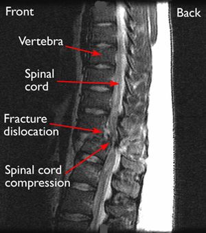
- Osteoporosis increases the risk for thoracic spinal fracture. A common type is a vertebral compression fracture (VCF) that can cause one or more bony bodies to flatten or become wedge shaped resulting in spinal cord and/or nerve compression. Sudden and acute back pain is associated with a VCF.[9]
- Different types of scoliosis—an abnormal side to side curvature of the spine—is well known to develop in the thoracic spine and may cause spinal deformity. eg Scheuermann’s disease, Posture related, Age related hyperkyphosis, Thoracic Hyperkyphosis, Forward Head posture,
- Ankylosing Spondylitis (Axial Spondyloarthritis)
- Spinal tumors can occur anywhere along the spine, but most occur in the thoracic (mid-to-upper back) region - About 70% of metastatic spinal tumors occur in the thoracic region, 20% occur in the lumbar (lower back) region, fewer occur in the cervical region.[10]
- More than half of spinal cord injuries occur in the cervical area, a third occur in the thoracic area, and the remainder occur mostly in the lumbar region.[11]
- T4 syndrome or upper thoracic syndrome was described as “a pattern that involves upper extremity paraesthesia”. It can be caused by thoracic hypomobility but can also have a sympathetic origin.
- The Slipping Rib Syndrome is an infrequent cause of thoracic and upper abdominal intermittent pain and is thought to arise from hypermobility of the rib cartilage of the false and floating ribs (these are the most involved in this syndrome)[12]
- Rib Stress Fracture is a frequently occurring pathology in the rower’s community, with an incidence of 6 to 12%.[13]

Final Remark[edit | edit source]

Now you know all about the anatomy, go on to look at this page Thoracic Examination
References[edit | edit source]
- ↑ 1.0 1.1 Othrobullets Thoracic spine Anatomy Available from:https://www.orthobullets.com/spine/2070/thoracic-spine-anatomy (last accessed 12.4.2020)
- ↑ 2.0 2.1 eOrthopod Thoracic spine anatomy Available from:https://eorthopod.com/thoracic-spine-anatomy/ (last accessed 12.4.2020)
- ↑ 3.0 3.1 Waxenbaum JA, Futterman B. Anatomy, Back, Thoracic Vertebrae.Available from:https://www.statpearls.com/kb/viewarticle/32138 (last accessed 12.4.2020)
- ↑ Safarini OA, Bordoni B. Anatomy, Thorax, Ribs. InStatPearls [Internet] 2019 Feb 19. StatPearls Publishing.Available from:https://www.ncbi.nlm.nih.gov/books/NBK538328/ (last accessed 14.4.2020)
- ↑ Teach me anatomy The Thoracic Spine Available from:https://teachmeanatomy.info/thorax/bones/thoracic-spine/ (last accessed 12.4.2020)
- ↑ David L. Felten MD, PhD, ... Mary Summo Maida PhD, in Netter's Atlas of Neuroscience (Third Edition), 2016 Available from:https://www.sciencedirect.com/topics/neuroscience/thoracic-nerves (last accessed 13.4.2020)
- ↑ Borstad JD, Ludewig PM. The effect of long versus short pectoralis minor resting length on scapular kinematics in healthy individuals. Journal of orthopaedic & sports physical therapy. 2005 Apr;35(4):227-38.Available from:https://www.ncbi.nlm.nih.gov/books/NBK538319/ (last accessed 13.4.2020)
- ↑ John-Michael Li, Jinny Tavee, in Handbook of Clinical Neurology, 2019 Available from:https://www.sciencedirect.com/topics/neuroscience/thoracic-nerves (last accessed 13.4.2020)
- ↑ Spine universe. Thoracic spine. Available from:https://www.spineuniverse.com/anatomy/thoracic-spine (last accessed 13.4.2020)
- ↑ Spine health. Metastatic tumours Available from:https://www.spine-health.com/conditions/spinal-tumor/metastatic-spine-tumors (last accessed 13.4.2020)
- ↑ Reeve foundation SCI Available from:https://www.christopherreeve.org/living-with-paralysis/health/causes-of-paralysis/spinal-cord-injury (last accessed 13.4.2020)
- ↑ Slipping Rib Syndrome: An elusive diagnosis; [/www.sciencedirect.com.ezproxy.vub.ac.be%3A2048/science/journal/15260542 Paediatric Respiratory Reviews] [/www.sciencedirect.com.ezproxy.vub.ac.be%3A2048/science/journal/15260542/22/supp/C Volume 22], March 2017, Pages 44–46
- ↑ Warden SJ, Gutschlag FR, Wajswelner H, et al. Aetiology of rib stress fractures in rowers. Sports Med 2002; 32 (13): 819-36 (last accessed 13.4.2020)

