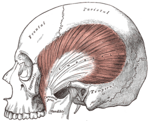
Temporalis
Original Editor - Carina Therese Magtibay
Top Contributors - Carina Therese Magtibay
Description[edit | edit source]
The temporalis muscle is one of the four primary muscles of mastication (chewing of food). It is a fan-shaped muscle with anterior fibres that have a vertical orientation, mid fibres have an oblique orientation, and posterior fibres have a more of horizontal orientation. [1]
Origin[edit | edit source]
Temporal bone, spanning from the temporal fossa to the inferior temporal line of the lateral skull.
Insertion[edit | edit source]
Coronoid process of the mandible
Nerve[edit | edit source]
Trigeminal nerve (CNV), deep temporal branch of mandibular division (CNV3).
Artery[edit | edit source]
Maxillary artery which is a branch of the external carotid artery.
Function[edit | edit source]
- Anterior and middle fibers: Elevates mandible
- Posterior fibers: Retracts the mandible
Assessment[edit | edit source]
Manual Muscle Testing[edit | edit source]
Jaw closure (Mandibular elevation)[2]
- Muscles assessed: Temporalis, Masseter and Medial pterygoid
- Test: Patient clenches jaws tightly
- Manual Resistance: The chin of the patient is grasped between the thumb and index finger of the examiner and held firmly in the thumb web. The other hand is placed on top of the head for stability. Resistance is given vertically downward in an attempt to open the closed jaw
- Instructions to Patient: "Clench (or hold) your teeth together as tightly as you can, keeping your lips relaxed. Hold it. Don't let me open your mouth."
- Criteria for Grading
- Functional: Patient closes mouth (jaw) tightly. Examiner should not be able to open the mouth.
- Weak Functional: Patient closes jaw, but examiner can open the mouth with less than maximal resistance.
- Non Functional: Patient closes mouth but tolerates no resistance. The masseter and temporalis muscles are palpated on both sides. The masseter is palpated under the zygomatic process on the lateral cheek above the angle of the jaw. The temporalis muscle is palpated over the temple at the hairline, anterior to the ear and superior to the zygomatic bone.
- 0: Patient cannot completely close the mouth. This is more of a cosmetic problem (drooling, for example) than a significant clinical one.
Note: In unilateral involvement, the jaw deviates to the strong side during attempts to close the mouth.
Clinical relevance[edit | edit source]
Masticatory muscle disorders to take note of:[1]
- Myofascial pain and dysfunction. Most common etiologies myofascial pain and dysfunction of masticatory muscles:
- Nocturnal bruxism
- Habitual clenching of the mouth
- Whiplash injuries during a trauma
- Temporomandibular joint (TMJ) dysfunction
- It can be caused by an imbalance of forces within the muscles of mastication.
- Nocturnal bruxism is a common cause of TMJ dysfunction.
- Trismus aka Muscle spasm of masticatory muscles
- It can be a symptom of tumor or infection. An infection like tetanus may present with "lockjaw" or trismus.
For more information about the conditions related to the temporalis muscle and its management, head over to:
Resources[edit | edit source]
- ↑ 1.0 1.1 Basit H, Tariq MA, Siccardi MA. Anatomy, head and neck, mastication muscles. InStatPearls [Internet] 2023 June 5. StatPearls Publishing. Available at https://www.ncbi.nlm.nih.gov/books/NBK541027/ (Last accessed 01/25/24)
- ↑ Hislop, H. J., & Montgomery, J. Daniels and Worthingham's Muscle Testing: Techniques of Manual Examination. 8th ed. St. Louis, Mo., Saunders, Elsevier. 2007.


