Sternum
Original Editor - Grace Barla
Top Contributors - Grace Barla, Kim Jackson, Vaibhav Panchal, Lucinda hampton and Vidya Acharya
Description[edit | edit source]
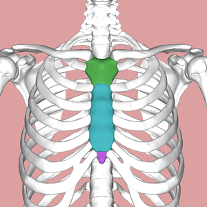
The sternum is a flat cancellous bone with a compact cortex, it is slightly convex anteriorly, with multiple indentations along its lateral borders (costal notches). It forms the anterior median part of the thoracic skeleton. It is commonly known as the breastbone. In shape it resembles like a short sword. The sternum is located along the midline of body in the anterior thoracic region just deep to the skin. It is a flat bone about six inches in length, around an inch wide, and only a fraction of an inch thick. it is longer in males than in females.[1][2]
Structure[edit | edit source]
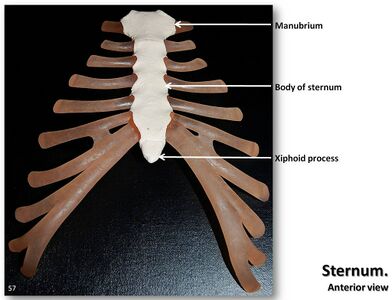
The sternum develops as three distinct parts:
- The manubrium
- The body of the sternum
- The xiphoid process
Manubrium[edit | edit source]
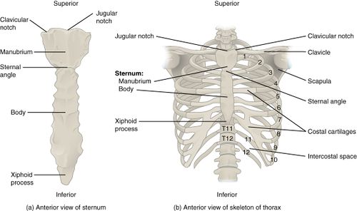
Manubrium is at the cephalad end of the sternum is the large, flat, wide. At the superior border of the bone there is jugular notch or suprasternal notch. The clavicular notches for the articulation of clavicles are projected upward and laterally on both sides of jugular notch.
The manubrium makes angle with the body, convex forwards, called as the sternal angle of Louis. certain events take place at this angle -
- Formation of cardiac plexus
- Upper limit of base of heart
- Arch of aorta starts here as continuation of ascending aorta.
- Arch of aorta ends here to continue as descending thoracic aorta.
- Trachea divides into 2 branches.
The body[edit | edit source]
- It is the longest region of the sternum and is roughly rectangular in shape. It is also known as gladiolus. It has a convex anterior surface, and a concave posterior surface.
- It has facets on each side for articulation with the costal cartilage from third to seventh ribs with part of second costal cartilage.
Xiphoid process[edit | edit source]
- The sternal body joins the xiphoid process at the xiphosternal junction.
- It is a small projection of bone which is usually pointed.
- It varies greatly in shape and may be bifid or perforated.
- It lies in the floor of the epigastric fossa.[1][3]
Function[edit | edit source]
When the need arises, inspiration and expiration can be increased greatly by actively changing the volume of the cylinder. When exercise, flight-or-fight responses, or other physiologic conditions demand increased respiratory performance, the accessory muscles of respiration respond to increase the diameter of the chest on inspiration and decrease the diameter on expiration. These changes, along with increased respiratory rate, allow a tremendous increase in respiratory gas exchange so the body can compensate for increased demand.
Changes in the position of the skeletal components of the chest wall cause the changes in the dimensions of the chest. Two very different types of motion of the ribs and sternum create these changes in diameter. Anteriorly, the ribs are connected to the sternum, which can be viewed as a handle on a mechanical water pump that is activated manually. High in the chest, at the manubrium, the ‘‘handle’’ has little motion, so its upward and anterior excursion is relatively limited. The joint between the manubrium and the body of the sternum allows the body and the xiphoid process to have a greater anterior and cephalad excursion, which causes the lower ribs to move more cephalad and anterior than the upper ribs, resulting in a greater increase in diameter in the lower chest. The accessory muscles of inspiration cause these increases in thoracic diameter, which result in forced inspiration. Conversely, the accessory muscles of expiration cause the sternum to move downward and inward, decreasing these diameters and causing forced expiration[3]
Articulations[edit | edit source]
The cartilages from ribs 1 and 2 articulate with the manubrium, but the cartilage from rib 2 also articulates with the body of the sternum. The main articulations with the body of the sternum are with cartilages 2, 3, 4, 5, 6, and 7. The upward and outward movement of these cartilages causes the greatest increase in the internal diameter of the chest. The lateral projections of the sternum show where the clavicle and all the cartilages from the ribs articulate with the sternum itself. The sternal movement is greatest at this joint, the angulation between the manubrium and the body of the sternum is called as sternal angle. This angulation, forms the ‘‘bucket handle’’ that moves upward and outward, with the superior end of the manubrium being relatively fixed.[3]
Muscle attachments[edit | edit source]
Manubrium[edit | edit source]
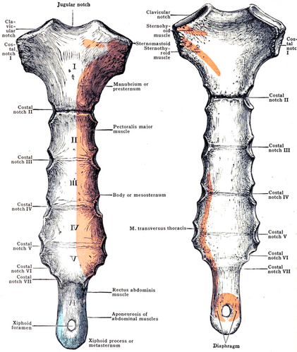
The anterior surface gives origin on either side to:
- a. The pectoralis major.
- b. The sternal head of the sternocleidomastoid
The posterior surface gives origin to:
- a. The sternohyoid in upper part (Fig. 13.12).
- b. The sternothyroid in lower part.
The suprasternal notch gives attachment to the lower fibres of the interclavicular ligament, and to the two subdivisions of the investing layer of cervical fascia. The margins of each clavicular notch give attachment to the capsule of the corresponding sternoclavicular joint.
The body[edit | edit source]
The anterior surface gives origin on either side to:
- the pectoralis major muscle.
The lower part of the posterior surface gives origin on either side to the sternocostalis muscle. Between the facets for articulation with the costal cartilages, the lateral borders provide attachment to:
- the external intercostal membranes
- internal intercostal muscles
The xiphoid[edit | edit source]
- The anterior surface provides insertion to the medial fibres of the rectus abdominis, and to the aponeuroses of the external and internal oblique muscles of the abdomen.
- The posterior surface gives origin to the diaphragm.
- The lateral borders of the xiphoid process give attachment to the aponeuroses of the internal oblique and transversus abdominis muscles.
- The upper end forms a primary cartilaginous joint with the body of the sternum.
- The lower end affords attachment to the linea alba.[1]
Blood supply[edit | edit source]
- The major source of branches to the sternum is the internal thoracic or mammary artery (ITA). It originates from the subclavian artery. It descends 1–2 cm from the lateral margin of the sternum adjacent to the posterior aspect of the chest wall, partly covered by the transversus thoracic muscle from the third to the sixth costal cartilage.
- Sternal branches are intersegmental, located in the intercostal spaces and form arcades at the lateral edge of the sternum. This reflects the intersegmental location of ossification centres that are supplied by sternal arteries.
- Arteries reach the anterior and posterior aspects of the sternum, feeding into dense periosteal plexuses, which are better developed on the posterior side. The plexuses are segmentally organized in infants, corresponding to sternebrae, but are confluent craniocaudally in adults.
- Harvesting the ITA for coronary bypass will disrupt sternal circulation to a variable extent, although this hypoperfusion may be temporary in most cases. When the ITA is dissected, any branches should be ligated as close as possible to the main vessel in order to preserve collateral branches. Likewise, sternal cerclages should be placed as close as possible to the sternal edge to preserve the arcades between sternal arteries. [5]
Clinical relevance[edit | edit source]
- Bone marrow for examination is usually obtained by manubriosternal puncture. It is done in its upper half to prevent injury to arch of aorta which lies behind its lower half.
- The slight movements that take place at the manubriosternal joint are essential for movements of the ribs.
- In the anomaly called ‘funnel chest’, the sternum is depressed in another anomaly called ‘pigeon chest’, there is forward projection of the sternum like the keel of a boat, and flattening of the chest wall on either side.
- For cardiac surgery, the manubrium and/or body of sternum need to be splitted in midline and the incision is closed with stainless steel wires.
- Sternum is protected from injury by attachment of elastic costal cartilages. Indirect violence may lead to fracture of sternum.
- Non-fusion of the sternal plates causes ectopia cordis, where the heart lies uncovered on the surface. Partial fusion of the plates may lead to the formation of sternal foramina, bifid xiphoid process.[1]
- Sternal fracture are associated with severe blunt trauma to the chest, such as in a vehicular accident.
- Due to direct connection and proximity, the ribs are also commonly fractured in the process.
- Costochondritis is the most common cause of sternal pain and occurs when the cartilage between the sternum and ribs become inflammed and/ or irritated.
- Tietzes syndrome usually affects the third, fourth and fifth costochondral joint. It causes severe pain when coughing and deep breathing.
References[edit | edit source]
- ↑ 1.0 1.1 1.2 1.3 Chaurasia BD. Human anatomy. CBS Publisher; 2004. 8th Edition. Vol.1. Pg 224.
- ↑ Drake R, Vogl AW, Mitchell AW, Tibbitts R, Richardson P. Gray's Atlas of Anatomy E-Book. Elsevier Health Sciences; 2020 Feb 27.
- ↑ 3.0 3.1 3.2 Graeber GM, Nazim M. The anatomy of the ribs and the sternum and their relationship to chest wall structure and function. Thoracic surgery clinics. 2007 Nov 1;17(4):473-89.
- ↑ Medical Lecture. Learn Movement in respiration,Pump handle & Bucket handle movement. Available from: https://www.youtube.com/watch?v=dxEDGK4nAZM [last accessed 11/28/2021]
- ↑ Neuhuber W, Lyer S, Alexiou C, Buder T. Anatomy and blood supply of the sternum. Deep sternal wound infections. 2016:7-12.


