Soleus
Original Editor Aarti Sareen
Top Contributors - Aarti Sareen, Laura Chimimba, Kim Jackson, Vanessa Rhule, Admin, Evan Thomas and Richard Benes
Introduction
[edit | edit source]
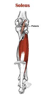
Located in superficial posterior compartment of the leg Soleus is a powerfull lower limb muscle which along with gastronemius and plantaris forms the calf muscle or triceps surae. It runs from back of knee to the ankle and is multipennate.
Anatomy[edit | edit source]
Origin[edit | edit source]
- posterior surface of the head and upper 1/3 of the shaft of the fibula;
- middle 1/3 of the medial border of the tibia, tendinous arch between tibia and fibula.
Insertion[edit | edit source]
Into calcaneus with gastrocnemius by way of achilles tendon
Action[edit | edit source]
- Plantar flexion of the foot at the ankle;
- Reversed origin insertion action: when standing, the calcanius becomes the fixed origin of the muscle;
- Soleus muslce stabilizes the tibia on the calcaneus limiting forward sway.
Nerve supply[edit | edit source]
Tibial nerve, L4, L5, S1 , S2
Synergists[edit | edit source]
Gastrocnemius, Plantaris, Tibialis posterior, Peroneus longus and Brevis, FHL and FDL.
Antagonists[edit | edit source]
Tibialis anterior
Blood supply[edit | edit source]
- Blood supply of the soleus muscle is from peroneal artery proximally and the posterior tibial artery distally;
- Muscle has a mixed blood supply;
- Vascular supply of the soleus is from popliteal, posterior tibial, & peroneal vascular pedicles to the proximal muscle, peroneal pedicles to distal lateral belly, and segmental posterior tibial pedicles to distal medial belly;
- With distal pedicles from the posterior tibial artery ligated & based on proximal pedicles from the posterior tibial and peroneal arteries, muscle can be transposed medially or laterally to cover defects in middle third of the leg;
- Proximal vasculature arises directly from the popliteal vessels and can reliably carry all but the distal 4 to 5 cm of the muscle;
- Intramuscularly, vasculature of the soleus divides into a bipenniform segmental pattern;
- w/ this vascular pattern, either half of the soleus muscle can be used, leaving a functional hemisoleus muscle intact
Function[edit | edit source]
Soleus has two major functions:
- To act as skeletal muscle: Along with other calf muscle it is powerfull plantarflexor and have major contribution in running,walking and dancing.In the seated calf raise (knees flexed approximately 90º), the gastrocnemius are virtually inactive while the load is borne almost entirely by the soleus. In moderate force, the soleus is preferentially activated in the concentric phase, whereas the gastrocnemius is preferentially activated in the eccentric phase [1].Human soleus muscle have majority of slow twitch fibers.Human soleus fiber composition is quite variable, containing between 60 and 100% slow fibers.[2][3][4].
- To act as muscle pump: along with other calf muscles it is known as peripheral heart as in uprite posture it enhance pumping the venous blood back into heart from periphery
Palpation[edit | edit source]
Accessory soleus muscle (ASM)
[edit | edit source]
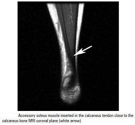
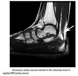
It is present in 0.7 to 5.5% of humans.[5]It is usually observed during the second or third decade of life and is more commonly seen in females than males at a ratio of 2:1. It is mostly unilateral.[6][7][8][9][10][11]. This supernumerary muscle is located under the gastrocnemius muscle, in the posterior upper third of the fibula, in the oblique soleus line, between the fibular head and the posterior part of the tibia. From its origin, the ASM runs anteriorly and medially until it reaches the Achilles tendon.[12]
Depending upon its insertion it is of 5 types, or in other words it can origninate from 5 sites
- Achilles tendon
- Upper calcaneus region
- Insertion in the upper calcaneus,
- Medial calcaneus region,
- Medial part of the calcaneus
Sometimes it is impossible to precisely identify the ASM origin and insertion, since the MRI fails to show details, depending on the slices.[13]Itmay cause pain on exercise. One may suspect a soft-tissue tumor, such as lipoma, hemangioma, and even sarcoma, but the anomalous muscle has a typical appearance on plain radiographs, and the appearance on computed tomography is diagnostic. If the patient is asymptomatic, no therapy is required, but if pain or other discomfort is provoked by exercise, exploration with fasciotomy or excision of the accessory muscle is recommended, as was done in six of our eleven patients who were seen between 1968 and 1985.[14]
Pathology[edit | edit source]
Strain/Rupture[edit | edit source]
Full or partial rupture of the soleus muscle usually occurs when the calf muscle becomes stretched while it is contracting (eccentric contraction). Partial ruptures represent the majority of the ruptures. The rupture occurs in many instances at the point of attachment of the soleus muscle to the Achilles tendon, which will often trigger an inflammation of the Achilles tendon as a result of the soleus rupture.
Symptoms: Pain when activating the calf muscle (running and jumping), when applying pressure on the Achilles tendon approx. 4 cm. above the anchor point on the heel bone or higher up in the calf muscle, and when stretching the tendon. Walking on tip-toe will aggravate the pain.
In all cases when there is a sense of a "crack", or sudden shooting pains in the Achilles tendon, medical attention should be sought as soon as possible. Ultrasound scanning or MRI examination is used to advantage when making the diagnosis, as even full ruptures can easily be overlooked by normal clinical examination. [15]
Further, about the soleus and calf strain can be read here
Whenever there occurs calf strain and the soleus is the muscle affected then there occur usually the medial or lateral pain i.e along the soleus fibres. And when the pain is in the bulk of the calf muscle then the gastrocnemius is the muscle affected,but, mostly both the muscles are affected together.
Soleus Syndrome
[edit | edit source]
Another cause of medial pain (posteriomedial aspect of ankle) just above the medial malleous is the soleus syndrome.[16] It occurs because of abnormal sliping of soleus muscle from its normal origin site and is similar to exertional campartment syndrome and commaly seen in dancers and athlets. Respond well to conservative treatment and if not then rarely fasciotomy of the soleus insertion may be required.[17]
References[edit | edit source]
- ↑ A Nardone, C Romanò,M Schieppati.Selective recruitment of high-threshold human motor units during voluntary isotonic lengthening of active muscles.J Physiol. 1989 February; 409: 451–471.
- ↑ Ariano MA, Armstrong RB, Edgerton VR (January 1973). "Hindlimb muscle fiber populations of five mammals". The Journal of Histochemistry and Cytochemistry 21 (1): 51–5.
- ↑ Burke RE, Levine DN, Salcman M, Tsairis P (May 1974). "Motor units in cat soleus muscle: physiological, histochemical and morphological characteristics". The Journal of Physiology 238 (3): 503–14.
- ↑ Gollnick PD, Sjödin B, Karlsson J, Jansson E, Saltin B (April 1974). "Human soleus muscle: a comparison of fiber composition and enzyme activities with other leg muscles". Pflügers Archiv 348 (3): 247–55
- ↑ Sookur PA, Naraghi AM, Bleakney RR, Jalan R, Chan O, White LM. Accessorymuscles: anatomy, symptoms and radiology evaluation. Radiographics. 2008;28(2):481-99.
- ↑ Crespo E, Minguez MF, Gascó J, Silvestre A, Jolín T, et al. Músculo sóleo accesorio como diagnóstico diferencial de un tumor de partes blandas del tobillo. Acta Ortop Castellano-Manch. 2004;(5):37-41.
- ↑ Romanus B, Lindahl S, Sterner B. Accessory soleus muscle. A clinical and radiographic presentation of eleven cases. J Bone Joint Surg Am. 1986; 68(5):731-4.
- ↑ Salomão O, Carvalho Junior AE, Fernandes TD, Romano D, Adachi PP, Sampaio Neto R. Músculo solear acessório: aspectos clínicos e achados cirúrgicos. Rev Bras Ortop. 1994;29(4):251-5.
- ↑ Leswick DA, Chow V, Stoneham GW. Resident's corner. Can Assoc Radiol J. 2003;54(5):313-5.
- ↑ Featherstone T. MRI diagnosis of accessory soleus muscle strain. Br J Sports Med. 1995;29(4):277-8.
- ↑ Doda N, Peh WC, Chawla A. Symptomatic accessory soleus muscle: diagnosis and follow-up on magnetic resonance imaging. Br J Radiol. 2006;79(946):e129-32.
- ↑ Flavio Belmont Del Nero; Cristiane Regina Ruiz; Roberto Aliaga Junior.The presence of accessory soleous muscle in humans.Einstein (São Paulo) vol.10 no.1 São Paulo Jan./Mar. 2012.
- ↑ Flavio Belmont Del Nero; Cristiane Regina Ruiz; Roberto Aliaga Junior.The presence of accessory soleous muscle in humans.Einstein (São Paulo) vol.10 no.1 São Paulo Jan./Mar. 2012.
- ↑ Romanus B, Lindahl S, Stener B.Accessory soleus muscle. A clinical and radiographic presentation of eleven cases.J Bone Joint Surg Am. 1986 Jun;68(5):731-4.
- ↑ citated from (sportnetdoc.com/foot-achilles/rupture-of-the-soleus-muscle) citated on august 31,2013.
- ↑ Michael RH, Holder LE: The soleus syndrome. Am J Sports Med 1985; 13:87.
- ↑ David A.Porter,Lew C.Schow.Baxter's Foot and Ankle in Sports.2nd Edition.Mosby Publication.


