Sacrotuberous Ligament
Description[edit | edit source]
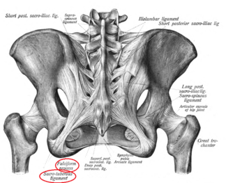
Sacrotuberous ligament is one of the pelvic ligaments, it is about 6,4-9,4 cm in length, located posterior inferiorly in the pelvis between the sacrum and ischial tuberosity, where most of its band inserts into ischial tuberosity forming with sacrospinous ligament the boundary to the greater and lesser sciatic notch. Some times it is described as an extension to the posterior sacroiliac ligament[1]. It has an attachment to the long head of biceps femoris, gluteus maximus, piriformis, and sacrospinous ligament, and is a part of the posterior sling.
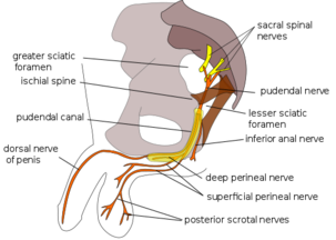
It is considered to be linked with pudendal nerve entrapment that thought to be compressed between the sacrotuberous and sacrospinous ligament at the pudendal canal, and pelvic girdle pain.
Attachments[edit | edit source]
The proximal broad base attaches to the posterior superior iliac spine PSIS, and long post sacroiliac ligament,
Descends inferiorly and laterally and spans to insert into the ischial tuberosity into its medial side. The insertion into the tuberosity has two parts: superior that attaches at the inferior part of the ischial tuberosity and inferior part, runs ventrally to attache at the superior part of the ischial tuberosity.
It continues with the ramus of ischium forming a membranous falciform process that lies deep to the pudendal canal.[1][2]
Function[edit | edit source]
Sacrotuberous ligament STL assists in pelvic stability, the obliquity arrangement of STL on both sides prevent the anterior tipping of sacrum by acting to control sacral nutation.
Connecting the lower limb with the trunk, biceps femoris, and perineum to the thoracolumbar fascia, and erector spinae.
STL and sacrospinous ligament stabilize the sacrum against twisting with the pelvis, and excessive side bending. Hence, an imbalance between STL on both sides cause pelvic rotation, SIJ strain, or LBP[3].
Clinical relevance[edit | edit source]
Pudendal nerve entrapment between the sacrotuberous and sacrospinous ligament, or through its existing the greater sciatic notch anterior to the STL, is one of the common causes of pudendal neurological manifestations that cause sensory more than motor deficits[5].
When the ligament becomes tighter than normal its pain pattern is referred down to the central aspect of the back of the thigh and in severe injuries or pain, it may go down to the calf and into the heal[6]. That interferes with hamstring referred pain pattern, if the referred pain is at the posterior thigh only.
One of the ways to differentiate between both STL and hamstring, with the patient lying flat and knee flexion about 90 degree and apply resistance to knee flexion, if this evoked pain at the posterior aspect of the thigh it is a hamstring origin because the resistance put stress on muscles and tendons, not ligaments.
The most common injury site to STL is the lateral margin of the sacrum[6].
If it is tight you may find by palpation the space of the top pf pelvis wider than the other side and the bottom of pelvis is closed as a result of this.
Assessment[edit | edit source]
Stand beside the patient, one hand reaches across to the opposite ischial tuberosity, the other to the coccyx, between both the sacrotuberous ligament runs. It is anterior to the medial margin of GMAX. lateral to the upper gluteal cleft not on the muscle itself.
Press into the ligament, you will feel roby / hard sensation underneath your fingers.
Compare both sides and stay longer on the side you feel that is more restrict or less bony space. During the assessment, you may experience numbness or peroneal pain you will need to concentrate on the mid part of the ligament where the pudendal nerve gets entraped[3].
Treatment[edit | edit source]
The treatment procedure as the assessment technique with applying firm, gentle, and mild resistance on the ligament avoid gliding or friction movement until you feel the tissue release underneath your finger.
References[edit | edit source]
- ↑ 1.0 1.1 Aldabe D, Hammer N, Flack NA, Woodley SJ. A systematic review of the morphology and function of the sacrotuberous ligament. Clinical Anatomy. 2019 Apr;32(3):396-407.
- ↑ https://www.kenhub.com/en/library/anatomy/ligaments-of-the-lower-limb
- ↑ 3.0 3.1 https://www.abmp.com/textonlymags/article.php?article=396
- ↑ PAINLess. Sarotuberous Ligament Anatomy. Available from: http://www.youtube.com/watch?v=Y6pKbnetuZU[last accessed 22/7/2020]
- ↑ Kaur J, Singh P. Pudendal Nerve Entrapment Syndrome. InStatPearls [Internet] 2019 Jun 21. StatPearls Publishing.
- ↑ 6.0 6.1 https://www.massagetoday.com/articles/14188/Pain-Caused-By-Low-Back-Ligaments
- ↑ AdvancedTrainings. Sacrotuberous Ligament - Advanced Myofascial Techniques DVD Series. Available from: http://www.youtube.com/watch?v=bj_z5zgNA3I[last accessed 22/7/2020]


