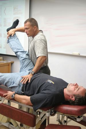
Resisted Isometric Movement Testing
Original Editor - Ashmita Patrao
Top Contributors - Ashmita Patrao, Lucinda hampton, Naomi O'Reilly and Kim Jackson
Introduction[edit | edit source]
Manual isometric muscle testing is a common clinical technique that is used to assess muscle strength. To provide the most accurate data for the test, the muscle being assessed should be at a length in which it produces maximum force.[1]
- The Resisted Isometric Movement testing was an examination developed by Cyriax. It was originally called resisted movements, and is sometimes known as resisted isometrics.
- Resisted Isometric Movement testing is a convenient and clinically useful technique to detect neuromuscular dysfunction and disease, and to track the progress of patients as they undergo rehabilitation[1].
- There is tremendous variability in the recommended positions and joint angles used to conduct these tests, with little apparent objective data used to position the joint such that muscle force production is greatest.[1]
Basics[edit | edit source]
Manual isometric muscle is the predominant method used to assess muscle strength in the clinical setting. There are generally two types of test procedures for isometric testing.
- A “make” test involves the patient exerting a maximum voluntary effort against fixed resistance provided by the examiner.
- A “break” test requires the patient to exert maximum voluntary effort against an increasing counterforce by the examiner to exceed or “break” the isometric force being generated by the patient.
Muscle strength
- Most frequently graded by the examiner on a six point subjective scale that ranges from no perceptible muscle contraction scored as a 0, to being able to resist the full counterforce of the examiner as a 5.
- More objective measurements can also be performed using a dynamometer.
While MMT is a commonly used and practical technique, there can be moderate to low inter-rater reliability in the subjective grading of muscle force production.[1]
Structures Assessed[edit | edit source]
If a joint is held at a mid range and the patient is asked to isometrically contract using a maximal effort, no strain would fall on the inert structures. eg if the elbow is flexed to 90 degrees and maximal resistance is applied to flexion, the load would fall on the biceps and brachialis. This helps in diagnosis of contractile or inert structures
- Contractile Structures: Structures that possess the ability to contract and relax. Eg: muscle, tendon and its attachment
- Inert Structures: Structures in the human body that cannot shorten or elongate in length. Eg: ligaments, capsule, bursa, nerve sheath, cartilage.
Technique[edit | edit source]
- The joint is positioned in mid range, keeping the inert tissues off of stretch and there must be no movement at the joint
- Muscles other than the testing muscles must not be included. Hence trick movements from surrounding muscles must be avoided.
- The muscle is positioned in a resting position which would be mostly a mid range position of the joint.
- The patient must be instructed to exert a maximal effort (isometric hold of grade 3 to grade 5) during the test.
- Examiner must be attentive in identifying pain or weakness that would be due to a nerve involvement
- There must not be any movement during this entire procedure, as
Weakness: Minor weakness need to be detected with the hands well positioned to offer resistance and counter pressure.
Interpretation[edit | edit source]
- Strong and Painless: No lesion in the contractile structure
- Strong and Painful: Minor lesion in a part f the muscle or tendon and its attachment.
- Weak and Painless: There could be a complete rupture of the muscle or tendon, but most commonly might be a malfunction of the nerves. This impaired function of the nerve leads to muscle weakness.
- Weak and Painful: Serious impairment, like a secondary deposit or a fracture might be present. However, if a patient is reluctant to replicate the severe pain it may appear as apparent weakness
- Painful on Repetition: Intermittent claudication could be the reason a the movement is painless and strong initially but hurts on repetition.
- All Resisted Movements Painful: A gross lesion lying proximally, which would mostly be a capsular lesion,
References[edit | edit source]
- ↑ 1.0 1.1 1.2 1.3 Garcia SC, Dueweke JJ, Mendias CL. Optimal joint positions for manual isometric muscle testing. Journal of sport rehabilitation. 2016 Nov 1;25(4).Available from: https://www.ncbi.nlm.nih.gov/pmc/articles/PMC5325820/(accessed 23.4.2021)
- Cyriax JH, Cyriax PJ.Cyriax's Illustrated Manual of Orthopaedic Medicine. 2nd edition: Buttrworth Heinemann. 1993.
- ↑ DJ Magee. Orthopaedic Physical Assessment. 6th Philadelphia: WB Saunders. 2014


