Remote Screening for Lumbar Spine Red Flags
- Please do not edit unless you are involved in this project, but please come back in the near future to check out new information!!
Introduction
Red flags are clinical findings that increase suspicion of a serious pathology (Finucane L. 2020), It is vital that practitioners are aware of these red flags as they form a key component of the assessment and management of low back pain whilst increasing patient safety (Ferguson, Morison and Ryan, 2015). Red flags are features from a patient's subjective and objective assessment which are thought to put them at a higher risk of serious pathology and warrant referral for further diagnostic testing (Delitto, George, and Godges. 2012).
Physiotherapists understanding of red flags for low back pain
The role of physiotherapists as primary identifiers of red flags has grown owing to the spread of self‐referral services (Holdsworth et al., 2006). Physiotherapists often exist without any medical input or review (Kersten et al., 2007; McPherson et al., 2006). Therefore, there is a need to ensure that physiotherapists have a good understanding of individual red flags, understand their importance, and can ask these questions in a clear and unambiguous manner. Similarly, physiotherapists must have a clear understanding and agreed pathways of care dependent on these findings. Failure to do so raises issues around patient safety and professional reputation (Ferguson, Morison and Ryan, 2015).
One study by Feguson et al. (2015) aimed to investigate the red flags that are routinely recorded by physiotherapists. This included: which red flags do they consider to be most important, how would they define each red flag and how they would ask each red‐flag question to a person with back pain. 98 physiotherapists responded to the survey, 84% worked exclusively in the National Health Service (NHS). They recorded that ‘Previous history of cancer’, ‘saddle anaesthesia’ and ‘difficulty with micturition’ were the red flags that raised suspicion of serious pathology the most. The physiotherapists involved in the study stated the following way to ask about red flags:
- 'History of cancer: ‘an individual who has previously been diagnosed with cancer’.
- 'Saddle anaesthesia: Since your symptoms commenced, have you noticed any pins and needles or numbness around your back passage or genital area’
Finally, limited consensus was found in how physiotherapists asked patients about red flags. However, one theme in particular emerged, which is the use of nebulous terminology - for example, the terms recent, weight loss and prolonged period.
COVID-19 & Remote Consultations
COVID-19 is an infectious respiratory disease, caused by the SARS-Cov-2 virus (Severe Acute Respiratory Syndrome Coronavirus Two) which spreads primarily through saliva and respiratory droplets when an infected person sneezes or coughs. Although the majority of people infected will only experience mild to moderate respiratory illness, the elderly and those with underlying health conditions are more likely to suffer from severe illness. There has been over 4.5 million confirmed cases which has resulted in over 300,000 deaths globally (WHO, 2020).
The reproduction number (R value) is a method used to identify the disease's ability to spread meaning, this value represents the number of people that one infected person will pass the virus on to, on average (Yi et al., 2020). Shim et al (2020) suggests that an R value >1.0 means the disease will spread exponentially whereas, an R value <1.0 means the disease will spread slowly and eventually die out. The R value is estimated to be between 1.4 and 2.5 which makes this disease significantly more contagious than the influenza virus (Liu et al., 2020). This has forced healthcare services to rapidly implement alternative models of care to avoid face-to-face contact between patient and clinician (Greenhalgh et al., 2020). Therefore, guidelines have been released for remote consultations based on current evidence evaluating its effectiveness (CSP, 2020; NHS, 2020).
A systematic review (SR) by Cottrell et al (2017) compared the effectiveness of 'telerehabilitation' (defined by phone call and/or video consultations) and face-to-face consultations within a physiotherapy musculoskeletal (MSK) setting. The results were equally effective on the physical function of patients (SMD MD 0.14, 95% CI -0.10-0.37) which is similar to the findings of another SR by Shukla et al (2016) which also reported upon the effectiveness of telehealth.
Qualitative studies have also provided an insight into patient and clinician experiences during remote consultations. Positive feedback included, time convenience, cost efficiency, easy to use technology, home environments, empowering self-management and taking away the stigmatisation of ‘hands on’ therapy for treating low back pain (Hinman et al., 2017; Synott et al., 2015).
This evidence supports the use of remote consultations. Therefore, it is important to discuss how MSK physiotherapists can successfully identify and screen patients for red flags when presenting with low back pain during remote consultations, given the current global pandemic.
Low Back Pain Red Flags
Back pain is very common in the UK. It has a lifetime chance of occurrence of 59% (GP online, 2008). The majority of back pain clears up quite quickly; however, back pain experienced along with ‘red flag’ symptoms may have a serious underlying cause.
This page discusses the clinical indicators for:
· Cauda Equina Syndrome
· Malignancy
· Vertebral Fractures
· Infection
Cauda Equina
Cauda equina syndrome (CES) is a rare but potentially devastating neurological condition affecting the nerve roots at the lower end of the spinal cord known as the cauda equina (CE). The CE is responsible for the innervation of the lower limbs, control of the anal sphincter, regulation and function of the bladder and distal bowel and sensation to the skin around the bottom and back passage (Shim, Park and Lee, 2009).
There is large debate over CES and consequently no agreed definition. However, the British Association of Spinal Surgeons (BASS) present a definition that may be useful in clinical practice (Tsiang et al. 2019):
'A patient presenting with acute back pain and/or leg pain with a suggestion of a disturbance of their bladder or bowel function and/or saddle sensory disturbance should be suspected of having a CES. Most of these patients will not have critical compression of the cauda equina. However, in the absence of reliably predictive symptoms and signs, there should be a low threshold for investigation with an emergency scan’ (Germon et al., 2015).
Epidemiology
There are many causes of CES with the most common being that of a lumbar spine disc herniation. It occurs most frequently between the ages of 31–50 (Fuso et al., 2013). Cauda equina compression usually occurs at the level L4/5 (Fraser et al., 2009). Although disc herniation is the most common mechanism, CES can be caused by any space-occupying lesion, such as spinal stenosis, tumour, cysts, infection, or bony ingress can narrow the spinal canal and cause compression of the cauda equina (Greenhalgh et al., 2018).
Published estimates of the incidence for CES are fewer than one per 100 000 population (Hurme et al., 1984; Podnar, 2007). However, in 2010–2011 in England, 981 decompression surgeries were performed for CES (The National Spinal Taskforce, 2013). At the time, the English population was 52 234 000 (Office for National Statistics, 2011) thus giving an incidence of 1.9 per 100 000.
In a primary care setting, Table 1 highlights the incidence of diagnosed patients with CES in the UK 2018/19 (Barnes, 2019).
| Condition | Number of primary diagnoses | % of total cases |
| Low Back Pain | 45,520 | 0.04% |
| Cauda Equina Syndrome | 170 | 0.0% |
| Malignant Neoplasm: Cauda Equina | 58 | 0.0% |
Table 1 - UK 2018/19 prevalence of Cauda Equina Syndrome
Prognosis
The prognosis for complete recovery is dependent upon many factors. The most important of these is the severity and duration of compression upon the damaged nerve(s). Generally, the longer the time before intervention to remove the compression causing nerve damage, the greater the damage caused to the nerve(s). Similarly, Kennedy et al. (1999) describes the most important factor identified in a series of predictors for favourable outcome in CES was an early diagnosis. This highlights to importance of understanding and identifying red flags.
Clinical Indicators
The subjective history is the most important aspect of the examination, particularly early in the presentation of a patient with CES as the imperceptible and possible vague symptoms related to early CES need to be identified using clear and unmistakable methods of communication (Bin et al., 2009; Sun et al., 2014).
Premkumar et al. (2018) and Tsiang et al. (2019) identified various clinical indicators of CES that should be screened for during the assessment of these patients. Premkumar et al. (2018) reported that the combination of recent loss of bladder control and recent loss of bowel control produced a specificity of 97.4%. Both studies highlighted that while the specificity was generally high, all red flag questions had poor sensitivity when identifying their diagnoses of interest.
| Indicator | Sensitivity (%) | Specificity (%) |
| Recent loss of bladder control | 22.2 | 90.4 |
| Recent loss of bowel control | 13.9 | 95 |
| Combination: Recent loss of bladder and bowel control | 8.3 | 97.2 |
Table 2 - Clinical Indicators from Premkumar et al. (2018).
| Indicator | Sensitivity (%) | Specificity (%) |
| Urinary retention | 30.3 | 96.2 |
| Incontinence | 50 | 86.5 |
| Saddle numbness | 0 | 94.7 |
| Weakness in limbs | 67.7 | 53.2 |
Table 3 - Clinical Indicators from Tsiang et al. (2019).
During screening for red flags, Greenhalgh et al. (2015) in their qualitative investigation of patient's experience of CES found that one of the key problems in communication was the technical/medical language used by clinicians. For example, ‘saddle numbness’ to a patient is not clearly understood. The patient participants in the study emphasised the need for clinicians to use clear and some would say ‘explicit language’ that can be readily understood during a consultation.
Malignancy
The spinal cord may be compressed due to tumours occupying space within the vertebral canal (Gilbert et al. 1978). This may then affect the neural function of the spinal cord causing unremitting pain, muscle power and sensation alteration, sexual dysfunction, bladder/bowel dysfunction and sleep disturbances (cancerresearchuk.org, 2020).
Epidemiology
Tumours are classified as primary, originating in the spine, and secondary, originating elsewhere in the body and spreading to the spine (cancerresearchuk.org). Secondary tumours are much more prevalent than primary tumours. They occur in approximately 70% of cancer patients (Ciftdemir et al. 2016) whereas primary tumours occur in approximately 0.07% of healthy people (Schellinger et al. 2008). The most common types of primary tumours are meningiomas (29%), nerve-sheath tumours (24%) and ependymomas (23%) (Schellinger et al. 2008). Secondary tumours can metastasise from many different areas of the body; most commonly they may spread from breast, lung and prostate primary tumours (John Hopkins Medicine, 2020).
In a primary care setting, malignancy is extremely rare. Table 4 highlights the incidence of primary diagnoses given that may result in low back pain within NHS primary care settings in the UK in 2018/19. 96,420,114 patients were seen in total (Barnes, 2019).
| Condition | Number of primary diagnoses | % of total cases |
| Low back pain | 45,520 | 0.04% |
| Malignant neoplasm: Vertebral column | 136 | 0.00% |
| Malignant neoplasm: Connective and soft tissue of trunk | 85 | 0.00% |
| Malignant neoplasm: Spinal meninges | 1 | 0.00% |
| Malignant neoplasm: CNS unspecified | 233 | 0.00% |
| Malignant neoplasm of other and ill-defined sites: Lower limb | 74 | 0.00% |
| Secondary malignant neoplasm of other unspecified parts of the nervous system | 158 | 0.00% |
| Secondary malignant neoplasm of bone and bone marrow | 17,629 | 0.02% |
Table 4 - UK 2018/19 prevalence of Malignancy.
Prognosis
Around 10-20% of patients diagnosed with spinal metastasis live for longer than two years after this diagnosis (Delank et al. 2011). Better prognoses and longer survival rates have been associated with earlier detection of the tumour (Ruckdeschel, 2005). Therefore, it is important to screen patients with low back pain for red flags associated spinal malignancy.
Clinical Indicators
When assessing patients with low back pain, there are a number of ‘red flags’ which may increase suspicion of spinal malignancy. Large scale studies by Henschke et al. (2013), Premkumar et al. (2018) and Tsiang et al. (2019) have identified numerous clinical indicators of malignancy that should be screened for during the assessment of these patients.
These studies accept that no single ‘red flag’ can be used in isolation to give a diagnosis of spinal malignancy. Instead, a combination may increase a clinician’s index of suspicion. The literature stated that patient-reported history of cancer, alongside low back pain, was identified as the most significant sign of spinal malignancy. Interestingly, Premkumar et al. (2018) reported that a past medical history of cancer, combined with unexplained weight loss, produced a specificity of 99.8%. The summary of findings from these studies is detailed in table 5 below.
| Indicator | Sensitivity (%) | Specificity (%) |
| Age >50 | 71.7 | 32.6 |
| Age >70 | 22.6 | 79.5 |
| Night pain | 54.2-55.4 | 41.8-49.6 |
| Unexplained weight loss | 8.2 | 95.6 |
| Pain at rest | 25 | 69.8 |
| Urinary retention | 4.2 | 95.8 |
| History of cancer | 32-75 | 78.7-95.6 |
| History of cancer + Unexplained weight loss | 2.5 | 99.8 |
Table 5 - Clinical Indicators from Premkumar et al. (2018) & Tsiang et al. (2019).
A Cochrane review carried out by Henschke et al. (2013) examining studies containing over 6000 patients emphasised the need for an affective diagnostic test to assist in the identification of spinal malignancy in patients with low back pain.
Vertebral fractures
Vertebral fractures can be caused by direct or indirect trauma and are more likely to occur in patients with decreased bone density (Amboss.com. 2020). Fractures may be classified as “stable” or if there is a risk of damage to the spinal cord “unstable”. A dorsal spine injury (vertebral arches, processes, and their ligaments) is always unstable and has a high probability of spinal cord injury (Amboss.com. 2020).
Types of vertebral fractures:
Vertebral compression fracture
- Loss of height of the vertebral body; due to trauma or pathological fracture
- Progressive thoracic kyphotic deformity if multiple vertebrae are affected
- Usually stable
- Wedge fracture (subtype)
Burst fracture
- Fracture of the vertebra in multiple locations
- Result of compression trauma with severe axial loading
- Possible displacement of bone fragments into the spinal canal
Fracture-dislocation
- Fractured vertebra and disrupted ligaments;
- Instability may cause spinal cord compression.
(Amboss.com. 2020).
Epidemiology
Of the 96,420,114 patients who were assessed in an NHS primary care setting in 2019, only 524 were diagnosed as having a Lumbar vertebral fracture. There were 498 diagnosed with a Fracture of other and unspecified parts of lumbar spine and pelvis, and 10 diagnosed with multiple fractures of Lumbar spine and pelvis (Barnes, 2019).
| Condition | Number of primary diagnoses | % of total cases |
| Low Back Pain | 45,520 | 0.04% |
| Fracture of lumbar vertebrae | 524 | 0.0% |
| Fracture of other and unspecified parts of lumbar spine and pelvis | 498 | 0.0% |
| Multiple fractures to lumbar spine and pelvis | 10 | 0.0% |
Table 6 - UK 2018/19 prevalence of Vertebrae Fractures
The actual incidence of vertebral fractures is likely much greater given the large number of vertebral fractures that go undetected. More than two-thirds of patients with Vertebral fractures are asymptomatic, and are diagnosed incidentally (Fink, 2005). Symptomatic patients may report abrupt onset of pain with position changes, coughing, sneezing, or lifting (Amboss.com. 2020). Physical examination findings are often 'normal' but may demonstrate kyphosis and midline spine tenderness (Amboss.com. 2020).
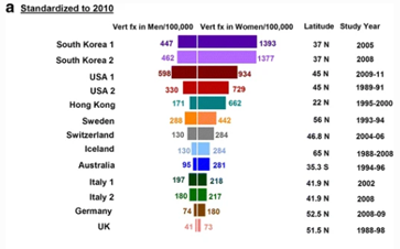
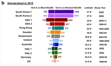
Ballane (2017) investigated the prevalence and incidence of vertebral fractures worldwide. This study incorporated 62 articles of fair to good quality and comparable methods for vertebral fracture identification. Graphs A and B display age-standardised incidence rates in men and women combining hospitalised and ambulatory vertebral fractures, ranked by descending incidence. Standardisation to 2010 UN population (a) and 2015 UN population (b).
Figure 1 - Epidemiology of vertebrae fractures from Ballane, 2017.
Rates partially depend on the definition of a vertebral fracture, clinical versus morphometric. The morphometric definition is not universal; at least seven methods have been used in different studies (included in Graphs A and B). Within the same method, varying decision thresholds (fracture grade or standard deviation from means) make the definition of vertebral fracture even more difficult (Ballane,. 2017). Different definitions include of vertebral fracture include:
- Morphometric method
- McCloskey method
- Eastell method
- Genant Method
- Davies method
- Melton method
- Denis Classification
(Ballane,. 2017)
Diagnosis
In the primary care setting, between 1% and 5% of all patients who present with LBP will have a serious spinal pathology which requires further assessment and often specific treatment (Deyo 1992; Henschke 2009). The most common of these serious spinal pathologies which initially manifests as LBP is vertebral fracture, followed by malignancy, infection, and inflammatory disease. The presence of a "red flag" should alert clinicians to the need for further examination and, in most cases, specific management (Waddell 2004). With respect to vertebral fractures, the typical “red flags” include >50 years of age, prolonged corticosteroid use, trauma and Osteoporosis. High quality evidence investigating the efficacy of these “red flags” correctly identifying serious pathologies has been investigated in studies such as Premkumar et al. (2018) and Tsiang et al. (2019).
| Indicator | Sensitivity (%) | Specificity (%) |
| Age of >50 years | 74 | 32.9 |
| Age of >70 years | 3.9 | 80 |
| Trauma | 24.7 | 88.6 |
| Combination 1: Trauma and age >50 years | 14.8 | 94.2 |
| Combination 2: Trauma and age >70 years | 5.2 | 98.2 |
Table 7 - Clinical Indicators from Premkumar et al. (2018).
| Indicator | Sensitivity (%) | Specificity (%) |
| Osteoporosis | 41.5 | 76.2 |
| Steroid use | 28.3 | 86.7 |
| Trauma | 22.6 | 93.8 |
Table 8 - Clinical Indicators from Tsiang et al. (2019).
The presence of both recent trauma and an age of >50 years carries a 13.1% probability of a vertebral fracture in the setting of low back pain; Tsiang et al. (2019). The presence of both recent trauma and an age of >70 years carries a 20.5% probability of vertebral fracture in the setting of low back pain. Tsiang et al. (2019).
Clinical implications
There is agreement in the current literature the findings give rise to a weak recommendation that a combination of a small subset of red flags may be useful to screen for vertebral fracture (Williams et al., 2013). It should also be noted that many red flags have high false positive rates; and if acted upon uncritically there would be consequences for the cost of management and outcomes of patients with LBP (Williams et al., 2013).
Infection
Infections can often masquerade as MSK conditions of the lower back (Kolinsky and Liang, 2018). Specifically, spinal infection (SI) is a term used to describe infectious diseases of the intervertebral discs, vertebral body and the paraspinal tissues (Nickerson and Sinha, 2016). SI can develop through open trauma, surrounding areas which are infected and through bacteria entering the blood circulation spreading throughout the body (Calhoun and Manring, 2005). Evidence has shown that infections can cause redness of the skin, swelling, pain, heat, raised temperature and raised vital signs (NICE, 2020).
Epidemiology
According to Duarte and Vaccaro (2013), SI accounts for 2-7% of all MSK infections. A Japanese national figure database estimated the incidence of vertebral osteomyelitis to range from 5.3/100,000 to 7.4/100,000 population per year between 2007 and 2010 (Akiyama et al., 2013). Postoperative SI is a common complication following spinal surgery. The incidence of SI post spinal surgery is reported to be between 0.1% to 6.7% (Dessy et al., 2017).
Similarly to CES and malignancy, spinal infections are exceptionally rare within the primary care setting. This is highlighted in Table 9 which demonstrates the incidence primary diagnoses of infections which can lead to low back pain within an NHS primary care setting in the UK in 2018/2019 (Barnes, 2019).
| Condition | Number of primary diagnoses | % of total cases |
| Low back pain | 45,520 | 0.00% |
| Osteomyelitis of vertebra | 63 | 0.00% |
| Osteomyelitis, unspecified | 1558 | 0.00% |
| Infection of intervertebral disc (pyogenic) | 6 | 0.00% |
| Discitis, unspecified | 175 | 0.00% |
| Infective myositis | 65 | 0.00% |
| Pyogenic arthritis, unspecified | 359 | 0.00% |
| Meningitis, unspecified | 75 | 0.00% |
| Infection following a procedure, not elsewhere classified | 398 | 0.00% |
| Intraspinal abscess and granuloma | 13 | 0.00% |
Table 9 - UK 2018/19 prevalence of Infection.
Prognosis
Untreated infections can cause serious life changing implications including severe neurological deficits (Lam and Webb, 2004; Inoue et al., 2013). Numerous studies have reported successful treatment rates of 50% to 91% when administered antibiotics however, mortality rate depends on the severity of co-morbidities and age (Kwon et al., 2017). By recognising the seriousness of implications if left untreated, it is of high importance that MSK physiotherapists are able to successfully screen patients for red flags associated with infection.
Clinical Indicators
It is vital for clinicians to be aware of the ‘red flags’ when assessing patients presenting with low back pain. Awareness of the clinical signs and symptoms of serious pathologies such as infections, will allow the clinician to ask the right questions. This will help rule out serious and sinister pathologies and aid your clinical decision for the most appropriate pathway for further investigation (Greenhalgh, Finucane, Mercer and Selfe, 2020).
Large retrospective studies identified various ‘red flags’ for SI and evaluated their diagnostic accuracy in low back pain patients (Galliker et al., 2020; Premkumar et al., 2018; Tsiang et al., 2019). These studies came to the same conclusions for SI as they did malignancy, fractures and CES. They concluded that due to the low sensitivity and specificity of ‘red flags’ it would be inappropriate to base your treatment decision on a singular ‘red flag’ in isolation. However, a combination should increase the clinicians index of suspicion and be used to probe further questioning or clinically reason further investigation. Premkumar et al (2018) provided further evidence to support the dismissal of using ‘red flags’ in isolation. Other results showed that negative responses to ’red flag’ questions did not decrease the likelihood of ‘red flag’ diagnoses.
Due to the low prevalence of infection, it was difficult to assess the diagnostic accuracy of their associated ‘red flags’ (Tsiang et al., 2019). In the Premkumar et al (2018) study, ‘red flag’ indicators for infection were based on published guidelines which included fever, chills, or sweating, recent infection, pain awakens from sleep and persistent sweating at night. Fever, chills, or sweating and recent infection all significantly increase the probability of SI. A summary of the findings from these studies are detailed in the table below.
| Indicator | Sensitivity (%) | Specificity (%) |
| Fever, chills, or sweating | 11.7 | 93.2 |
| Pain awakens from sleep | 57.5 | 41.8 |
| Persistent sweating at night | 17.5 | 86.1 |
| Recent infection | 24.2 | 97.4 |
| Night pain | 37.5 | 49.1 |
| Pain at rest | 12.5 | 69.7 |
| Fever | 25.0 | 97.6 |
Table 10 - Clinical Indicators from Premkumar et al. (2018) & Tsiang et al. (2019).
Yusuf et al (2019) conducted a descriptive review reporting on the characteristics of 2224 patients with SI. Interestingly, the most common clinical features were spinal pain (72%), fever (55%) and neurological dysfunction (33%). It was also found that SI was common in immunosuppressed patients due to other health conditions. The most common determinants were identified as diabetes (18%), IV drug use (9%) and recent surgery (6%). Although the diagnostic accuracy could not be tested, these findings within a large cohort are important to acknowledge to support your clinical reasoning and increase your index of suspicion.
Ultimately, screening for ‘red flag’ indicators for SI does not change when assessing patients remotely. These screening questions are part of a subjective assessment therefore, can be conducted over a number of remote models of care. Finally, an accumulation of ‘red flags’ should lead the clinicians management of these patients and prompt further questioning and investigation when needed.
Screening Tool
The Lenton Lumbar Spine Red Flag Screening Tool was developed after compiling all of the evidence discussed in the previous sections. It can be used to help physiotherapists and other healthcare professionals screen patients who present with red flag symptoms.
It can be accessed via the link below:
References
Akiyama, T., Chikuda, H., Yasunaga, H., Horiguchi, H., Fushimi, K. and Saita, K., 2013. Incidence and risk factors for mortality of vertebral osteomyelitis: a retrospective analysis using the Japanese diagnosis procedure combination database. BMJ Open, 3(3), p.e002412
Amboss.com. 2020. Vertebral Fractures – Knowledge For Medical Students And Physicians. [online] Available at: <https://www.amboss.com/us/knowledge/Vertebral_fractures> [Accessed 22 May 2020].
Ballane, G., Cauley, J., Luckey, M. and El-Hajj Fuleihan, G., 2017. Worldwide prevalence and incidence of osteoporotic vertebral fractures. Osteoporosis International, 28(5), pp.1531-1542.
Barnes, M., 2019. NHS Digital, Hospital Episode Statistics For England. Outpatient Statistics, 2018 - 2019.. Primary Diagnosis by Attendance Type. NHS Digital.
Bin, M.A., Hong, W.U., Jia, L.S., Wen, Y.U.A.N., Shi, G.D. and Shi, J.G., 2009. Cauda equina syndrome: a review of clinical progress. Chinese medical journal, 122(10), pp.1214-1222.
Calhoun, J. and Manring, M., 2005. Adult Osteomyelitis. Infectious Disease Clinics of North America, 19(4), pp.765-786.
Cancerresearchuk.org. 2020. Spinal Cord Compression | Cancer In General | Cancer Research UK. [online] Available at: <https://www.cancerresearchuk.org/about-cancer/coping/physically/spinal-cord-compression/about> [Accessed 20 May 2020].
Cancerresearchuk.org. 2020. Spinal Cord Tumours (Primary) | Cancer Research UK. [online] Available at: <https://www.cancerresearchuk.org/about-cancer/brain-tumours/types/treatment-spinal-cord-tumours> [Accessed 20 May 2020].
Ciftdemir, M., Kaya, M., Selcuk, E. and Yalniz, E., 2016. Tumors of the spine. World journal of orthopedics, 7(2), p.109.
Cottrell, M., Galea, O., O’Leary, S., Hill, A. and Russell, T., 2017. Real-time telerehabilitation for the treatment of musculoskeletal conditions is effective and comparable to standard practice: a systematic review and meta-analysis. Clinical Rehabilitation, 31(5), pp.625-638.
Delank, K.S., Wendtner, C., Eich, H.T. and Eysel, P., 2011. The treatment of spinal metastases. Deutsches Aerzteblatt International, 108(5), p.71.
Delitto, A., George, S. and Godges. J, 2012. Low Back Pain Clinical Practice Guidelines Linked to the International Classification of Functioning, Disability, and Health from the Orthopaedic Section of the American Physical Therapy Association. Journal of orthopaedics and sports physical therapy. 42(4), pp. 57
Dessy*, A., Yuk*, F., Maniya, A., Connolly, J., Nathanson, J., Rasouli, J. and Choudhri, T., 2017. Reduced Surgical Site Infection Rates Following Spine Surgery Using an Enhanced Prophylaxis Protocol. Cureus,
Deyo RA, Rainville J, Kent DL. What can the history and physical examination tell us about low back pain?. Journal of the American Medical Association 1992;268(6):760‐5.
Duarte, R. and Vaccaro, A., 2013. Spinal infection: state of the art and management algorithm. European Spine Journal, 22(12), pp.2787-2799.
Ferguson, F.C., Morison, S. and Ryan, C.G., 2015. Physiotherapists' understanding of red flags for back pain. Musculoskeletal care, 13(1), pp.42-50.
Fink HA, Milavetz DL, Palermo L, et al.; Fracture Intervention Trial Research Group. What proportion of incident radiographic vertebral deformities is clinically diagnosed and vice versa? J Bone Miner Res. 2005;20(7):1216–1222
Finucane L. 2020. An Introduction to Red Flags in Serious Pathology.
Fraser, S., Roberts, L. and Murphy, E., 2009. Cauda equina syndrome: a literature review of its definition and clinical presentation. Archives of physical medicine and rehabilitation, 90(11), pp.1964-1968.
Fuso, F.A.F., Dias, A.L.N., Letaif, O.B., Cristante, A.F., Marcon, R.M. and de Barros Filho, T.E.P., 2013. Epidemiological study of cauda equina syndrome. Acta ortopedica brasileira, 21(3), p.159.
Galliker, G., Scherer, D., Trippolini, M., Rasmussen-Barr, E., LoMartire, R. and Wertli, M., 2020. Low Back Pain in the Emergency Department: Prevalence of Serious Spinal Pathologies and Diagnostic Accuracy of Red Flags. The American Journal of Medicine, 133(1), pp.60-72.e14
Germon, T., Ahuja, S., Casey, A.T., Todd, N.V. and Rai, A., 2015. British Association of Spine Surgeons standards of care for cauda equina syndrome. The Spine Journal, 15(3), pp.S2-S4.
Gilbert, R.W., Kim, J.H. and Posner, J.B., 1978. Epidural spinal cord compression from metastatic tumor: diagnosis and treatment. Annals of Neurology: Official Journal of the American Neurological Association and the Child Neurology Society, 3(1), pp.40-51.
GP online. 2008. Red flag symptoms: Back pain [Online]. Available from: https://www.gponline.com/red-flag-symptoms-back-pain/musculoskeletal-disorders/musculoskeletal-disorders/article/798743 [Accessed 20/05/20]
Greenhalgh, S., Finucane, L., Mercer, C. and Selfe, J., 2018. Assessment and management of cauda equina syndrome. Musculoskeletal Science and Practice, 37, pp.69-74.
Greenhalgh, S., Finucane, L., Mercer, C. and Selfe, J., 2020. Safety netting; best practice in the face of uncertainty. Musculoskeletal Science and Practice, 48, p.102179.
Greenhalgh, S., Truman, C., Webster, V. and Selfe, J., 2015. An investigation into the patient experience of Cauda Equina Syndrome: A qualitative study. Physiotherapy Practice and Research, 36(1), pp.23-31.
Greenhalgh, T., Wherton, J., Shaw, S. and Morrison, C., 2020. Video consultations for covid-19. BMJ, p.m998.
Henschke, N., Maher, C.G., Ostelo, R.W., de Vet, H.C., Macaskill, P. and Irwig, L., 2013. Red flags to screen for malignancy in patients with low‐back pain. Cochrane database of systematic reviews, (2).
Hinman, R., Nelligan, R., Bennell, K. and Delany, C., 2017. “Sounds a Bit Crazy, But It Was Almost More Personal:” A Qualitative Study of Patient and Clinician Experiences of Physical Therapist-Prescribed Exercise For Knee Osteoarthritis Via Skype. Arthritis Care & Research, 69(12), pp.1834-1844
Holdsworth, L.K., Webster, V.S., McFadyen, A.K. and Scottish Physiotherapy Self-Referral Study Group, 2006. Self-referral to physiotherapy: deprivation and geographical setting: is there a relationship? Results of a national trial. Physiotherapy, 92(1), pp.16-25.
Hurme, M., Alaranta, H., Törmä, T. and Einola, S., 1983. Operated lumbar disc herniation: epidemiological aspects. In Annales chirurgiae et gynaecologiae. Vol. 72, No. 1, pp. 33-36.
Inoue, S., Moriyama, T., Horinouchi, Y., Tachibana, T., Okada, F., Maruo, K. and Yoshiya, S., 2013. Comparison of clinical features and outcomes of staphylococcus aureus vertebral osteomyelitis caused by methicillin-resistant and methicillin-sensitive strains. SpringerPlus, 2(1).
John Hopkins Medicine. 2020. Spinal Cancer And Spinal Tumors. [online] Available at: <https://www.hopkinsmedicine.org/health/conditions-and-diseases/spinal-cancer-and-spinal-tumors> [Accessed 20 May 2020].
Kennedy, J.G., Soffe, K.E., McGrath, A., Stephens, M.M., Walsh, M.G. and McManus, F., 1999. Predictors of outcome in cauda equina syndrome. European Spine Journal, 8(4), pp.317-322.
Kersten, P., McPherson, K., Lattimer, V., George, S., Breton, A. and Ellis, B., 2007. Physiotherapy extended scope of practice–who is doing what and why?. Physiotherapy, 93(4), pp.235-242.
Kolinsky, D. and Liang, S., 2018. Musculoskeletal Infections in the Emergency Department. Emergency Medicine Clinics of North America, 36(4), pp.751-766.
Kwon, J., Hyun, S., Han, S., Kim, K. and Jahng, T., 2017. Pyogenic Vertebral Osteomyelitis: Clinical Features, Diagnosis, and Treatment. Korean Journal of Spine, 14(2), pp.27-34.
Lam, K. and Webb, J., 2004. Discitis. Hospital Medicine, 65(5), pp.280-286.
Liu, Y., Gayle, A., Wilder-Smith, A. and Rocklöv, J., 2020. The reproductive number of COVID-19 is higher compared to SARS coronavirus. Journal of Travel Medicine, 27(2).
McPherson, K., Kersten, P., George, S., Lattimer, V., Breton, A., Ellis, B., Kaur, D. and Frampton, G., 2006. A systematic review of evidence about extended roles for allied health professionals. Journal of health services research & policy, 11(4), pp.240-247.
NHS.uk. 2020. GP Online And Video Consultations. [online] Available at: <https://www.nhs.uk/using-the-nhs/nhs-services/gps/gp-online-and-video-consultations/> [Accessed 20 April 2020].
NICE.org.uk. 2020. Recommendations | Sepsis: Recognition, Diagnosis And Early Management | Guidance | NICE. [online] Available at: <https://www.nice.org.uk/guidance/ng51/chapter/recommendations> [Accessed 20 May 2020].
Nickerson, E. and Sinha, R., 2016. Vertebral osteomyelitis in adults: an update. British Medical Bulletin, 117(1), pp.121-138.
Office for National Statistics, 2011. ANNUAL Mid-year Population Estimates 2010. Office for National Statistics.
Patel U, Skingle S, Campbell GA, Crisp AJ, Boyle IT. Clinical profile of acute vertebral compression fractures in osteoporosis. Br J Rheumatol. 1991;30(6):418–421
Podnar, S., 2007. Epidemiology of cauda equina and conus medullaris lesions. Muscle & Nerve: Official Journal of the American Association of Electrodiagnostic Medicine, 35(4), pp.529-531.
Premkumar, A., Godfrey, W., Gottschalk, M.B. and Boden, S.D., 2018. Red flags for low Back pain are not always really red: a prospective evaluation of the clinical utility of commonly used screening questions for low Back pain. JBJS, 100(5), pp.368-374.
Ruckdeschel, J.C., 2005. Early detection and treatment of spinal cord compression. Oncology, 19(1).
Schellinger, K.A., Propp, J.M., Villano, J.L. and McCarthy, B.J., 2008. Descriptive epidemiology of primary spinal cord tumors. Journal of neuro-oncology, 87(2), pp.173-179.
Shim, E., Tariq, A., Choi, W., Lee, Y. and Chowell, G., 2020. Transmission potential and severity of COVID-19 in South Korea. International Journal of Infectious Diseases, 93, pp.339-344.
Shim, J.H., Park, C.K. and Lee, J.H., 2009. European spine journal: official publication of the European Spine Society, the European Spinal Deformity Society, and the European Section of the Cervical Spine Research Society. Eur Spine J, 18(10)), pp.1423-1430.
Shukla, H., Nair, S. and Thakker, D., 2016. Role of telerehabilitation in patients following total knee arthroplasty: Evidence from a systematic literature review and meta-analysis. Journal of Telemedicine and Telecare, 23(2), pp.339-346
Sun, J.C., Xu, T., Chen, K.F., Qian, W., Liu, K., Shi, J.G., Yuan, W. and Jia, L.S., 2014. Assessment of cauda equina syndrome progression pattern to improve diagnosis. Spine, 39(7), pp.596-602.
Synnott, A., O’Keeffe, M., Bunzli, S., Dankaerts, W., O'Sullivan, P. and O'Sullivan, K., 2015. Physiotherapists may stigmatise or feel unprepared to treat people with low back pain and psychosocial factors that influence recovery: a systematic review. Journal of Physiotherapy, 61(2), pp.68-76.
The National Spinal Taskforce. 2013. Commissioning spinal services - getting the service back on track.
Todd, N.V., 2017. Guidelines for cauda equina syndrome. Red flags and white flags. Systematic review and implications for triage. British journal of neurosurgery, 31(3), pp.336-339.
Tsiang, J.T., Kinzy, T.G., Thompson, N., Tanenbaum, J.E., Thakore, N.L., Khalaf, T. and Katzan, I.L., 2019. Sensitivity and specificity of patient-entered red flags for lower back pain. The Spine Journal, 19(2), pp.293-300.
Verhagen, A.P., Downie, A., Popal, N., Maher, C. and Koes, B.W., 2016. Red flags presented in current low back pain guidelines: a review. European Spine Journal, 25(9), pp.2788-2802.
Waddell G. The Back Pain Revolution. 2nd Edition. London: Churchill Livingstone, 2004.
Williams, C., Henschke, N., Maher, C., van Tulder, M., Koes, B., Macaskill, P. and Irwig, L., 2013. Red flags to screen for vertebral fracture in patients presenting with low-back pain. Cochrane Database of Systematic Reviews,.
Yi, Y., Lagniton, P., Ye, S., Li, E. and Xu, R., 2020. COVID-19: what has been learned and to be learned about the novel coronavirus disease. International Journal of Biological Sciences, 16(10), pp.1753-1766.


